
Calcified foci in continuation in liver on usg
calcified foci liver ultrasound
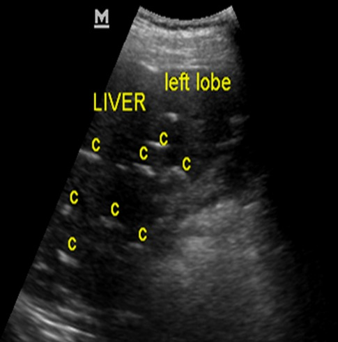
**Imaging Modality:** Abdominal Ultrasound (B-mode) **Anatomical Region:** Upper abdomen, specifically the left lobe of the liver. **Observed Pathology:** Multiple intrahepatic calcifications (labeled 'C'). **Characteristic Visual Features:** The image demonstrates a heterogeneous liver parenchyma containing numerous discrete, brightly hyperechoic foci. These lesions exhibit a linear or curvilinear morphology and are associated with distinct posterior acoustic shadowing, indicating a high-density, calcified composition. The distribution of these calcifications is multifocal and results in a visible distortion of the normal, fine-grained homogeneous echo texture of the hepatic architecture. No discrete cystic components or solid masses are visible in the immediate field of view. **Key Diagnostic Features:** - **Hyperechoic foci:** High-amplitude reflections consistent with mineralized deposits. - **Posterior acoustic shadowing:** Pathognomonic feature of calcification or gallstones, where the ultrasound beam is absorbed or reflected, creating a dark void deep to the lesion. - **Parenchymal distortion:** Evidence of chronic or extensive involvement affecting the organ's structural integrity. **Clinical Context:** These findings are consistent with sequelae of chronic infections (e.g., granulomatous disease), hepatolithiasis, or parasitic infestations such as calcified hydatid cysts or schistosomiasis.
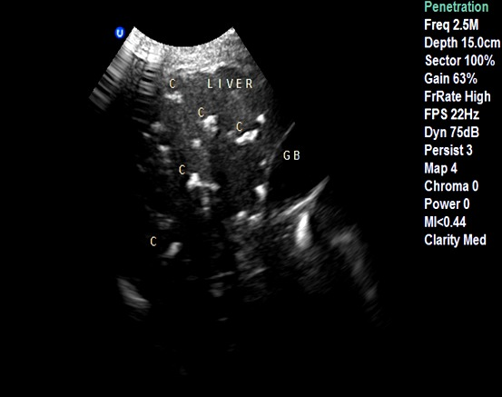
**Imaging Modality:** Abdominal Ultrasound (B-mode) **Anatomical Region:** Upper abdomen, specifically the liver parenchyma and adjacent gallbladder (labeled "GB"). **Observed Pathology:** Multiple intrahepatic calcifications. **Characteristic Visual Features:** The ultrasound image displays numerous, discrete, highly echogenic (bright) foci distributed throughout the hepatic parenchyma. These lesions exhibit classic posterior acoustic shadowing, characterized by dark vertical bands extending deep to the echogenic structures due to the attenuation of the sound beam. The distribution is multifocal and irregular, resulting in a distorted parenchymal echo pattern. The adjacent gallbladder is visible but partially obscured by the shadowing artifacts from overlying calcified structures. **Key Diagnostic Features:** - **Hyper-echogenicity:** Distinct white foci indicating mineralized or dense tissue. - **Posterior Acoustic Shadowing:** Confirms the solid, calcified nature of the lesions. - **Anatomical Distortion:** Visible alteration of the normal homogeneous liver texture by multiple shadowing calcifications. **Clinical Significance:** This pattern is characteristic of disseminated intrahepatic calcifications, which may be secondary to chronic granulomatous disease, healed infections (e.g., tuberculosis or histoplasmosis), or biliary calculi.
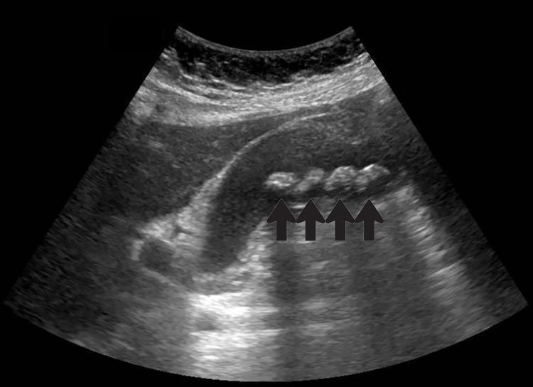
This diagnostic ultrasound image in the left lateral decubitus position demonstrates the gallbladder and adjacent liver parenchyma. The gallbladder lumen contains multiple curvilinear hyperechoic foci, indicated by four black arrows, consistent with cholelithiasis (gallstones). These stones exhibit distinct posterior acoustic shadowing, a hallmark sonographic sign of solid calcified structures. The gallbladder wall appears diffusely thickened, measuring approximately 6.3 mm, which is suggestive of cholecystitis. Trace amounts of anechoic pericholecystic fluid are visible, further indicating local inflammatory changes. The surrounding liver parenchyma displays a normal homogeneous echotexture. This image serves as a clinical example of symptomatic gallbladder disease, illustrating key diagnostic features used to identify acute cholecystitis and cholelithiasis during a transabdominal ultrasound examination.
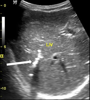
**Imaging Modality:** Abdominal Ultrasound (B-mode) **Anatomical Region:** Right lobe of the liver, specifically focusing on the intrahepatic biliary system. **Observed Pathology:** Intrahepatic biliary calcification. **Characteristic Visual Features:** The image demonstrates a focused area of linear and curvilinear hyperechoic structures within the intrahepatic ducts. These echogenic foci exhibit distinct posterior acoustic shadowing, a hallmark of dense calcification. The distribution is localized to the biliary tree of the right hepatic lobe. The surrounding hepatic parenchyma (labeled "LIV") appears relatively homogeneous but shows subtle coarse echotexture. **Clinical Context and Differential:** The morphology of the hyperechoic shadows is suggestive of calcified parasitic remains (such as *Ascaris lumbricoides* or liver flukes) within the biliary tract. This appearance is a key diagnostic feature for chronic or inactive biliary ascariasis, where dead worms undergo calcification. **Key Diagnostic Cues:** - Strong hyperechoic signal with clean posterior shadowing. - Tubular or curvilinear shape conforming to the ductal anatomy. - Localization within the intrahepatic ducts rather than the gallbladder.
Calcified Foci "In Continuation" in the Liver on Ultrasound
What Does "In Continuation" Mean?
Sonographic Appearance
- Highly echogenic (bright white) foci
- With posterior acoustic shadowing - dark bands deep to the lesion caused by sound beam attenuation
- This confirms solid, calcified (dense) composition
- The pattern helps distinguish them from gas (which shows dirty, reverberating shadowing and moves with respiration/position)
"US clearly demonstrates focal calcification, with increased reflectivity and a posterior acoustic corridor, but this feature alone does not always allow distinction from focal gas."
Differential Diagnosis - Organized by Pattern
1. Calcifications Along Portal Tracts (Most Important "In Continuation" Pattern)
Schistosomiasis (top differential for contiguous/linear periportal calcification)
- Schistosoma japonicum and S. mansoni produce calcified eggs deposited along portal tracts
- Imaging shows the classic "turtle-back" appearance - clusters of calcified eggs along portal triads
- Also produces "pipestem" (clay pipestem) fibrosis - periportal collagen deposition seen on USG as echodense circles/rings surrounding portal vessels
- The calcified foci follow the portal branching pattern - hence appearing "in continuation"
-
- Goldman-Cecil Medicine, Echinococcosis/Schistosomiasis chapter
Clonorchis / Opisthorchis (Liver Flukes)
- Calcified dead flukes within intrahepatic bile ducts
- Linear/curvilinear hyperechoic structures with shadowing following ductal anatomy
- Tubular shape conforming to biliary tree distribution
Ascariasis (biliary)
- Dead, calcified worms in intrahepatic ducts
- Long tubular echogenic structures with acoustic shadow
2. Scattered/Multifocal Calcifications (Granulomatous Disease)
| Cause | Key Feature |
|---|---|
| Tuberculosis | Healed caseating granulomas, often discrete foci; may affect liver + spleen simultaneously |
| Histoplasmosis | Granulomas healing with calcification, especially in endemic regions; liver + spleen + lungs |
| Sarcoidosis | Rare hepatic calcification from healed granulomas |
| Pneumocystis jirovecii (in immunocompromised) | Widespread multifocal "starfield" calcifications throughout liver and spleen |
"Similar to tuberculosis, healing of granulomas can result in calcification, especially in lymph nodes and the liver and spleen."
3. Calcification Within Hepatic Lesions
| Lesion | Pattern |
|---|---|
| Hydatid cyst (Echinococcus) | Peripheral/eggshell calcification; may be ring-like or curvilinear |
| Healed pyogenic abscess | Irregular dystrophic calcification within scar |
| Healed amoebic abscess | Post-inflammatory dystrophic calcification |
| Giant hemangioma | Phlebolith-type calcification |
| Hepatoblastoma / Fibrolamellar HCC | Primary tumor with focal calcification |
| Calcified metastases | Especially mucinous colorectal adenocarcinoma - often ill-defined; can be multiple and in continuity if along vascular routes |
"Benign parenchymal calcification may occur following focal insults such as tuberculosis, Pneumocystis infection, sarcoidosis, pyogenic abscess and parenchymal haematoma... Focal calcification also occurs within benign lesions (giant haemangioma) and malignant lesions, particularly mucin-secreting adenocarcinoma of the colon."
4. Vascular/Biliary Calcification
- Hepatic artery atherosclerosis - linear calcification following vessel course
- Portal vein thrombosis with calcification - rare, contiguous along portal vein
- Biliary calculi (hepatolithiasis) - calcified stones within intrahepatic ducts; arranged along biliary tree
Key Distinguishing Features on USG
| Feature | Calcification | Gas (Pneumobilia) |
|---|---|---|
| Echogenicity | Bright, clean | Bright, "dirty" |
| Posterior shadow | Clean acoustic shadow | Reverberant/dirty shadow |
| Movement | Fixed | Moves with gravity/respiration |
| Distribution | Variable | Follows bile ducts, non-dependent |
USG Images
Summary - Most Likely Diagnoses for "Calcified Foci in Continuation"
- Schistosomiasis - periportal calcification following portal tract branching ("turtle-back"/pipestem pattern) - the classic cause of contiguous calcified foci
- Intrahepatic biliary calculi / biliary parasites (ascaris, clonorchis) - calcifications following bile duct distribution
- Calcified granulomas (TB, histoplasmosis) - if multifocal but less organized
- Calcified metastases - if patient has history of mucinous colorectal carcinoma
- Grainger & Allison's Diagnostic Radiology, Liver Calcification section
- Goldman-Cecil Medicine, Schistosomiasis chapter
- Sleisenger & Fordtran's Gastrointestinal and Liver Disease, Pipestem Fibrosis section