
23 year old female having fungal infection at abdominal area and back of leg
tinea corporis fungal skin infection abdomen
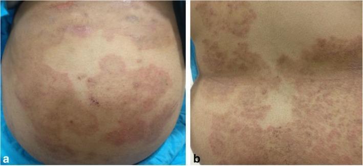
Two color clinical photographs (a and b) show extensive skin lesions on a patient's abdomen and back, characteristic of Tinea corporis. Panel (a) displays the abdominal region with large, well-defined erythematous (reddish) plaques. These plaques exhibit irregular, polycyclic margins and some central clearing, creating a mosaic-like pattern interspersed with normal-appearing skin. Panel (b) shows the back, where the lesions are more confluent and densely packed, presenting as diffuse erythematous areas with scaling and a plaque-like texture. The morphology is consistent with a dermatophyte infection, demonstrating classic fungal skin manifestations including circular or annular arrangements and progressive spread. These images are used in medical education to teach the clinical recognition of extensive fungal dermatosis and its potential systemic or ocular associations, such as retinochoroiditis in immunocompromised or non-compliant patients.
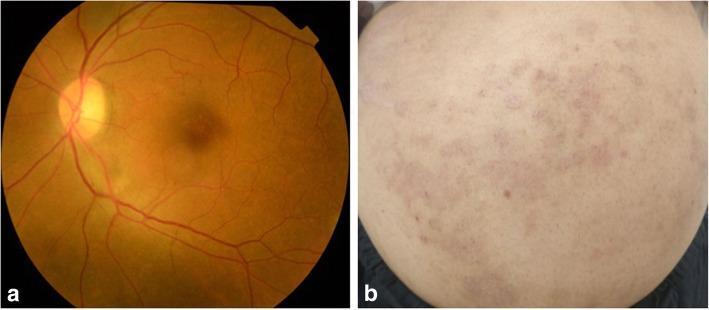
This composite clinical figure demonstrates the multisystemic resolution of a fungal infection (Tinea corporis) after two weeks of antifungal treatment. Image A is a fundus photograph of the left eye showing the posterior pole and mid-periphery. It reveals a resolving retinochoroiditis lesion, characterized by a diffuse, faint yellowish patch with irregular margins located along the inferotemporal vascular arcade. The optic disc, macula, and remaining vasculature appear stable. Image B is a clinical photograph of the abdomen showing multiple resolving skin lesions. These present as scattered, pinkish-red erythematous patches and plaques with less distinct borders compared to the acute phase, set against otherwise normal-appearing skin. The figure highlights the clinical correlation between ophthalmologic findings and dermatologic manifestations in systemic or co-occurring fungal disease, illustrating the typical visual progression of healing during therapeutic intervention.
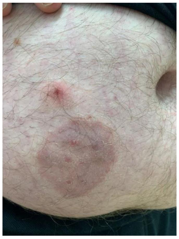
This clinical photograph displays a dermatological presentation of tinea corporis (zoonotic dermatophytosis) on the abdomen of an adult male. The primary feature is a well-circumscribed, annular, erythematous plaque approximately 4-5 cm in diameter. The lesion exhibits a classic 'ringworm' morphology with a slightly raised, scaly inflammatory border and a partially cleared central area containing subtle papular elements. Superior to the primary plaque is a secondary, smaller, raised erythematous papule with a central punctum or crust. The surrounding skin shows mild diffuse erythema and scattered smaller inflammatory macules. Hair follicles are visible throughout the affected and unaffected regions. This image serves as an educational example of fungal infection (likely Trichophyton mentagrophytes) transmitted from a pet (guinea pig) to a human host, emphasizing the visual diagnostic features of annular inflammatory skin lesions.
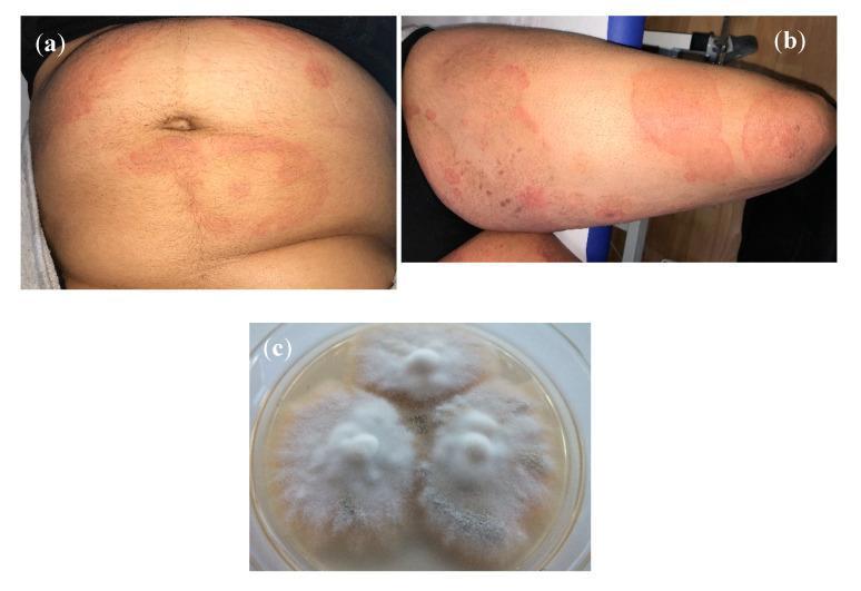
This composite figure provides a clinical and laboratory overview of a dermatophytosis infection caused by Trichophyton mentagrophytes Type VIII. Images (a) and (b) are clinical photographs displaying classic signs of tinea corporis. Photograph (a) shows the abdomen with multiple erythematous, annular (ring-shaped) lesions featuring raised, active red borders and central clearing. Photograph (b) depicts large, coalescing, arcuate plaques on the thigh with peripheral scaling and varying degrees of erythema. Image (c) is a diagnostic microbiology photograph showing a subculture of the isolate on Sabouraud's dextrose agar. The fungal colonies are fast-growing, characterized by a white, granular texture with flat, radiating margins. This visual set demonstrates the correlation between dermatological manifestations and mycological identification for an emerging anthropophilic fungal variant. The content is suitable for medical education regarding fungal skin infections, diagnostic dermatology, and medical mycology.
tinea corporis treatment antifungal
Fungal Skin Infection - 23-Year-Old Female (Abdomen & Back of Leg)
Most Likely Diagnosis: Tinea Corporis (Ringworm)
Classic Features to Look For
- Annular (ring-shaped) lesions with central clearing and a raised, scaly, erythematous border
- Pruritus (itching) and burning sensation
- Lesions spread centrifugally (outward) from the point of skin invasion
- Can be arcuate, concentric, or oval in shape
- Pustules within the active border are a strong clue for tinea
Diagnosis
- Scrape the leading (active) edge of the lesion onto a slide
- Add 2 drops of KOH solution, place coverslip
- Examine under microscope for branching hyphae and arthroconidia
- Sensitivity 77-88%, specificity 62-95%
Common Causes / Risk Factors
| Route | Details |
|---|---|
| Person-to-person | Including autoinoculation from tinea pedis or capitis |
| Animal-to-human | Household pets (cats, dogs), farm animals |
| Fomites | Contaminated towels, clothing, furniture |
| Environment | Gymnasiums, locker rooms, swimming pools |
Treatment
1. Topical Antifungals (First-Line for Localized Infection)
| Drug Class | Examples | Regimen |
|---|---|---|
| Allylamines (preferred) | Terbinafine 1% cream | Once daily x 7-14 days |
| Azoles | Clotrimazole, Miconazole, Ketoconazole | Twice daily x 2-4 weeks |
| Azoles | Econazole | Once or twice daily x 2-4 weeks |
Key evidence: Terbinafine 1% cream achieved mycologic cure of 84.2% vs 23.3% with placebo. Allylamines (terbinafine) show greater effectiveness than azoles (clotrimazole) for tinea corporis. (Textbook of Family Medicine 9e)
2. Oral Antifungals (Systemic)
- Infection covers large areas of skin (as in this case with both abdomen and legs)
- Lesions are recalcitrant to topical treatment
- Involves hairy skin areas
| Drug | Dose | Duration |
|---|---|---|
| Terbinafine (preferred) | 250 mg once daily | 2-4 weeks |
| Itraconazole | 100-200 mg daily | 2-4 weeks |
| Griseofulvin | 500 mg daily | 4-6 weeks (less preferred) |
Important 2025 update: A systematic review (PMID 40704771) flags emerging terbinafine-resistant T. indotineae as a serious concern, particularly in South Asia. If standard treatment fails, consider itraconazole or voriconazole with susceptibility testing.
Hygiene & Prevention Measures
- Avoid sharing towels, clothing, bedsheets with others
- Keep the affected skin clean and dry - moisture promotes fungal growth
- Wear loose, breathable (cotton) clothing, especially in the abdominal area
- Wash clothing at high temperature
- Avoid scratching to prevent autoinoculation to other body sites
- Check and treat household pets if animal transmission is suspected
- Inform close contacts to monitor for similar lesions
Important Considerations for This Patient
- Extent matters - Since infection involves two distinct body areas (abdomen AND back of leg), this may warrant oral antifungal therapy in addition to topical treatment.
- Rule out tinea incognito - If she has previously used topical steroids on the rash, the classic ring appearance may be absent (borders become blurred, more diffuse).
- Blood sugar check - Recurrent or extensive fungal infections in a young woman warrant ruling out underlying diabetes mellitus.
- Immunosuppression - If infections are extensive or recurrent, check for immunodeficiency.
Disclaimer: This information is educational only. The patient should be evaluated by a dermatologist or physician for an accurate diagnosis and individualized treatment plan.