
Skin examination For RNVV
primary skin lesions macule papule plaque vesicle pustule diagram
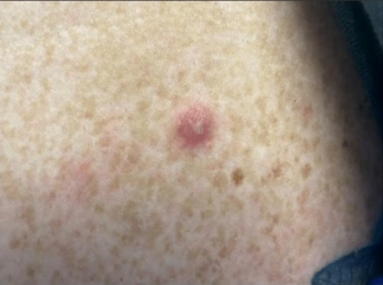
Clinical photograph of a characteristic vesiculopustular skin lesion associated with disseminated gonococcal infection (DGI). The primary lesion is a solitary, erythematous papule, approximately 3-5 mm in diameter, with a centralized greyish-white pustule/vesicle and a dusky, necrotic appearance at the core. The surrounding skin shows generalized solar lentigines (freckling) but no acute inflammatory changes. This visual finding is a hallmark of the arthritis-dermatitis syndrome seen in Neisseria gonorrhoeae dissemination, typically presenting as sparse, painless, non-pruritic lesions on distal extremities. This image serves as an educational reference for identifying cutaneous manifestations of systemic sexually transmitted infections in a clinical setting.
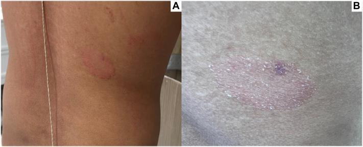
Two-panel clinical photograph displaying primary skin lesions on the mid-right back, characteristic of pityriasis rosea. Panel A shows a 2.5-cm, well-demarcated, annular red plaque on the torso, along with a secondary, smaller, ill-defined erythematous papule located superiorly. Panel B provides a high-detail close-up of the primary plaque, illustrating its morphology: it features a raised, erythematous border and a slightly depressed center with fine, surface scaling. A focal hyperpigmented macule is visible within the center of the lesion, marking the site of a prior punch biopsy. These visual findings—specifically the larger herald-style plaque with centrifugal scaling—are key clinical indicators of spongiotic dermatitis and help differentiate it from other papulosquamous disorders such as tinea corporis or nummular eczema.
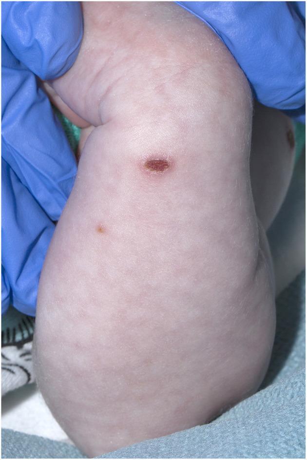
This clinical photograph displays skin lesions on the arm of a 21-day-old neonate, characteristic of the healing phase of varicella-zoster virus (VZV) infection. The primary finding is a centrally located, oval, crusted lesion showing a thick, dark-reddish-brown scab, indicating a dried vesicle or pustule. A second, smaller, and flatter brownish papule is visible proximally. The surrounding skin exhibits 'cutis marmorata,' a reticulated or mottled vascular pattern commonly seen in neonates due to vasomotor instability, but also noted here in the clinical context of neonatal varicella. No significant surrounding erythema or edema is observed at this stage of the lesions. This image serves as a pedagogical example of neonatal dermatology and the clinical progression of viral exanthems from vesicular to crusted stages. The presence of blue medical gloves indicates a clinical examination setting.
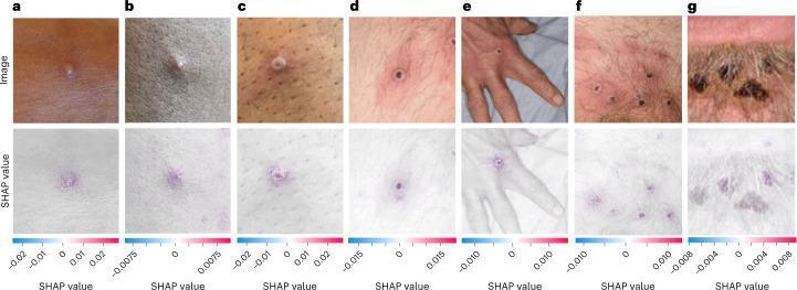
A clinical comparison chart and diagnostic visualization illustrating the progression of mpox (MPXV) skin lesions. The top row features seven photographic images (a-g) documenting the dermatological evolution: an early-stage vesicle (a), a small pustule (b), an umbilicated pustule with a central depression (c), a papule with central necrosis (d), an ulcerated lesion on a hand (e), multiple ulcerated lesions in a pubic region (f), and late-stage crusted plaques (g). The lesions exhibit characteristic features of orthopoxvirus infection, including perilesional erythema and central umbilication. The bottom row displays a corresponding SHAP (SHapley Additive exPlanations) analysis overlaid on each image. Positive SHAP values (indicated in red/purple) represent discriminative regions—specifically the primary lesions and surrounding inflammatory areas—used by a convolutional neural network (CNN) for diagnostic classification. Negative SHAP values (in blue) highlight healthy skin areas that detract from the viral prediction. This image serves as a clinical reference for disease staging and AI-assisted diagnostic transparency in infectious disease management.
Skin Examination - RNVV
1. Ideal Conditions
- Bright, natural or well-directed artificial light - subtle findings are missed in poor lighting
- Patient fully undressed (gown + draping for modesty)
- Systematic head-to-toe approach - uncover one area at a time
- Additional tools: magnifier/dermatoscope, Wood lamp (365 nm), penlight for mucosal sites
"Without good lighting, subtle but important details may be missed." - Fitzpatrick's Dermatology
2. Technique of Examination
| Step | Action |
|---|---|
| 1. General impression | Observe from distance - asymmetry, cachexia, jaundice, body habitus |
| 2. Systematic inspection | Head to toe, uncovering one area at a time |
| 3. Lighting adjustment | Side-lighting to reveal border depth and surface texture |
| 4. Palpation | Determine soft/firm/tender/fluid-filled character |
| 5. Dermoscopy | Especially for pigmented lesions - identify concerning features |
| 6. Mucosal sites | Penlight/flashlight for oral, genital mucosa |
| 7. Documentation | Body map or descriptive record; photography for suspicious lesions |
3. Morphology - Reading the Skin
PRIMARY LESIONS
Flat (Non-palpable)
| Lesion | Size | Description |
|---|---|---|
| Macule | < 1 cm (Fitzpatrick) / < 2 cm (Harrison's) | Flat color change only; not palpable (e.g., freckle, petechiae) |
| Patch | ≥ 1 cm (Fitzpatrick) / > 2 cm (Harrison's) | Flat color change, differs from macule only in size |
Raised (Palpable) - Solid
| Lesion | Size | Description |
|---|---|---|
| Papule | < 1 cm | Elevated or depressed, solid; may be sessile, pedunculated, dome-shaped, flat-topped, filiform, umbilicated |
| Plaque | ≥ 1 cm | Large flat-topped raised lesion; edges distinct (psoriasis) or indistinct (eczema) |
| Nodule | ≥ 1 cm | Firm, raised; solid or fluid (e.g., large dermal nevus) |
| Tumor | > 5 cm | Solid raised growth |
Raised (Palpable) - Fluid-filled
| Lesion | Size | Contents |
|---|---|---|
| Vesicle | < 1 cm | Serum, blood, or lymph - translucent (e.g., contact dermatitis) |
| Bulla | ≥ 1 cm | Same as vesicle, larger (e.g., bullous pemphigoid) |
| Pustule | < 1 cm | Pus (leukocytes) - note: does NOT necessarily indicate infection |
| Wheal | Variable | Erythematous, edematous; short-lived vasodilation/permeability (urticaria) |
Depressed
| Lesion | Description |
|---|---|
| Erosion | Loss of epidermis only; no dermal loss |
| Ulceration | Loss of epidermis AND at least part of dermis |
"Flat-topped or planar papules tend to be processes affecting the epidermis and superficial dermis, while dome-shaped/nodular lesions often exhibit deeper infiltration." - Fitzpatrick's
SECONDARY LESIONS (Epidermal Changes)
| Lesion | Description |
|---|---|
| Scale | Excessive accumulation of stratum corneum |
| Crust | Dried exudate; serous (yellow) or hemorrhagic (red) |
| Lichenification | Thickened skin with accentuated skin fold markings (chronic rubbing) |
| Fissure | Linear crack; occurs where stratum corneum is thick and least expandable (palms/soles) |
| Excoriation | Linear, angular erosions from scratching; may be crusted |
| Erosion | Loss of epidermis |
| Ulcer | Full-thickness epidermal + dermal loss |
| Atrophy | Depression with intact epidermis (dermal/subcutaneous loss) OR shiny/wrinkled skin (epidermal atrophy) |
| Scar | Secondary to trauma/inflammation; erythematous, hypo/hyperpigmented; destroys hair follicles |
| Telangiectasia | Dilated superficial blood vessel |
4. Color Assessment
| Color | Cause | Example |
|---|---|---|
| Brown | Melanin (epidermal or dermal), hemosiderin, amyloid | Nevus, melanoma, seborrheic keratosis |
| Gray-blue | Dermal melanin - Tyndall effect (longer wavelengths transmitted through dermis) | Blue nevus, Mongolian spot |
| Red/Erythema | Vascular dilation, inflammation | Cellulitis, rosacea |
| White | Melanin absence, scar, fibrosis | Vitiligo, scar |
| Yellow | Carotenoids, bilirubin, lipid | Jaundice, xanthoma |
| Purple/Violaceous | Hemorrhage (non-blanching), lichen planus | Purpura, lichen planus |
5. Shape & Configuration
| Term | Description | Example |
|---|---|---|
| Annular | Ring-shaped; paler center | Tinea corporis, granuloma annulare |
| Targetoid | 3 zones - dark center, pale middle, dark rim | Erythema multiforme |
| Atypical target | 2 zones - dark center, pale rim | Stevens-Johnson syndrome |
| Stellate | Star-shaped, multiple angulated edges | Calciphylaxis |
| Serpiginous | Snake-like, wandering | Cutaneous larva migrans |
| Whorled | Marble-cake pattern (2 colors interleaved) | Incontinentia pigmenti, hypomelanosis of Ito |
| Reticular | Net-like | Livedo racemosa |
6. Configuration of Multiple Lesions
| Term | Description |
|---|---|
| Dermatomal/Zosteriform | Unilateral, in dermatomal distribution |
| Grouped/Herpetiform | Clustered together (herpes simplex vesicles) |
| Scattered | Sparse, irregular distribution |
| Polycyclic | Coalescing circles or incomplete rings (urticaria, subacute cutaneous lupus) |
| Linear | Along lines of Blaschko or other lines |
7. Special Features to Note on Palpation
- Soft/firm/fluctuant - differentiates cyst vs lipoma vs abscess
- Tender - inflammatory vs non-inflammatory
- Warmth - infection, inflammation
- Mobility - fixed to deep structures vs freely mobile
- Skin turgor - pinch skin (forearm/abdominal); normal = immediate recoil; tenting = dehydration/decreased elasticity
8. Reaction Patterns (Diagnostic Categories)
| Reaction Pattern | Key Morphology | Common Diagnoses |
|---|---|---|
| Papulosquamous | Circumscribed papule/plaque + scale | Psoriasis, lichen planus, tinea |
| Eczematous | Vesicles, oozing, crusting, lichenification | Atopic/contact dermatitis |
| Vesiculobullous | Blisters | Pemphigus, bullous pemphigoid, herpes |
| Pustular | Pustules | Folliculitis, impetigo, acne |
| Vascular/Purpuric | Purpura, petechiae, non-blanching | Vasculitis, DIC |
| Nodular/Tumoral | Nodules, tumors | BCC, SCC, lymphoma |
9. Documentation
- Document type of lesions and locations - descriptively or on a body map
- Photograph suspicious lesions with triangulation from anatomic landmarks (for biopsy localization)
Classify according to primary secondary and special type
skin lesion classification chart primary secondary special types diagram
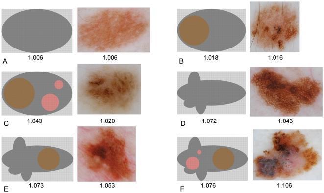
A comparison chart illustrating the classification of pigmented skin lesions through schematic representations and clinical dermoscopic photographs. The figure is organized into six pairs (A-F), demonstrating a progression of morphological irregularity. On the left of each pair, a gray schematic diagram represents the lesion's lacunarity and geometric properties; on the right, a corresponding clinical dermoscopic image shows real-world examples. The progression moves from (A) 'No irregularity'—a smooth oval with homogenous pigmentation—to (B) simple internal asymmetry, (C) hierarchical internal asymmetry with multiple colored internal regions, and (D-F) increasing boundary asymmetry with lobulated borders and complex internal architectural patterns. Key dermoscopic features visible in the clinical images include reticular networks, globules, and varied pigmentation ranging from light tan to dark brown and reddish-purple. The figure serves as a diagnostic educational tool for identifying risk factors in melanocytic lesions, highlighting how mathematical lacunarity values correlate with increasing clinical and structural complexity.
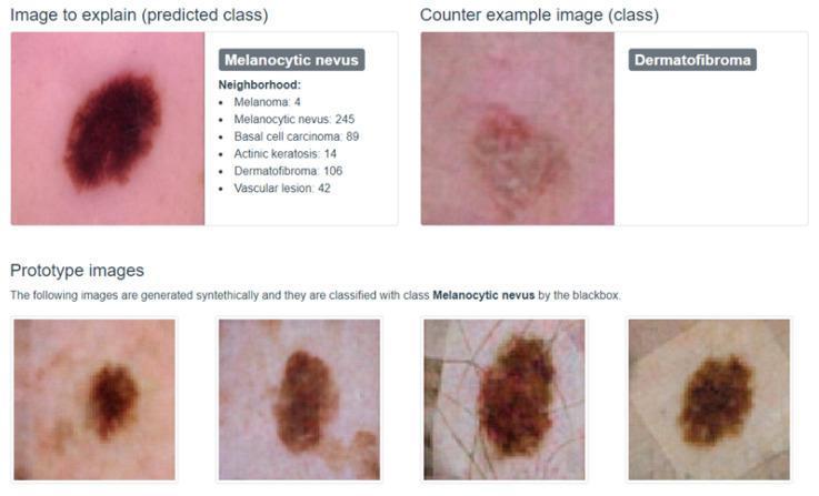
This clinical dermatological comparison chart showcases visual differentiation between types of skin lesions as used in machine learning classification models. The upper section features a 'Clinical Photograph' of a melanocytic nevus (the primary case), appearing as a dark brown, well-circumscribed, oval macule with uniform pigmentation and smooth texture. It is positioned adjacent to a 'Counter example image' of a dermatofibroma, which displays a lighter, reddish-brown hue, less distinct borders, and a subtle central firm or dimpled texture characteristic of this benign fibrous tumor. The lower section presents four 'Prototype images' (exemplars) of melanocytic nevi, demonstrating natural variability in lesion morphology. These variations include differences in color intensity (ranging from tan to deep brown), border regularity, and the visibility of surrounding skin markings such as fine lines and hair follicles. This visual tool is designed for medical education and AI model interpretability, helping to distinguish between benign melanocytic proliferations and non-melanocytic skin lesions like dermatofibromas based on morphology, color distribution, and border characteristics.
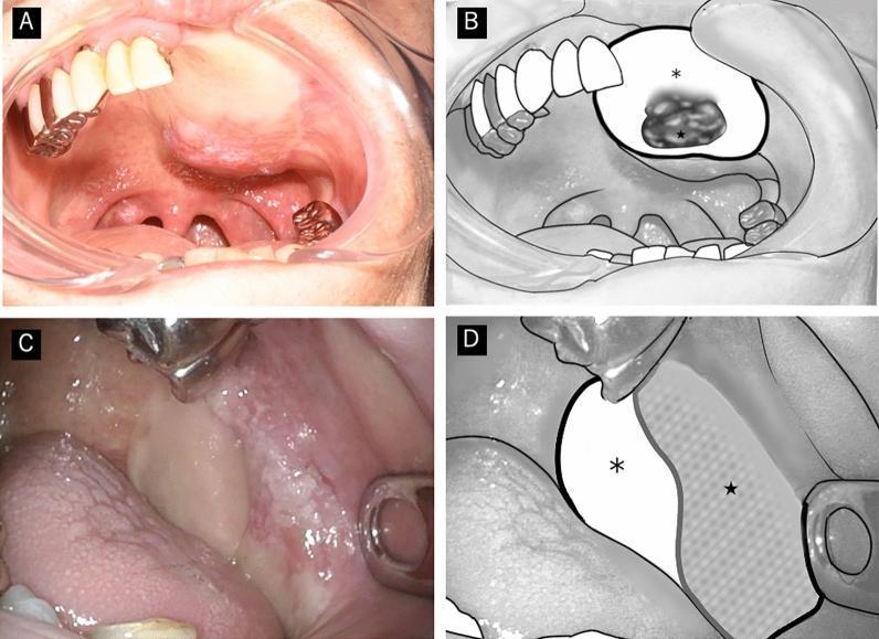
This composite educational image illustrates macroscopic findings and schematic representations of secondary neoplasms arising on reconstructive flap skin paddles in the oral cavity. Panels A and B present a clinical photograph and corresponding diagram of a flap neoplasm located centrally on the skin paddle. The lesion is characterized by focal erythema and a protruding, partially elevated morphology. Panels C and D show a clinical photograph and diagram of a similar lesion that demonstrates erythema and partial elevation extending toward the skin-mucosa junction, representing a peripheral or junctional presentation. These images demonstrate the typical macroscopic features of 'flap cancers,' which in these clinical cases were histologically confirmed as well-differentiated squamous cell carcinomas. The visual focus is on the distinguishing features of secondary primary neoplasms arising in surgical flaps, highlighting the importance of monitoring skin paddles for new lesions such as erythema, papillary projections, or non-ulcerative protrusions.
Classification of Skin Lesions
A. PRIMARY LESIONS
Lesions that arise de novo from the disease process itself - the "nouns" of dermatologic description.
I. Flat (Non-palpable) - Color change only, no elevation
| Lesion | Size | Key Feature | Example |
|---|---|---|---|
| Macule | < 1 cm | Flat, color change only; not palpable | Freckle, petechiae, café-au-lait |
| Patch | ≥ 1 cm | Flat; differs from macule only in size | Vitiligo, Mongolian spot, port wine stain |
II. Raised (Palpable) - Solid contents
| Lesion | Size | Topography | Example |
|---|---|---|---|
| Papule | < 1 cm | Elevated or depressed; solid or cystic | Acne comedone, molluscum, wart |
| Plaque | ≥ 1 cm | Flat-topped, raised; distinct or indistinct edges | Psoriasis, lichen planus |
| Nodule | ≥ 1 cm | Raised; firm; solid or fluid-filled | Dermatofibroma, lipoma |
| Tumor | > 5 cm | Solid, raised growth | SCC, BCC |
Papule subtypes by surface: sessile, pedunculated, dome-shaped, flat-topped, filiform, mammillated, acuminate (conical), umbilicated
III. Raised (Palpable) - Fluid-filled
| Lesion | Size | Contents | Example |
|---|---|---|---|
| Vesicle | < 1 cm | Serum, blood, or lymph; translucent | Herpes simplex, contact dermatitis, chickenpox |
| Bulla | ≥ 1 cm | Same as vesicle, larger | Bullous pemphigoid, pemphigus vulgaris |
| Pustule | < 1 cm | Pus (leukocytes ± debris); may be sterile | Acne, folliculitis, impetigo |
| Wheal | Variable | Edematous papule/plaque; short-lived vasodilation | Urticaria, insect bite |
Vesicle/bulla cleavage level: intraepidermal (flaccid, e.g., pemphigus) vs subepidermal (tense, e.g., bullous pemphigoid)
IV. Depressed (Below skin surface)
| Lesion | Contents | Key distinction | Example |
|---|---|---|---|
| Erosion | N/A | Loss of epidermis only; heals without scarring | Ruptured vesicle, aphthous ulcer |
| Ulceration | N/A | Loss of epidermis + at least part of dermis; heals with scarring | Venous ulcer, pressure sore |
Note: Erosion and ulceration can be primary or secondary lesions.
B. SECONDARY LESIONS (Epidermal / Surface Changes)
Develop as the disease evolves or result from scratching, rubbing, infection, or treatment. Overlaid on primary lesions.
| Lesion | Definition | Example / Clinical Correlation |
|---|---|---|
| Scale | Excess accumulation of stratum corneum (macroscopic dead skin flakes) | Psoriasis (silvery), pityriasis rosea, tinea versicolor |
| Crust | Dried exudate (serum = yellow/honey-colored; blood = red/hemorrhagic) | Impetigo (honey crust), herpes (crusted vesicle) |
| Lichenification | Thickened skin with accentuated skin-fold markings from chronic rubbing | Chronic atopic dermatitis, lichen simplex chronicus |
| Fissure | Linear crack through epidermis ± dermis; occurs where stratum corneum is thick and least expandable | Cheilitis, palmar/plantar eczema |
| Excoriation | Linear, angular erosions from scratching; may be crusted | Atopic dermatitis, neurotic excoriation |
| Scar | Permanent fibrous replacement after trauma/inflammation; erythematous → hypopigmented over time; destroys hair follicles | Post-surgical, healed burn |
| Keloid | Scar extending beyond original wound margins | Keloid on chest/ear lobe |
| Atrophy | Acquired loss of tissue - dermal/subcutaneous (depression + intact epidermis) OR epidermal (shiny, wrinkled, cigarette-paper skin) | Striae, topical steroid atrophy, lichen sclerosus |
| Eschar | Specialized crust - dry, dark red-purple/brown/black; signals skin necrosis | Calciphylaxis, necrotizing fasciitis, anthrax |
Scale Subtypes (Fitzpatrick's Table 1-4)
| Scale Type | Description |
|---|---|
| Craquelé/xerotic | Dried cracked appearance like a dry riverbed |
| Cutaneous horn | Conical compact stratum corneum projection |
| Exfoliative/desquamative | Splits off in sheets or fine flakes |
| Follicular | Keratotic plugs or spines |
| Lamellar | Large, plate-like |
| Micaceous | Silvery, mica-like (psoriasis) |
| Ostraceous | Oyster-shell-like (psoriasis) |
| Pityriasiform | Fine, bran-like (pityriasis) |
C. SPECIAL / TERTIARY LESIONS
Lesions that do not fit neatly into primary or secondary categories - they are unique structural findings with specific diagnostic significance.
| Lesion | Definition | Significance / Example |
|---|---|---|
| Comedo | Plug of desquamated epithelium and sebum in a hair follicle; open (blackhead) or closed (whitehead) | Acne vulgaris |
| Milia | Small (1-2 mm), firm, white/yellow inclusion cysts; trapped keratin | Neonatal milia, post-blister milia |
| Burrow | Threadlike, serpiginous tunnel in the stratum corneum made by a parasite | Pathognomonic of scabies (Sarcoptes scabiei) |
| Telangiectasia | Permanently dilated superficial dermal blood vessel | Rosacea, hereditary hemorrhagic telangiectasia, ataxia-telangiectasia, systemic sclerosis |
| Petechiae | Pinpoint (< 3 mm) non-blanching red-purple macule from capillary hemorrhage | Thrombocytopenia, meningococcemia, vasculitis |
| Purpura | Larger non-blanching hemorrhage (3 mm - 1 cm) | Vasculitis, scurvy, DIC |
| Ecchymosis | Large (> 1 cm) non-blanching bruise/extravasated blood | Trauma, coagulopathy |
| Vegetation | Irregular, rough, warty or cauliflower-like surface growth | Condylomata acuminata, verrucous carcinoma |
| Abscess | Localized collection of pus within the dermis/subcutis | Furuncle, carbuncle, hidradenitis suppurativa |
| Cyst | Epithelium-lined cavity containing fluid, semi-solid, or solid matter | Epidermal inclusion cyst, pilar cyst |
| Sinus | Channel/tract communicating with skin surface | Pilonidal sinus, hidradenitis |
| Poikiloderma | Triad: atrophy + telangiectasia + pigmentary change (hyper + hypo) | Dermatomyositis, mycosis fungoides, chronic radiation dermatitis |
| Xeroderma | Pathological skin dryness with fine scaling | Ichthyosis, hypothyroidism |
Summary Chart
SKIN LESIONS
│
├── PRIMARY (Arise de novo)
│   ├── Flat (Non-palpable)
│   │   ├── Macule (< 1 cm)
│   │   └── Patch (≥ 1 cm)
│   ├── Raised - Solid
│   │   ├── Papule (< 1 cm)
│   │   ├── Plaque (≥ 1 cm)
│   │   ├── Nodule (≥ 1 cm)
│   │   └── Tumor (> 5 cm)
│   ├── Raised - Fluid
│   │   ├── Vesicle (< 1 cm, clear)
│   │   ├── Bulla (≥ 1 cm, clear)
│   │   ├── Pustule (pus)
│   │   └── Wheal (edema)
│   └── Depressed
│       ├── Erosion (epidermis only)
│       └── Ulceration (epidermis + dermis)
│
├── SECONDARY (Evolve from primary)
│   ├── Scale (excess stratum corneum)
│   ├── Crust (dried exudate)
│   ├── Lichenification (chronic rubbing)
│   ├── Fissure (linear crack)
│   ├── Excoriation (scratch mark)
│   ├── Scar / Keloid
│   ├── Atrophy (epidermal or dermal)
│   └── Eschar (necrotic crust)
│
└── SPECIAL (Unique structural / vascular)
    ├── Comedo, Milia, Cyst
    ├── Burrow (scabies)
    ├── Telangiectasia
    ├── Petechiae / Purpura / Ecchymosis
    ├── Vegetation / Abscess / Sinus
    └── Poikiloderma