
Layer of scaple
Reading File
Finding Sources
Searching the Web
layers of scalp anatomy SCALP mnemonic clinical significance
Finding Sources
Reading File
Reading File
Loading Image
Here is a complete breakdown of the layers of the SCALP, sourced from Gray's Anatomy for Students:
Layers of the Scalp - Mnemonic: SCALP
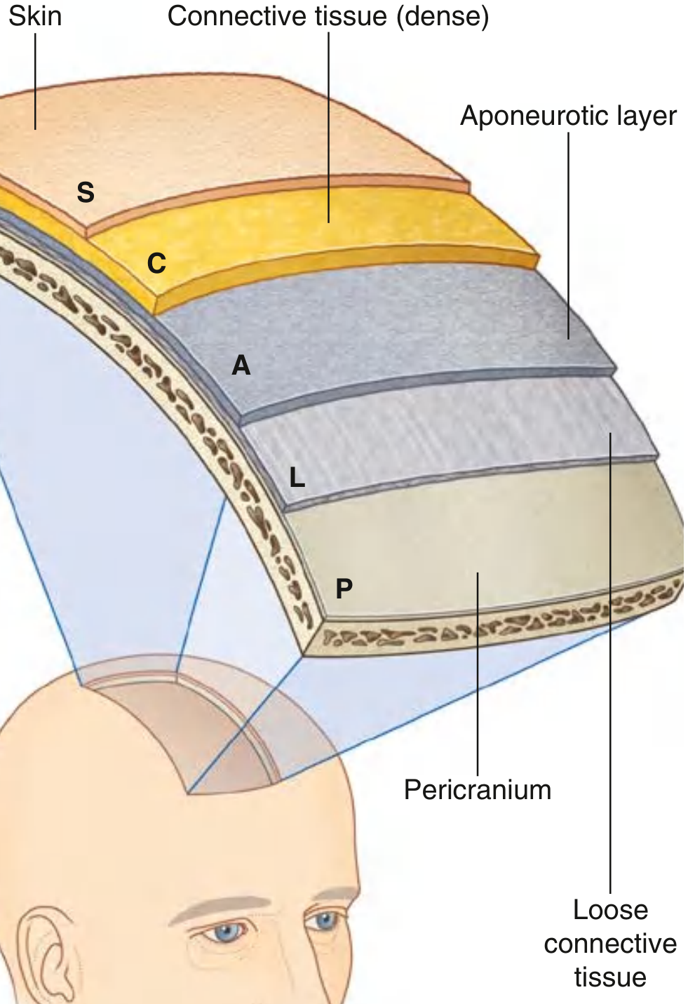
S - Skin
- The outermost layer, structurally similar to skin elsewhere on the body.
- Contains hair follicles, sebaceous glands, and sweat glands.
- Hair is present over most of its surface.
C - Connective Tissue (Dense)
- Deep to the skin; anchors the skin firmly to the layer below.
- Contains arteries, veins, and nerves supplying the scalp.
- Clinically important: when the scalp is cut, the dense connective tissue holds cut vessels open, preventing them from contracting - this causes profuse bleeding from even minor scalp wounds.
A - Aponeurotic Layer (Galea Aponeurotica / Epicranial Aponeurosis)
- The deepest of the top three "firmly attached" layers.
- Consists of the occipitofrontalis muscle - with a frontal belly anteriorly and an occipital belly posteriorly - connected by the epicranial aponeurosis (galea aponeurotica).
- The first three layers (S, C, A) are tightly bound together as a single unit, called the "scalp proper" - the part avulsed in traumatic scalping injuries.
L - Loose Connective Tissue
- Separates the aponeurotic layer from the pericranium.
- Allows the scalp proper to move freely over the skull (why you can wrinkle your forehead).
- Called the "danger zone" of the scalp: infections here spread easily through this plane and can track intracranially via emissary veins (valveless veins connecting extracranial to intracranial dural venous sinuses), potentially causing meningitis or cavernous sinus thrombosis.
- Hematomas in this layer ("subaponeurotic hematoma") spread widely across the scalp.
P - Pericranium
- The deepest layer; the periosteum of the outer surface of the calvaria.
- Firmly attached to skull bones but loosely attached at suture lines.
- Subperiosteal hematomas (cephalohematomas in neonates) are limited by suture lines because the pericranium is tightly adherent there.
Key Clinical Points
| Layer | Clinical Relevance |
|---|---|
| Dense connective tissue (C) | Profuse bleeding from scalp cuts (vessels can't retract) |
| Loose connective tissue (L) | "Danger zone" - spread of infection intracranially; wide hematoma spread |
| Pericranium (P) | Cephalohematoma limited by suture lines |
Gray's Anatomy for Students, Chapter 8 (Head)
Below scalpe layers
Finding Sources
Searching the Web
layers below scalp skull meninges anatomy from superficial to deep
Finding Sources
Finding Sources
Reading File
Reading File
Loading Image
Finding Sources
Here is the complete picture of everything below the scalp, from superficial to deep - all the way down to the brain:
Layers Below the Scalp (Superficial to Deep)
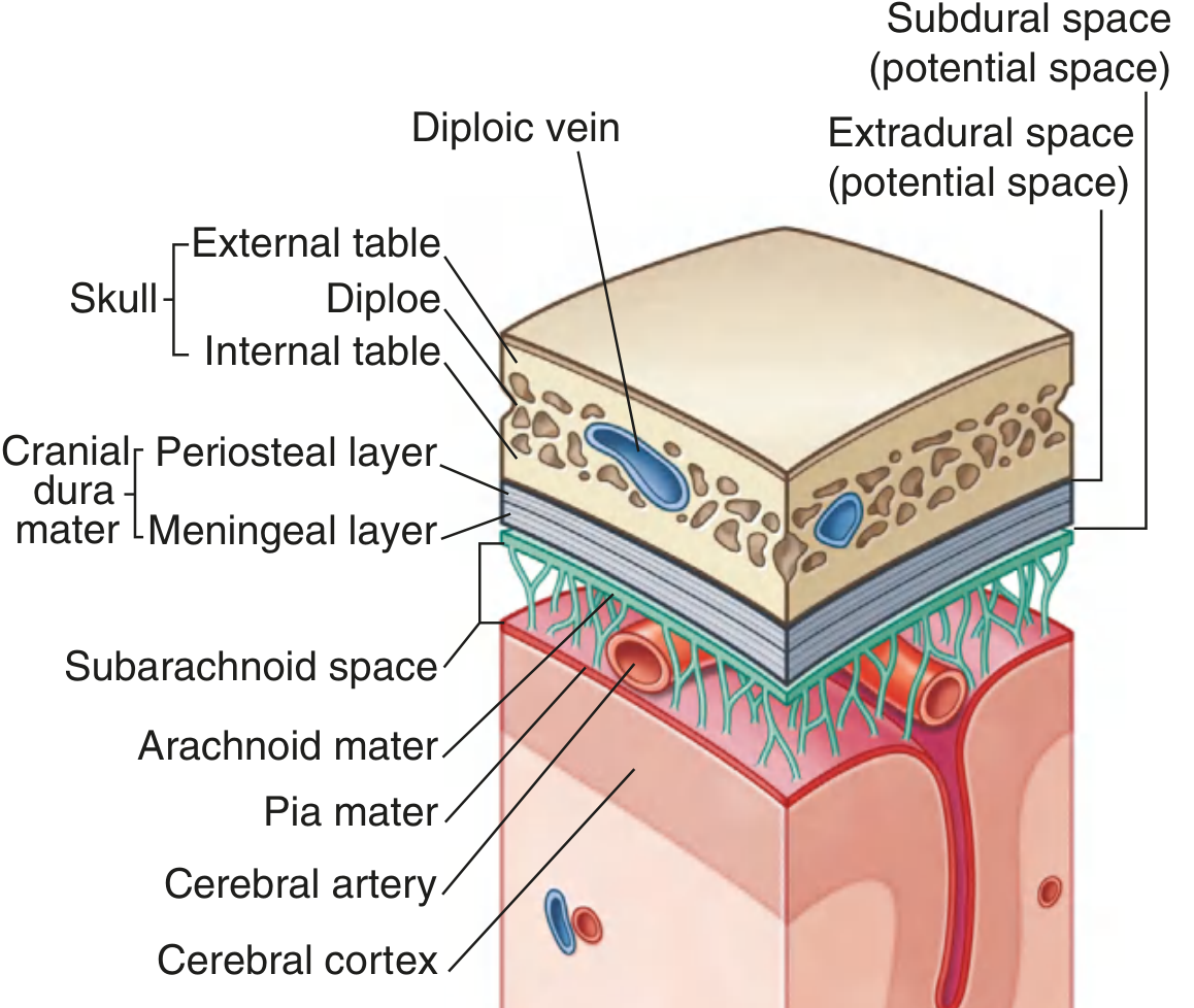
1. Skull (Calvaria) - 3 Sub-layers
The skull bones consist of three distinct layers (like a sandwich):
| Sub-layer | Description |
|---|---|
| Outer table | Dense compact (cortical) bone |
| Diploe | Cancellous (spongy) bone containing diploic veins and red marrow |
| Inner table | Dense compact bone (thinner and more brittle than outer table) |
The inner table fractures more easily than the outer table - this is why intracranial injury can occur even without obvious outer scalp damage.
2. Cranial Dura Mater - 2 Sub-layers
The dura is the outermost, thickest, and toughest meningeal covering. The cranial dura (unlike spinal dura) has two layers:
| Sub-layer | Description |
|---|---|
| Periosteal layer (outer) | Firmly fused to the inner surface of the skull; acts as the inner periosteum of the cranial cavity; contains meningeal arteries |
| Meningeal layer (inner) | Closely applied to arachnoid mater; continues through foramen magnum as spinal dura |
Between the skull and dura = Extradural (epidural) space - a potential space. In epidural haemorrhage, blood (typically from rupture of the middle meningeal artery) accumulates here, stripping the dura from the skull.
Between the meningeal dura and arachnoid = Subdural space - another potential space. In subdural haemorrhage, blood collects here (typically from tearing of bridging veins).
The two dural layers separate to form:
- Dural folds/partitions - falx cerebri, tentorium cerebelli, falx cerebelli, diaphragma sellae
- Dural venous sinuses - superior sagittal sinus, transverse sinus, etc.
3. Arachnoid Mater
- A thin, delicate, avascular membrane applied to the inner surface of the dura.
- Spider-web-like trabeculae extend from it down to the pia mater.
- No blood vessels in the arachnoid itself.
- Between arachnoid and pia = Subarachnoid space, filled with cerebrospinal fluid (CSF) and containing cerebral arteries and veins.
4. Pia Mater
- The innermost, most delicate meningeal layer.
- Closely adherent to the surface of the brain, following every gyrus and sulcus.
- Highly vascular - carries blood vessels that dip into the brain substance.
- Cannot be separated from the brain surface without causing damage.
5. Brain (Cerebral Cortex)
The outermost layer of the brain proper, consisting of grey matter (neuronal cell bodies).
Complete Sequence: Scalp to Brain
SCALP:
  S - Skin
  C - Connective tissue (dense)
  A - Aponeurosis (galea)
  L - Loose areolar tissue  ← "danger zone"
  P - Pericranium (outer periosteum of skull)

SKULL:
  - Outer table
  - Diploe (with diploic veins)
  - Inner table

MENINGES:
  - Extradural space (potential)
  - Dura mater:  Periosteal layer + Meningeal layer
  - Subdural space (potential)
  - Arachnoid mater
  - Subarachnoid space (contains CSF)
  - Pia mater

BRAIN:
  - Cerebral cortex (grey matter)
Clinical Correlation: Haematomas at Each Level
| Space | Haematoma | Cause |
|---|---|---|
| Subgaleal (loose areolar layer) | Subgaleal/subaponeurotic haematoma | Blunt trauma, vacuum delivery |
| Subperiosteal | Cephalhaematoma (neonate) | Birth trauma - limited by sutures |
| Extradural | Extradural haemorrhage | Middle meningeal artery rupture (temporal bone fracture) |
| Subdural | Subdural haemorrhage | Tearing of bridging veins (acceleration-deceleration injury) |
| Subarachnoid | Subarachnoid haemorrhage | Berry aneurysm rupture, trauma |
Source: Gray's Anatomy for Students, Chapters 1 and 8
This is a shared conversation. Sign in to Orris to start your own chat.