
Tissue is supplied by what?
What Supplies Tissue?
1. Blood Vessels (Primary Supply)
- Oxygen (O2) - diffuses from blood into cells
- Nutrients (glucose, amino acids, fatty acids, vitamins)
- Hormones and signaling molecules
2. Diffusion (for Avascular Tissues)
| Avascular Tissue | Source of Diffusion |
|---|---|
| Cartilage | Capillaries in the perichondrium |
| Articular cartilage | Synovial fluid |
| Cornea | Aqueous humor + tear film |
| Lens | Aqueous humor |
| Intervertebral disc | Surrounding vertebral endplates |
3. Lymphatic Vessels
Summary
| Supply Route | What It Delivers | Tissue Type |
|---|---|---|
| Capillaries / blood vessels | O2, nutrients, hormones | Most vascularized tissues |
| Diffusion | O2, nutrients | Avascular tissues (cartilage, cornea, lens) |
| Lymphatics | Fluid drainage, immune surveillance | All tissues |
Cell level bv is?
capillary structure endothelial cells blood vessel diagram
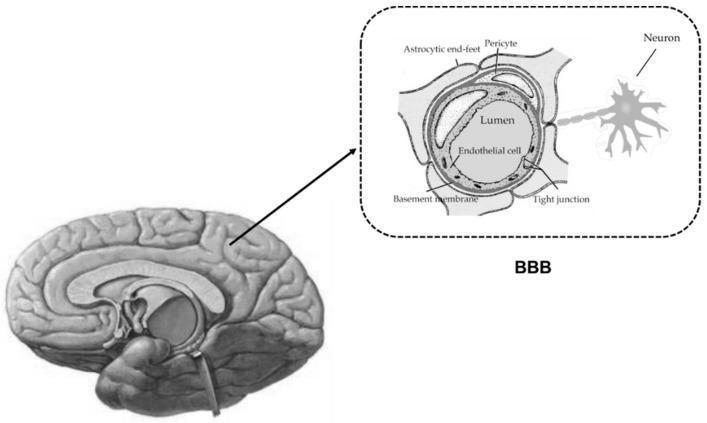
This medical illustration depicts the anatomy and cellular organization of the Blood-Brain Barrier (BBB) and the Neurovascular Unit (NVU). The primary figure shows a sagittal view of the human brain, with an inset focusing on a microscopic cross-section of a brain capillary. The capillary is lined by endothelial cells containing tight junctions, which serve as a restrictive physical barrier between the vessel lumen and the brain parenchyma. Surrounding the endothelial layer is a basement membrane and closely associated pericytes. The entire vessel structure is unsheathed by astrocytic end-feet, which facilitate metabolic support and barrier maintenance. A neuron is also shown with its terminals in close proximity to the astrocytic end-feet, illustrating the functional coupling between neural activity and blood flow within the NVU. This diagram is designed for intermediate to advanced neurobiology or pharmacology education, highlighting the structural basis of central nervous system homeostasis and selective permeability.
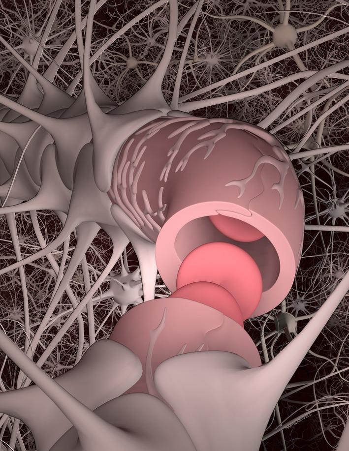
An anatomical diagram illustrating the components of the neurovascular unit (NVU) within the central nervous system. The central focus is a brain capillary formed by brain microvascular endothelial cells (BMECs), shown as a pink cylindrical structure with red blood cells visible in the lumen. Branching pericytes are depicted adhering to the abluminal surface of the capillary. The vessel is almost entirely enveloped by astrocyte end-feet, represented as light-colored, broad expansions from star-shaped glial cells. In the background, a dense network of neuronal processes and other glial cells represents the brain parenchyma and extracellular matrix. This illustration highlights the structural relationship between the vascular system and the neural environment, demonstrating the basis of the blood-brain barrier (BBB) and the cooperative regulation of brain homeostasis through cellular interactions between neurons, astrocytes, pericytes, and endothelial cells.
At the Cell Level: the Capillary
Structure of a Capillary
"The capillaries are thin-walled structures lined with a single layer of endothelial cells, which is surrounded by a basal lamina. Capillaries are the site where nutrients, gases, water, and solutes are exchanged between the blood and the tissues."
"True capillaries (inner radius, 2 to 5 μm) consist of a single layer of endothelial cells surrounded by a basement membrane, a fine network of reticular collagen fibers, and - in some tissues - pericytes."
Key Structural Components
| Component | Detail |
|---|---|
| Endothelial cells | Single cell-thick lining; the actual "wall" of the capillary |
| Basement membrane | Thin supporting layer around endothelium |
| Pericytes | Contractile cells that wrap around the capillary; regulate diameter |
| Lumen diameter | 2-5 μm (RBCs pass through in single file) |
How Exchange Occurs Across the Capillary Wall
| Substance | Route |
|---|---|
| O2, CO2 (lipid-soluble) | Dissolve directly through endothelial cell membranes |
| Water, ions (water-soluble) | Pass through intercellular clefts or fenestrations |
| Large molecules | Transcytosis via pinocytic vesicles / caveolae |
The Microcirculation Hierarchy
- Costanzo Physiology, 7th Ed.
- Medical Physiology (Boron & Boulpaep)
Tissue level?
At the Tissue Level: the Arteriole
Why the Arteriole?
"The arterioles are the smallest branches of the arteries. Their walls have an extensive development of smooth muscle, and they are the site of highest resistance to blood flow."
"Arterioles are not only the site of highest resistance in the vasculature, but they are also the site where resistance can be changed by alterations in sympathetic nerve activity, by circulating catecholamines, and by other vasoactive substances."
How Arterioles Control Tissue Supply
| Mechanism | Effect on Tissue Blood Flow |
|---|---|
| Sympathetic (α1 activation) - vasoconstriction | Reduces blood flow to tissue |
| Sympathetic (β2 activation) - vasodilation | Increases blood flow (e.g. skeletal muscle) |
| Local metabolites (CO2, H+, adenosine) | Dilate arterioles → increases supply to active tissue |
| Precapillary sphincters | Open/close to route blood to specific capillaries |
The Hierarchy in Context
| Level | Vessel | Function |
|---|---|---|
| Organ level | Artery | Conducts blood to the organ |
| Tissue level | Arteriole | Regulates flow into capillary beds |
| Cell level | Capillary | Exchange of O2, nutrients, waste |
- Costanzo Physiology, 7th Ed.