
How cancer cell die by necrosis
Necrosis in cancer cells is a form of uncontrolled, passive cell death triggered by external injury - distinct from the programmed death of apoptosis. It typically occurs when cells are deprived of oxygen (ischemia), exposed to toxins, physical trauma, or extreme temperature changes. The cell swells, its membrane ruptures, and cellular contents spill out, triggering inflammation in surrounding tissue.
Searching sources...
Reading File
Searching the Web
cancer cell death by necrosis mechanisms pathophysiology
Now let me search the medical library for detailed textbook content on necrosis in cancer cells.
Finding Sources
Searching Images
necrosis cancer cell death histology diagram
Reading File
Finding Sources
I found a great diagram. Let me fetch it and also get the Robbins pathology content on tumor cell death evasion.
Loading Image
Reading File
Finding Sources
I now have excellent textbook content and the diagram. Let me compile the full, detailed answer.
How Cancer Cells Die by Necrosis
Necrosis is a nonprogrammed, pathological form of cell death - it is uncontrolled and occurs when a cancer cell is exposed to overwhelming injury that destroys its ability to maintain homeostasis. Unlike apoptosis (the cell's orderly "suicide program"), necrosis is chaotic and destructive.
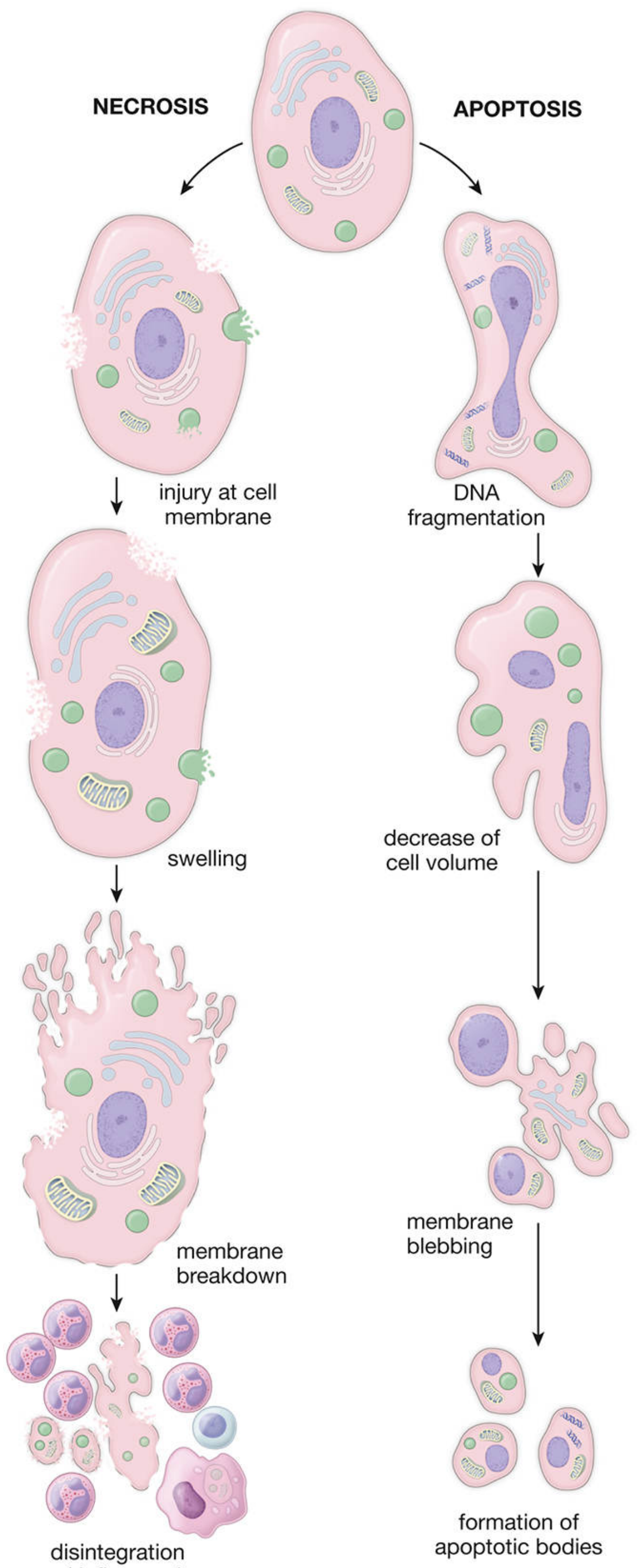
Triggers / Causes in Cancer
Cancer cells undergo necrosis when subjected to:
- Ischemia (hypoxia) - rapidly growing tumors outgrow their blood supply; inner tumor regions starve of oxygen and glucose
- Toxins (e.g., certain chemotherapy agents that exert direct cytotoxicity)
- Bacterial or viral infections that damage cell membranes
- Physical injury (trauma, extreme temperature, radiation at high doses)
- Osmotic shock
Step-by-Step Mechanism
The sequence proceeds as follows (from Histology: A Text and Atlas):
Step 1 - Membrane damage
The triggering insult (ischemia, toxin, etc.) damages the plasma membrane. Ion pumps fail, and the membrane can no longer maintain the selective barrier between cytoplasm and extracellular space.
Step 2 - Ion influx and cell swelling
Extracellular ions, especially Ca²+, flood into the cell. Water follows osmotically. The cell and its organelles (mitochondria, rough ER, nucleus) begin to swell - this is visible as cellular edema.
Step 3 - Organelle dysfunction
Swelling causes irreversible dysfunction of mitochondria and other organelles. ATP production halts completely. The lysosomal membrane also ruptures, releasing proteolytic enzymes (proteases, RNAases, DNAases, phosphatases) directly into the cytosol. These enzymes autodigest the cell from within.
Step 4 - Membrane rupture (cell lysis)
The swollen cell membrane ultimately ruptures (lyses), spilling all intracellular contents - including lysosomal enzymes, inflammatory mediators, and DAMPs (Damage-Associated Molecular Patterns) - into the surrounding extracellular space.
Step 5 - Inflammatory response
Released cell contents trigger an intense local inflammatory reaction. Immune cells (neutrophils, macrophages) are recruited. This inflammation causes collateral damage to neighboring cancer cells and normal tissue alike. This is in sharp contrast to apoptosis, which causes NO inflammation.
Necrosis vs. Apoptosis in Cancer: Key Contrast
| Feature | Necrosis | Apoptosis |
|---|---|---|
| Cell volume | Swells | Shrinks |
| Membrane integrity | Ruptures | Maintained |
| Organelles | Swell and disintegrate | Remain intact initially |
| Contents released? | Yes - spills out | No - packaged into apoptotic bodies |
| Inflammation | Intense | None |
| Controlled? | No (accidental/pathological) | Yes (programmed) |
| Phagocytosis | Poor (debris) | Efficient (apoptotic bodies) |
As Robbins Pathologic Basis of Disease notes, cancer cells are under intense pressure to evade programmed apoptosis (via BCL2 overexpression, TP53 mutations, etc.) - which makes necrosis, from ischemia and chemotherapy-induced direct cytotoxicity, a significant alternative death route in tumors.
Necroptosis - "Programmed Necrosis"
A newer, important concept is necroptosis - a regulated, programmed form of cell death that has the morphological features of necrosis but is molecularly controlled. It is triggered by:
- Tumor necrosis factor (TNF) family members
- Caspase-8 inhibition (often occurring in cancer cells that block apoptosis)
- Assembly of the necrosome complex (RIPK1-RIPK3 signaling)
The result still looks like necrosis - cell swelling, membrane rupture, inflammation - but it follows a defined signaling pathway. Research shows necroptosis plays roles in tumor metastasis and drug resistance, making it a therapeutic target of interest.
Why Necrosis Matters in Cancer Biology
- Tumor center necrosis - Large, poorly vascularized tumors routinely show a necrotic core on imaging and histology, a sign of aggressive, fast-growing disease.
- Immunogenicity - Necrotic cancer cells release DAMPs that can actually stimulate anti-tumor immune responses, which is why some researchers are exploring necrosis-inducing strategies as cancer therapy.
- Chemotherapy - Some agents kill cancer cells through direct membrane cytotoxicity (necrosis-like death), not just apoptosis.
- Prognostic marker - The extent of necrosis in tumor specimens is used histologically to grade tumor aggressiveness (e.g., in sarcomas and CNS tumors).
Sources:
- Histology: A Text and Atlas with Correlated Cell and Molecular Biology (Eroschenko), pp. 275-277 - Nonprogrammed Cell Death: Necrosis
- Robbins, Cotran & Kumar Pathologic Basis of Disease - Evasion of Cell Death, Chapter 7
- StatPearls - Necrosis Pathology for cellular mechanism detail
- Nature Reviews - Regulated Cell Death in Cancer for necroptosis context
This is a shared conversation. Sign in to Orris to start your own chat.