
Three-dimensional hermetic obturation is one of the main factors of the long-term success of endodontic treatment [1], while adequate root canal filling is proportionally correlated with the long-term dimensional stability of the root canal sealer. Nowadays, calcium silicate-based sealers (CSBSs) meet those needs and are experiencing an increase in clinical use, especially with new premixed materials, due to their simple handling. Other than their biocompatibility [2], the dimensional stability and bioactivity of CSBSs after setting is the main reason why they could potentially outperform standard epoxy based sealers. Furthermore, the literature shows a low percentage of porosity [3] and a setting reaction achieved in the presence of moisture, finally forming hydroxyapatite at the interface and creating a chemical bond to dentin [4,5]. Bioengineering 2026, 13, 675 https://doi.org/10.3390/bioengineering13060675 Bioengineering 2026, 13, 675 2 of 11 However, studies up to now have not shown homogeneous results when evaluating the bond strength (BS) of CSBSs and epoxy resin-based sealer. Although certain studies claim higher BS values of CSBSs [6,7], a number of studies in the last five years claim that epoxy resin-based sealers outperform CSBSs in terms of BS [8–10]. Since the chemical–physical properties and bioactivity of CSBS are clinically relevant, it is of interest to enhance the BS to establish the full potential of the material. According to the scientific information to date, laser-assisted protocols are emerging as a potential solution, since findings show their efficacy in irrigation fluid activation, biofilm removal, debris and vital tissue removal, and root canal filling remnant removal [11–14]. Laser assisted protocols have also been proven to change the intracanal dentinal surface, which could potentially affect the bonding of materials, as shown in studies claiming composite bonding enhancement [15,16]. Erbium:yttrium–aluminum–garnet (Er:YAG) lasers are used in endodontic treatment with the novel mode shock wave-enhanced emission of photoacoustic streaming (SWEEPS). Due to the fact that its wavelength matches the absorption peak of water, it can be absorbed by irrigation fluids and hydroxyapatite [17]. SWEEPS was created in order to increase the efficiency of the existing PIPS mode. The main mechanism involves delivering pairs of ultra-short pulses which create bubbles. An additional second bubble accelerates the collapse of the first one, forming a violent collapse and emitting a shock wave [18]. Studies showed that SWEEPS can cause canal wall erosion [19], while a group of authors reported that hydroxyapatite crystals in mineralized dentin tissue remain intact with laser irradiation of no higher than 19.11 J/cm2 [20]. Furthermore, laser irradiation was noticed to improve the crystallinity of lamella hydroxyapatite and rearrange its growth orientations [20]. On the other hand, diode lasers’ antibacterial efficacy has been proven [21], but other than antimicrobial efficacy, diode laser protocols have also shown melting of intracanal dentin [22,23]. Still, the use of diode lasers has been recommended due to their low costs compared to other lasers [21]. Studies have already revealed that BS is influenced by the type of irrigation [24,25], and certain studies have shown that laser-assisted protocols [26] positively affect the adhesion and BS of CSBS to the root canal dentin walls. Nevertheless, there are still few studies examining laser-assisted protocols and, according to our knowledge, none using SWEEPS technology to enhance the BS of bioceramic sealer. Addressing this gap, the aim of this study was to compare the BS and porosity of a premixed CSBS (BioRoot Flow, Septodont, Saint Maur Des Fosses, France) used with a single-cone obturation technique (SC) after SWEEPS-mode Er:YAG laser-activated irrigation, diode laser root canal irradiation, and conventional irrigation (CI). The null hypothesis of the study was that there would be no difference in the BS and porosity of the root canal filling with the CSBS after the different final root canal disinfection protocols matter into images with liitle matter
Scientific infographic summarizing a dental endodontics research paper about calcium silicate-based sealers (CSBSs) and laser-assisted root canal treatment. Include: a cross-section diagram of a tooth root canal showing obturation/filling with sealer, icons representing Er:YAG SWEEPS laser and diode laser protocols, comparison chart between CSBS and epoxy resin-based sealers for bond strength, hydroxyapatite crystal formation at dentin interface, porosity percentage indicator, and key concepts: biocompatibility, dimensional stability, bioactivity, bond strength. Clean scientific poster style with labeled diagrams and minimal text callouts.
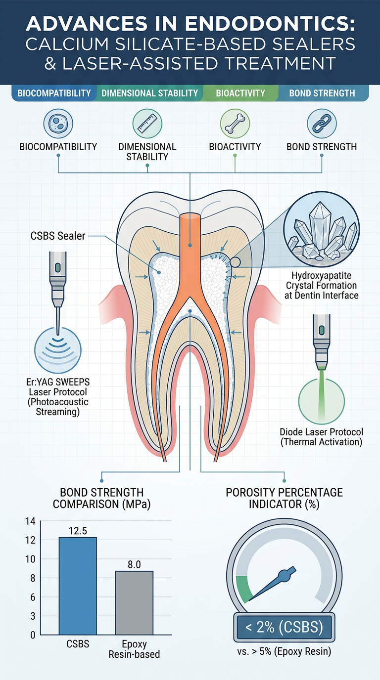
- Root canal obturation - the goal of 3D hermetic sealing for long-term endodontic success
- Calcium silicate-based sealers (CSBSs) vs. epoxy resin-based sealers - bond strength comparison
- Bioactivity - hydroxyapatite formation at the dentin interface, creating a chemical bond
- Laser protocols compared:
- Er:YAG SWEEPS - uses paired ultra-short pulses, bubble-collapse shock waves, matches water/hydroxyapatite absorption peak
- Diode laser - proven antibacterial efficacy, causes intracanal dentin melting, lower cost
- Conventional irrigation (CI) - the control group
- Study material: BioRoot Flow (premixed CSBS, Septodont) with single-cone obturation technique
- Outcome measures: Bond strength (BS) and porosity of the root canal filling
Three-dimensional hermetic obturation is one of the main factors of the long-term success of endodontic treatment [1], while adequate root canal filling is proportionally correlated with the long-term dimensional stability of the root canal sealer. Nowadays, calcium silicate-based sealers (CSBSs) meet those needs and are experiencing an increase in clinical use, especially with new premixed materials, due to their simple handling. Other than their biocompatibility [2], the dimensional stability and bioactivity of CSBSs after setting is the main reason why they could potentially outperform standard epoxy based sealers. Furthermore, the literature shows a low percentage of porosity [3] and a setting reaction achieved in the presence of moisture, finally forming hydroxyapatite at the interface and creating a chemical bond to dentin [4,5]. Bioengineering 2026, 13, 675 https://doi.org/10.3390/bioengineering13060675 Bioengineering 2026, 13, 675 2 of 11 However, studies up to now have not shown homogeneous results when evaluating the bond strength (BS) of CSBSs and epoxy resin-based sealer. Although certain studies claim higher BS values of CSBSs [6,7], a number of studies in the last five years claim that epoxy resin-based sealers outperform CSBSs in terms of BS [8–10]. Since the chemical–physical properties and bioactivity of CSBS are clinically relevant, it is of interest to enhance the BS to establish the full potential of the material. According to the scientific information to date, laser-assisted protocols are emerging as a potential solution, since findings show their efficacy in irrigation fluid activation, biofilm removal, debris and vital tissue removal, and root canal filling remnant removal [11–14]. Laser assisted protocols have also been proven to change the intracanal dentinal surface, which could potentially affect the bonding of materials, as shown in studies claiming composite bonding enhancement [15,16]. Erbium:yttrium–aluminum–garnet (Er:YAG) lasers are used in endodontic treatment with the novel mode shock wave-enhanced emission of photoacoustic streaming (SWEEPS). Due to the fact that its wavelength matches the absorption peak of water, it can be absorbed by irrigation fluids and hydroxyapatite [17]. SWEEPS was created in order to increase the efficiency of the existing PIPS mode. The main mechanism involves delivering pairs of ultra-short pulses which create bubbles. An additional second bubble accelerates the collapse of the first one, forming a violent collapse and emitting a shock wave [18]. Studies showed that SWEEPS can cause canal wall erosion [19], while a group of authors reported that hydroxyapatite crystals in mineralized dentin tissue remain intact with laser irradiation of no higher than 19.11 J/cm2 [20]. Furthermore, laser irradiation was noticed to improve the crystallinity of lamella hydroxyapatite and rearrange its growth orientations [20]. On the other hand, diode lasers’ antibacterial efficacy has been proven [21], but other than antimicrobial efficacy, diode laser protocols have also shown melting of intracanal dentin [22,23]. Still, the use of diode lasers has been recommended due to their low costs compared to other lasers [21]. Studies have already revealed that BS is influenced by the type of irrigation [24,25], and certain studies have shown that laser-assisted protocols [26] positively affect the adhesion and BS of CSBS to the root canal dentin walls. Nevertheless, there are still few studies examining laser-assisted protocols and, according to our knowledge, none using SWEEPS technology to enhance the BS of bioceramic sealer. Addressing this gap, the aim of this study was to compare the BS and porosity of a premixed CSBS (BioRoot Flow, Septodont, Saint Maur Des Fosses, France) used with a single-cone obturation technique (SC) after SWEEPS-mode Er:YAG laser-activated irrigation, diode laser root canal irradiation, and conventional irrigation (CI). The null hypothesis of the study was that there would be no difference in the BS and porosity of the root canal filling with the CSBS after the different final root canal disinfection protocols matter into 4 presentation images with clearly
Presentation slide image 1 of 4: "The Case for Calcium Silicate-Based Sealers (CSBSs)" - Clean academic slide style with white background. Show a labeled cross-section diagram of a tooth root canal with sealer filling. Include bullet points: 3D hermetic obturation = key to long-term endodontic success; CSBSs: biocompatible, dimensionally stable, bioactive; Setting reaction in moisture → forms hydroxyapatite → chemical bond to dentin; Low porosity. Add a small diagram showing hydroxyapatite crystal formation at the dentin-sealer interface. Bold title at top, clear readable fonts, blue and white color scheme.
Presentation slide image 2 of 4: "The Problem: Inconsistent Bond Strength Results" - Clean academic slide style. Title at top in bold. Show a comparison bar chart: CSBS vs Epoxy Resin-Based Sealer bond strength, with some studies favoring CSBS and recent studies (last 5 years) favoring epoxy resin sealers - show this as two conflicting bar groups labeled "Some studies" and "Recent studies (last 5 years)". Add a question mark icon and the research gap statement: Bond strength of CSBSs remains inconsistent - how can it be enhanced? Blue, red and white color scheme, clear labels, academic poster style.
Presentation slide image 3 of 4: "Laser-Assisted Protocols as a Solution" - Clean academic presentation slide. Title at top. Two columns side by side: Left column "Er:YAG SWEEPS Laser" with bullet points: wavelength matches water and hydroxyapatite absorption; paired ultra-short pulses create bubbles; second bubble collapses first → shock wave emitted; improves hydroxyapatite crystallinity; safe below 19.11 J/cm2. Right column "Diode Laser" with bullets: proven antibacterial efficacy; causes intracanal dentin melting; low cost. Include small diagrams: a laser beam hitting a tooth canal on left, and a diode laser icon on right. Blue and green color scheme, clean fonts.
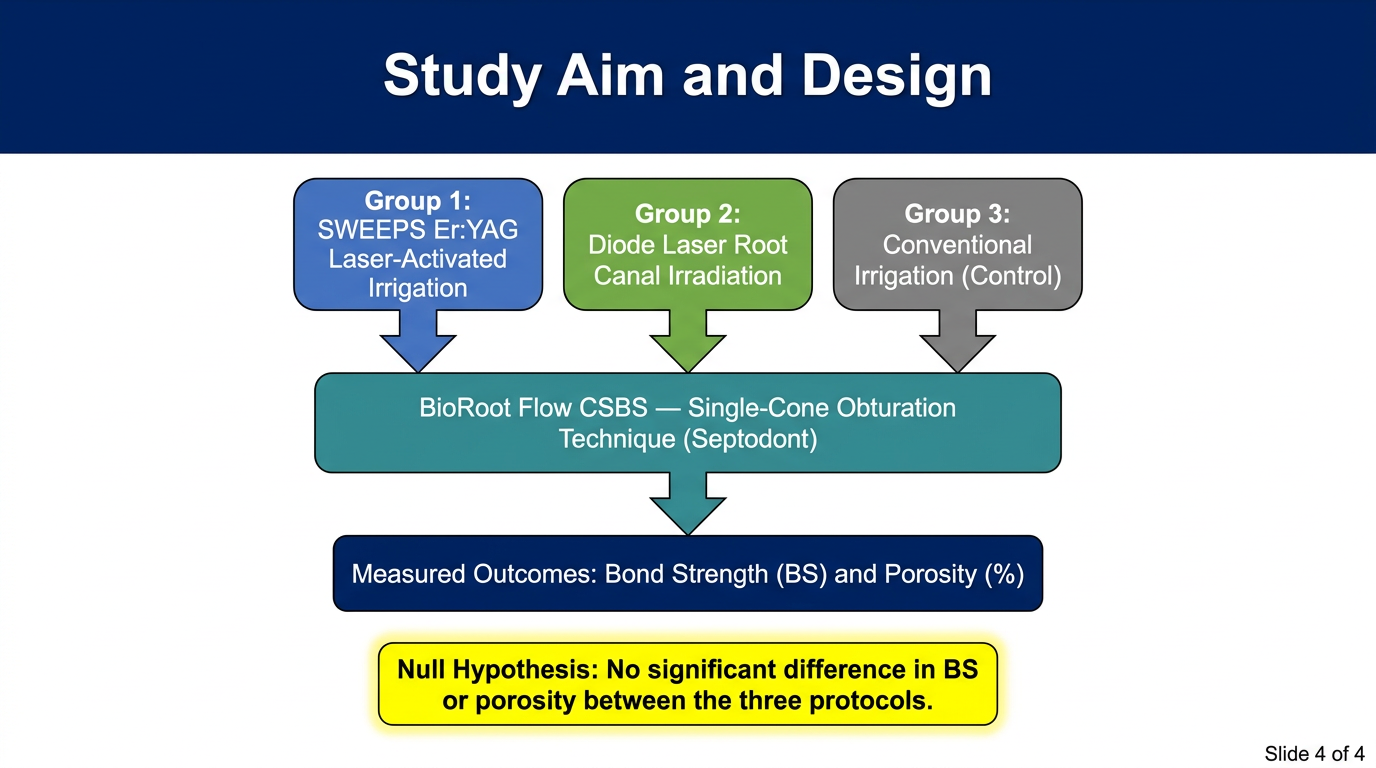
Presentation slide image 4 of 4: "Study Aim and Design" - Clean academic presentation slide. Title at top in bold. Show a clear flowchart: 3 groups of extracted teeth → Group 1: SWEEPS Er:YAG laser-activated irrigation + BioRoot Flow CSBS; Group 2: Diode laser root canal irradiation + BioRoot Flow CSBS; Group 3: Conventional irrigation (control) + BioRoot Flow CSBS. Arrow pointing down to: Outcome measures: Bond Strength (BS) and Porosity. Add text box: "Null Hypothesis: No difference in BS or porosity between the three protocols." Include BioRoot Flow label and single-cone obturation technique note. Blue and white academic color scheme, bold readable text.
Error calling model 'gemini-3-pro-image' (RESOURCE_EXHAUSTED): 429 RESOURCE_EXHAUSTED. {'error': {'code': 429, 'message': 'Resource has been exhausted (e.g. check quota).', 'status': 'RESOURCE_EXHAUSTED'}}
We encountered an error while processing your request. Please try again or reach out to us for help.
Three-dimensional hermetic obturation is one of the main factors of the long-term success of endodontic treatment [1], while adequate root canal filling is proportionally correlated with the long-term dimensional stability of the root canal sealer. Nowadays, calcium silicate-based sealers (CSBSs) meet those needs and are experiencing an increase in clinical use, especially with new premixed materials, due to their simple handling. Other than their biocompatibility [2], the dimensional stability and bioactivity of CSBSs after setting is the main reason why they could potentially outperform standard epoxy based sealers. Furthermore, the literature shows a low percentage of porosity [3] and a setting reaction achieved in the presence of moisture, finally forming hydroxyapatite at the interface and creating a chemical bond to dentin [4,5]. Bioengineering 2026, 13, 675 https://doi.org/10.3390/bioengineering13060675 Bioengineering 2026, 13, 675 2 of 11 However, studies up to now have not shown homogeneous results when evaluating the bond strength (BS) of CSBSs and epoxy resin-based sealer. Although certain studies claim higher BS values of CSBSs [6,7], a number of studies in the last five years claim that epoxy resin-based sealers outperform CSBSs in terms of BS [8–10]. Since the chemical–physical properties and bioactivity of CSBS are clinically relevant, it is of interest to enhance the BS to establish the full potential of the material. According to the scientific information to date, laser-assisted protocols are emerging as a potential solution, since findings show their efficacy in irrigation fluid activation, biofilm removal, debris and vital tissue removal, and root canal filling remnant removal [11–14]. Laser assisted protocols have also been proven to change the intracanal dentinal surface, which could potentially affect the bonding of materials, as shown in studies claiming composite bonding enhancement [15,16]. Erbium:yttrium–aluminum–garnet (Er:YAG) lasers are used in endodontic treatment with the novel mode shock wave-enhanced emission of photoacoustic streaming (SWEEPS). Due to the fact that its wavelength matches the absorption peak of water, it can be absorbed by irrigation fluids and hydroxyapatite [17]. SWEEPS was created in order to increase the efficiency of the existing PIPS mode. The main mechanism involves delivering pairs of ultra-short pulses which create bubbles. An additional second bubble accelerates the collapse of the first one, forming a violent collapse and emitting a shock wave [18]. Studies showed that SWEEPS can cause canal wall erosion [19], while a group of authors reported that hydroxyapatite crystals in mineralized dentin tissue remain intact with laser irradiation of no higher than 19.11 J/cm2 [20]. Furthermore, laser irradiation was noticed to improve the crystallinity of lamella hydroxyapatite and rearrange its growth orientations [20]. On the other hand, diode lasers’ antibacterial efficacy has been proven [21], but other than antimicrobial efficacy, diode laser protocols have also shown melting of intracanal dentin [22,23]. Still, the use of diode lasers has been recommended due to their low costs compared to other lasers [21]. Studies have already revealed that BS is influenced by the type of irrigation [24,25], and certain studies have shown that laser-assisted protocols [26] positively affect the adhesion and BS of CSBS to the root canal dentin walls. Nevertheless, there are still few studies examining laser-assisted protocols and, according to our knowledge, none using SWEEPS technology to enhance the BS of bioceramic sealer. Addressing this gap, the aim of this study was to compare the BS and porosity of a premixed CSBS (BioRoot Flow, Septodont, Saint Maur Des Fosses, France) used with a single-cone obturation technique (SC) after SWEEPS-mode Er:YAG laser-activated irrigation, diode laser root canal irradiation, and conventional irrigation (CI). The null hypothesis of the study was that there would be no difference in the BS and porosity of the root canal filling with the CSBS after the different final root canal disinfection protocols matter convert into 4 images for presentation
Presentation slide image titled "Why Calcium Silicate-Based Sealers (CSBSs)?" Academic style, clean white background, blue accents. Show a labeled cross-section diagram of a tooth root canal filled with sealer. Key points listed clearly: 3D hermetic obturation is critical for long-term endodontic success; CSBSs are biocompatible, dimensionally stable, and bioactive; Setting reaction occurs in moisture, forming hydroxyapatite crystals at dentin interface; Creates chemical bond to dentin; Low porosity percentage. Small inset diagram showing hydroxyapatite crystal bonding to dentin wall. Bold readable title, clean academic fonts.
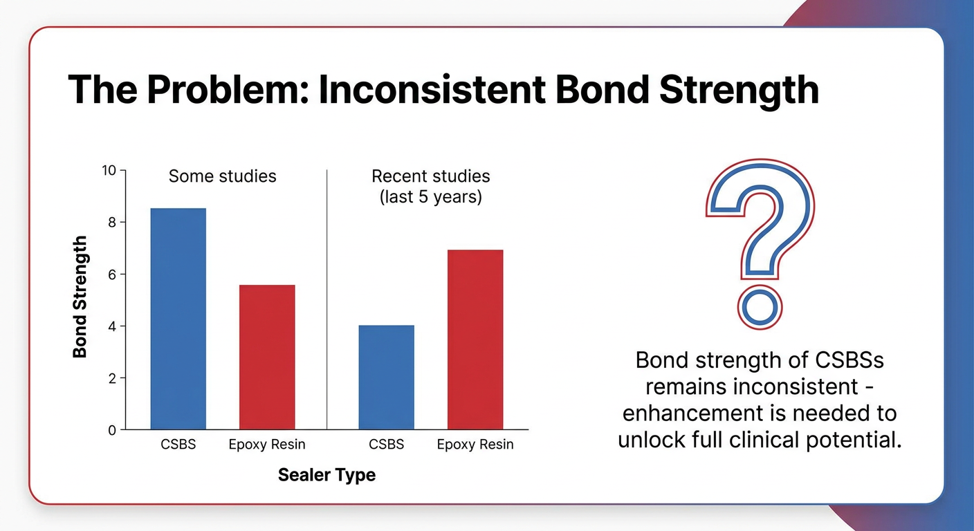
Presentation slide titled "The Problem: Inconsistent Bond Strength" Academic clean white background with red and blue accents. Show a bar chart comparing bond strength: left bars labeled "Some studies" showing CSBS higher than epoxy resin; right bars labeled "Recent studies (last 5 years)" showing epoxy resin higher than CSBS. Add a large question mark graphic and text: "Bond strength of CSBSs remains inconsistent - enhancement is needed to unlock full clinical potential." Clear axis labels: Y-axis = Bond Strength, X-axis = Sealer Type. Bold readable title at top. Simple clean academic design.
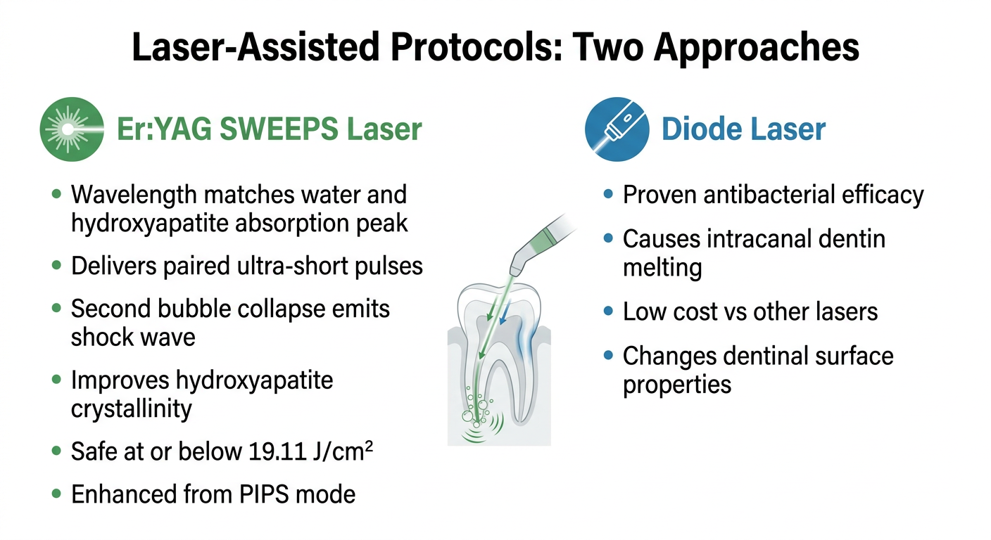
Presentation slide titled "Laser-Assisted Protocols: Two Approaches" Clean academic white background, green and blue color scheme. Two side-by-side columns with clear headers. Left column header "Er:YAG SWEEPS Laser" with icon of laser beam, bullet points: Wavelength matches water and hydroxyapatite absorption peak; Delivers paired ultra-short pulses; Second bubble collapse emits shock wave; Improves hydroxyapatite crystallinity; Safe at or below 19.11 J/cm2; Enhanced from PIPS mode. Right column header "Diode Laser" with icon, bullet points: Proven antibacterial efficacy; Causes intracanal dentin melting; Low cost vs other lasers; Changes dentinal surface properties. Small diagram of laser entering root canal in center. Bold readable title at top.
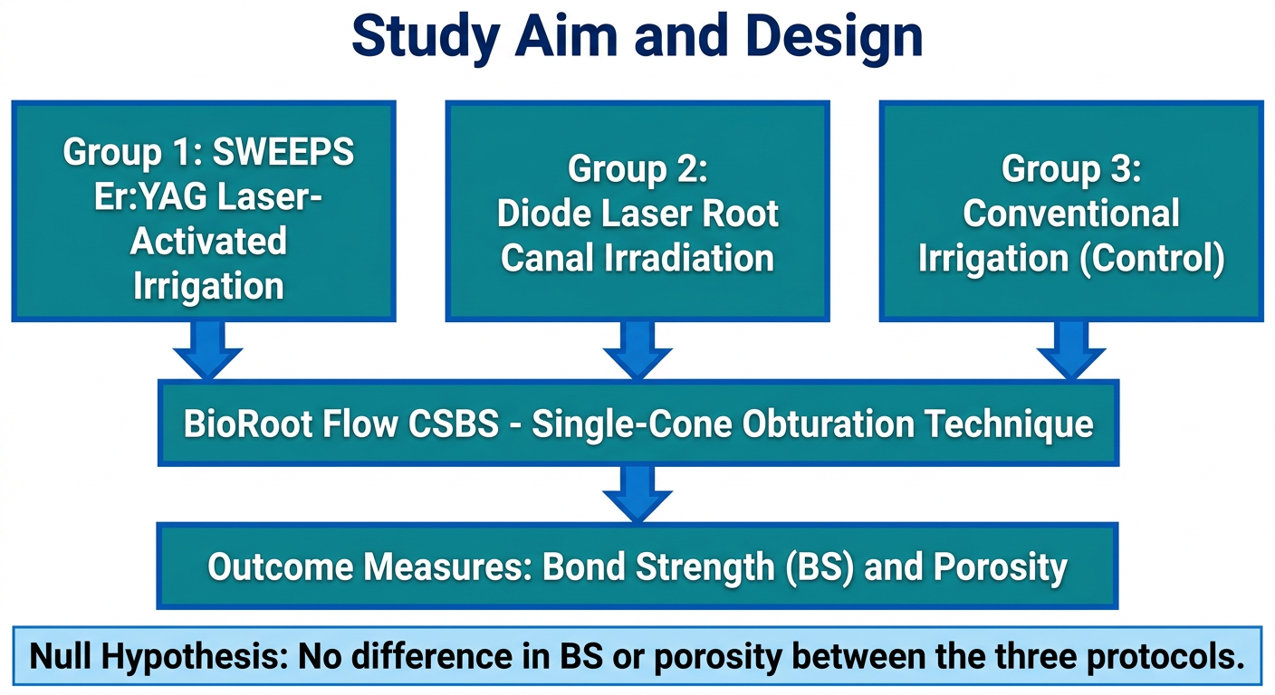
Presentation slide titled "Study Aim and Design" Clean academic white background, blue and teal color scheme. Show a clear vertical flowchart with three parallel branches: Branch 1 box: "Group 1: SWEEPS Er:YAG Laser-Activated Irrigation"; Branch 2 box: "Group 2: Diode Laser Root Canal Irradiation"; Branch 3 box: "Group 3: Conventional Irrigation (Control)". All three branches point down with arrows to a single box: "BioRoot Flow CSBS - Single-Cone Obturation Technique". That box points down to: "Outcome Measures: Bond Strength (BS) and Porosity". Below that, a highlighted text box: "Null Hypothesis: No difference in BS or porosity between the three protocols." Bold readable fonts, clean academic design, clearly labeled boxes and arrows.
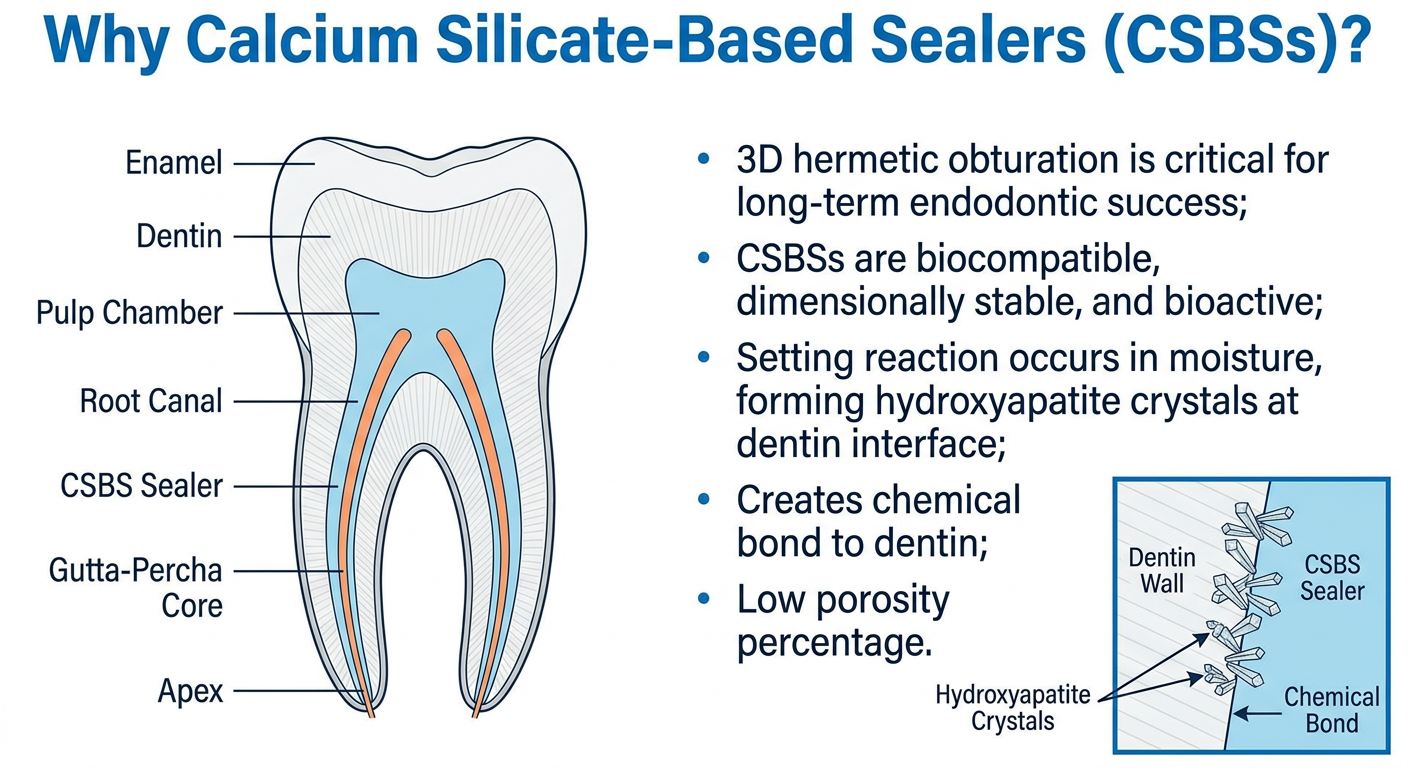
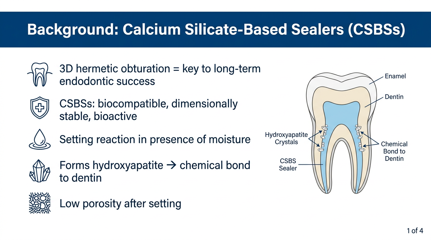
- Slide 1 - Why CSBSs are clinically valuable (background rationale)
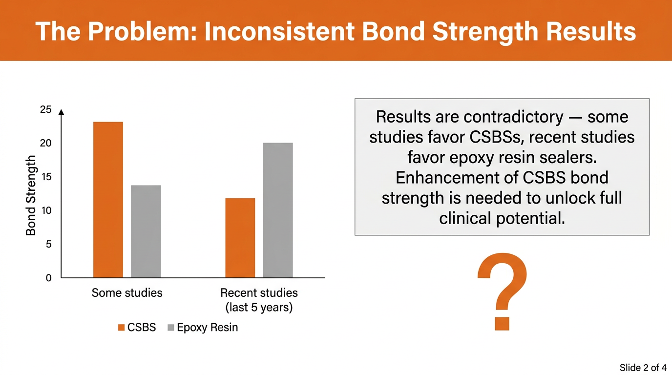
- Slide 2 - The inconsistency problem with bond strength (research gap)
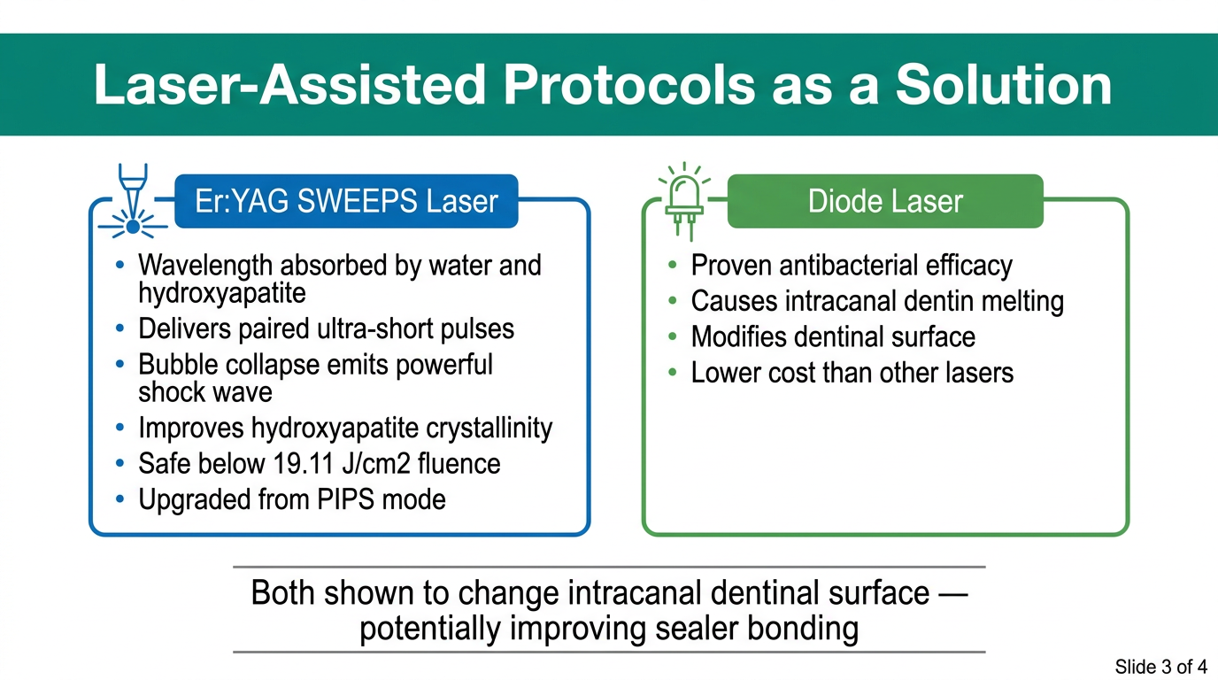
- Slide 3 - The two laser types being investigated (Er:YAG SWEEPS vs. Diode)
- Slide 4 - The study design, three groups, and null hypothesis
convert this into separate images for my presentation:Three-dimensional hermetic obturation is one of the main factors of the long-term success of endodontic treatment [1], while adequate root canal filling is proportionally correlated with the long-term dimensional stability of the root canal sealer. Nowadays, calcium silicate-based sealers (CSBSs) meet those needs and are experiencing an increase in clinical use, especially with new premixed materials, due to their simple handling. Other than their biocompatibility [2], the dimensional stability and bioactivity of CSBSs after setting is the main reason why they could potentially outperform standard epoxy based sealers. Furthermore, the literature shows a low percentage of porosity [3] and a setting reaction achieved in the presence of moisture, finally forming hydroxyapatite at the interface and creating a chemical bond to dentin [4,5]. Bioengineering 2026, 13, 675 https://doi.org/10.3390/bioengineering13060675 Bioengineering 2026, 13, 675 2 of 11 However, studies up to now have not shown homogeneous results when evaluating the bond strength (BS) of CSBSs and epoxy resin-based sealer. Although certain studies claim higher BS values of CSBSs [6,7], a number of studies in the last five years claim that epoxy resin-based sealers outperform CSBSs in terms of BS [8–10]. Since the chemical–physical properties and bioactivity of CSBS are clinically relevant, it is of interest to enhance the BS to establish the full potential of the material. According to the scientific information to date, laser-assisted protocols are emerging as a potential solution, since findings show their efficacy in irrigation fluid activation, biofilm removal, debris and vital tissue removal, and root canal filling remnant removal [11–14]. Laser assisted protocols have also been proven to change the intracanal dentinal surface, which could potentially affect the bonding of materials, as shown in studies claiming composite bonding enhancement [15,16]. Erbium:yttrium–aluminum–garnet (Er:YAG) lasers are used in endodontic treatment with the novel mode shock wave-enhanced emission of photoacoustic streaming (SWEEPS). Due to the fact that its wavelength matches the absorption peak of water, it can be absorbed by irrigation fluids and hydroxyapatite [17]. SWEEPS was created in order to increase the efficiency of the existing PIPS mode. The main mechanism involves delivering pairs of ultra-short pulses which create bubbles. An additional second bubble accelerates the collapse of the first one, forming a violent collapse and emitting a shock wave [18]. Studies showed that SWEEPS can cause canal wall erosion [19], while a group of authors reported that hydroxyapatite crystals in mineralized dentin tissue remain intact with laser irradiation of no higher than 19.11 J/cm2 [20]. Furthermore, laser irradiation was noticed to improve the crystallinity of lamella hydroxyapatite and rearrange its growth orientations [20]. On the other hand, diode lasers’ antibacterial efficacy has been proven [21], but other than antimicrobial efficacy, diode laser protocols have also shown melting of intracanal dentin [22,23]. Still, the use of diode lasers has been recommended due to their low costs compared to other lasers [21]. Studies have already revealed that BS is influenced by the type of irrigation [24,25], and certain studies have shown that laser-assisted protocols [26] positively affect the adhesion and BS of CSBS to the root canal dentin walls. Nevertheless, there are still few studies examining laser-assisted protocols and, according to our knowledge, none using SWEEPS technology to enhance the BS of bioceramic sealer. Addressing this gap, the aim of this study was to compare the BS and porosity of a premixed CSBS (BioRoot Flow, Septodont, Saint Maur Des Fosses, France) used with a single-cone obturation technique (SC) after SWEEPS-mode Er:YAG laser-activated irrigation, diode laser root canal irradiation, and conventional irrigation (CI). The null hypothesis of the study was that there would be no difference in the BS and porosity of the root canal filling with the CSBS after the different final root canal disinfection protocols matter into 4 presentation images with clearly Searched 4 sources ! Error calling model 'gemini-3-pro-image' (RESOURCE_EXHAUSTED): 429 RESOURCE_EXHAUSTED. {'error': {'code': 429, 'message': 'Resource has been exhausted (e.g. check quota).', 'status': 'RESOURCE_EXHAUSTED'}} We encountered an error while processing your request. Please try again or reach out to us for help. 🔗 Contact Support ✉️ hi@orris.care Three-dimensional hermetic obturation is one of the main factors of the long-term success of endodontic treatment [1], while adequate root canal filling is proportionally correlated with the long-term dimensional stability of the root canal sealer. Nowadays, calcium silicate-based sealers (CSBSs) meet those needs and are experiencing an increase in clinical use, especially with new premixed materials, due to their simple handling. Other than their biocompatibility [2], the dimensional stability and bioactivity of CSBSs after setting is the main reason why they could potentially outperform standard epoxy based sealers. Furthermore, the literature shows a low percentage of porosity [3] and a setting reaction achieved in the presence of moisture, finally forming hydroxyapatite at the interface and creating a chemical bond to dentin [4,5]. Bioengineering 2026, 13, 675 https://doi.org/10.3390/bioengineering13060675 Bioengineering 2026, 13, 675 2 of 11 However, studies up to now have not shown homogeneous results when evaluating the bond strength (BS) of CSBSs and epoxy resin-based sealer. Although certain studies claim higher BS values of CSBSs [6,7], a number of studies in the last five years claim that epoxy resin-based sealers outperform CSBSs in terms of BS [8–10]. Since the chemical–physical properties and bioactivity of CSBS are clinically relevant, it is of interest to enhance the BS to establish the full potential of the material. According to the scientific information to date, laser-assisted protocols are emerging as a potential solution, since findings show their efficacy in irrigation fluid activation, biofilm removal, debris and vital tissue removal, and root canal filling remnant removal [11–14]. Laser assisted protocols have also been proven to change the intracanal dentinal surface, which could potentially affect the bonding of materials, as shown in studies claiming composite bonding enhancement [15,16]. Erbium:yttrium–aluminum–garnet (Er:YAG) lasers are used in endodontic treatment with the novel mode shock wave-enhanced emission of photoacoustic streaming (SWEEPS). Due to the fact that its wavelength matches the absorption peak of water, it can be absorbed by irrigation fluids and hydroxyapatite [17]. SWEEPS was created in order to increase the efficiency of the existing PIPS mode. The main mechanism involves delivering pairs of ultra-short pulses which create bubbles. An additional second bubble accelerates the collapse of the first one, forming a violent collapse and emitting a shock wave [18]. Studies showed that SWEEPS can cause canal wall erosion [19], while a group of authors reported that hydroxyapatite crystals in mineralized dentin tissue remain intact with laser irradiation of no higher than 19.11 J/cm2 [20]. Furthermore, laser irradiation was noticed to improve the crystallinity of lamella hydroxyapatite and rearrange its growth orientations [20]. On the other hand, diode lasers’ antibacterial efficacy has been proven [21], but other than antimicrobial efficacy, diode laser protocols have also shown melting of intracanal dentin [22,23]. Still, the use of diode lasers has been recommended due to their low costs compared to other lasers [21]. Studies have already revealed that BS is influenced by the type of irrigation [24,25], and certain studies have shown that laser-assisted protocols [26] positively affect the adhesion and BS of CSBS to the root canal dentin walls. Nevertheless, there are still few studies examining laser-assisted protocols and, according to our knowledge, none using SWEEPS technology to enhance the BS of bioceramic sealer. Addressing this gap, the aim of this study was to compare the BS and porosity of a premixed CSBS (BioRoot Flow, Septodont, Saint Maur Des Fosses, France) used with a single-cone obturation technique (SC) after SWEEPS-mode Er:YAG laser-activated irrigation, diode laser root canal irradiation, and conventional irrigation (CI). The null hypothesis of the study was that there would be no difference in the BS and porosity of the root canal filling with the CSBS after the different final root canal disinfection protocols
Professional presentation slide, wide 16:9 format, white background with dark blue header bar. Title: "Background: Calcium Silicate-Based Sealers (CSBSs)". Content in clean bullet points with icons: tooth icon - "3D hermetic obturation = key to long-term endodontic success"; shield icon - "CSBSs: biocompatible, dimensionally stable, bioactive"; water droplet icon - "Setting reaction in presence of moisture"; crystal icon - "Forms hydroxyapatite → chemical bond to dentin"; low porosity icon - "Low porosity after setting". Right side: simple labeled diagram of a tooth cross-section showing root canal filled with sealer and hydroxyapatite crystals at dentin wall. Slide number 1 of 4 in corner. Clean, modern, academic style.
Professional presentation slide, wide 16:9 format, white background with orange header bar. Title: "The Problem: Inconsistent Bond Strength Results". Left side: two-group bar chart comparing CSBS vs Epoxy Resin sealer bond strength. Group 1 labeled "Some studies" shows CSBS bar taller than Epoxy bar (CSBS wins). Group 2 labeled "Recent studies (last 5 years)" shows Epoxy bar taller than CSBS bar (Epoxy wins). Y-axis labeled "Bond Strength". Right side: text box with key message: "Results are contradictory — some studies favor CSBSs, recent studies favor epoxy resin sealers. Enhancement of CSBS bond strength is needed to unlock full clinical potential." Large question mark graphic below. Slide number 2 of 4. Clean modern academic style.
Professional presentation slide, wide 16:9 format, white background with teal green header bar. Title: "Laser-Assisted Protocols as a Solution". Two clearly divided columns. Left column with blue border, header "Er:YAG SWEEPS Laser", laser beam icon, bullet points: "Wavelength absorbed by water and hydroxyapatite"; "Delivers paired ultra-short pulses"; "Bubble collapse emits powerful shock wave"; "Improves hydroxyapatite crystallinity"; "Safe below 19.11 J/cm2 fluence"; "Upgraded from PIPS mode". Right column with green border, header "Diode Laser", diode icon, bullet points: "Proven antibacterial efficacy"; "Causes intracanal dentin melting"; "Modifies dentinal surface"; "Lower cost than other lasers". Center bottom note: "Both shown to change intracanal dentinal surface — potentially improving sealer bonding". Slide number 3 of 4. Clean modern academic design.
Professional presentation slide, wide 16:9 format, white background with dark navy blue header bar. Title: "Study Aim and Design". Center of slide: clean horizontal flowchart. Three boxes at the top in a row: Box 1 (blue): "Group 1: SWEEPS Er:YAG Laser-Activated Irrigation"; Box 2 (green): "Group 2: Diode Laser Root Canal Irradiation"; Box 3 (grey): "Group 3: Conventional Irrigation (Control)". All three boxes have arrows pointing down to one shared box (teal): "BioRoot Flow CSBS — Single-Cone Obturation Technique (Septodont)". Arrow pointing further down to outcome box (dark blue): "Measured Outcomes: Bond Strength (BS) and Porosity (%)". Below that, a highlighted yellow box: "Null Hypothesis: No significant difference in BS or porosity between the three protocols." Slide number 4 of 4. Clean, modern, academic diagram style with clear labels.
| Slide | Topic | Key Content |
|---|---|---|
| 1 | Background | Why CSBSs are clinically valuable - biocompatibility, hydroxyapatite bonding, low porosity |
| 2 | The Problem | Contradictory bond strength data between CSBSs and epoxy resin sealers |
| 3 | The Solution | Er:YAG SWEEPS vs. Diode laser - mechanisms, effects, and cost |
| 4 | Study Design | 3 groups, BioRoot Flow, single-cone technique, null hypothesis |
CONVERT THIS INTO IMAGE FOR MY PRESENTATION: Sample Selection The protocol of this ex vivo study was approved by the local Ethics Committee (number of approval 003-01/26-05-04). Power analysis using the chi-squared test (a = 0.05 and b = 0.95) was performed, and a minimum of eight canals per test group was determined. Single-rooted, single-canal extracted human premolars from patients aged between 25 and 35 years old (collected at the Department of Oral Surgery School of Dental Medicine) were collected and stored in 0.1% water solution of chloramine-T trihydrate and in distilled water two hours before procedures. The inclusion criteria for the samples were as follows: one root and one round root canal, with an initial patency file corresponding to ISO size https://doi.org/10.3390/bioengineering13060675 Bioengineering 2026, 13, 675 3 of 11 15 or 20 (confirmed with K-file #15 or #20). The exclusion criteria were as follows: oval canals, previous endodontic treatment, calcified root canals, class II carious lesions, external resorption, and internal resorption. All collected teeth were subjected to CBCT scanning, and, out of a pool of 72 teeth, 40 samples were confirmed to have one round canal (Cranex 3DX; Soredex, Tuusula, Finland, field of view 50 × 50 mm; ENDO, 85 µm; 6.3 mA; 90 kV; 8.7 s; 450.3 mGycm2). Samples were selected and standardized to a working length of 20 mmwithout decoronation, but the crowns were slightly polished while preserving the proximal, buccal, and oral walls
Professional presentation slide, wide 16:9 format, white background with dark blue header bar. Title: "Sample Selection" with a small tooth icon. Content organized in clean sections: TOP ROW - three info boxes side by side: Box 1 (blue): "Ethics & Power Analysis" - Ethics approval: 003-01/26-05-04; Chi-squared test (α=0.05, β=0.95); Minimum 8 canals per group Box 2 (teal): "Sample Source" - Single-rooted human premolars; Patients aged 25–35 years; Stored in 0.1% chloramine-T trihydrate; Distilled water 2h before procedures Box 3 (green): "Initial Pool → Final Sample" - Large funnel diagram: 72 teeth scanned by CBCT → 40 confirmed round single canals selected MIDDLE SECTION - two columns: Left column (green checkmark header) "Inclusion Criteria": One root, one round canal; Patency file ISO size 15 or 20; Confirmed with K-file #15 or #20 Right column (red X header) "Exclusion Criteria": Oval canals; Previous endodontic treatment; Calcified root canals; Class II carious lesions; External or internal resorption BOTTOM BAR: "Standardization: Working length = 20 mm | Crowns slightly polished, proximal/buccal/oral walls preserved | CBCT: Cranex 3DX, 85µm, 90kV" Clean modern academic design, clear readable fonts, well-spaced layout.
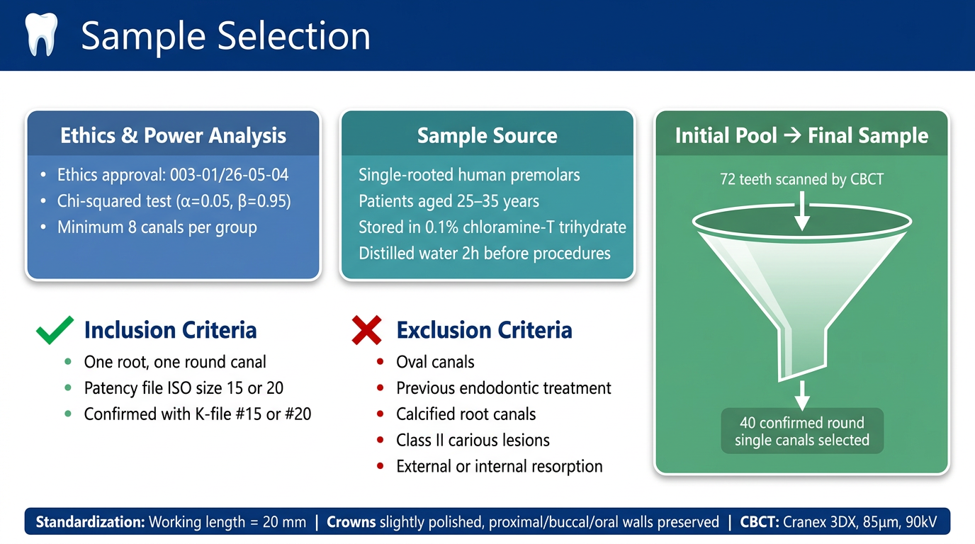
- Ethics & Power Analysis - approval number and statistical design
- Sample Source - who donated the teeth and storage conditions
- 72 → 40 funnel - CBCT screening process at a glance
- Inclusion vs. Exclusion Criteria - side-by-side green/red layout for instant readability
- Standardization bar at the bottom - working length, crown polishing, CBCT parameters
convert this into for my presentation
yes
convert this into for my presentation: Sample Preparation The same operator (GKP) performed root canal preparation, experimental protocols and obturation. Traditional access cavities were prepared using a water-cooled diamond fissure No. 016 (Komet, Rock Hill, SC, USA). Canal patency was confirmed according to the inclusion criteria. The working length (WL) was determined when the K-file was visible at the apical foramen under 6.5× magnification (Orascoptic eye zoom max Dragonfly pro, Orascoptic, Madison, WI, USA), and 0.5 mm was subtracted from the measured length. The apical foramina of all selected roots were sealed with hot glue and embedded in polyvinylsiloxane (Exaflex putty, GC, Lucerne, Switzerland) to create a closed system [27]. Root canals were instrumented using a reciprocating engine-driven system up to ISO size 50/0.05 (Reciproc Blue RB50, VDW Dental, München, Germany) using a motor set at reciprocation motion (Reciproc Gold, VDW Dental, München, Germany). During the instrumentation, 5 mL of 3.5% sodium hypochlorite (NaOCL) was used per canal, using a 31-gauge needle (SteriTips, DiaDent, Burnaby, BC, Canada) and 2 mL syringe
convert this into images for my presentation: Sample Preparation The same operator (GKP) performed root canal preparation, experimental protocols and obturation. Traditional access cavities were prepared using a water-cooled diamond fissure No. 016 (Komet, Rock Hill, SC, USA). Canal patency was confirmed according to the inclusion criteria. The working length (WL) was determined when the K-file was visible at the apical foramen under 6.5× magnification (Orascoptic eye zoom max Dragonfly pro, Orascoptic, Madison, WI, USA), and 0.5 mm was subtracted from the measured length. The apical foramina of all selected roots were sealed with hot glue and embedded in polyvinylsiloxane (Exaflex putty, GC, Lucerne, Switzerland) to create a closed system [27]. Root canals were instrumented using a reciprocating engine-driven system up to ISO size 50/0.05 (Reciproc Blue RB50, VDW Dental, München, Germany) using a motor set at reciprocation motion (Reciproc Gold, VDW Dental, München, Germany). During the instrumentation, 5 mL of 3.5% sodium hypochlorite (NaOCL) was used per canal, using a 31-gauge needle (SteriTips, DiaDent, Burnaby, BC, Canada) and 2 mL syringe. After the instrumentation, the root canals were dried using corresponding paper points, and the samples were randomly assigned to one of three test groups or one control group using a random number table as follows: Group 1. SWEEPS (n =10) The samples were treated using an erbium-doped yttrium–aluminum–garnet (Er:YAG) laser (LightWalker AT, Fotona, Ljubljana, Slovenia) with a radial laser tip (600 µm, 9 mm, Fotona, Ljubljana, Slovenia). During activation, the irrigant was constantly delivered in the access cavity using a 31-gauge needle (DiaDent), and the radial laser tip was inserted into the access cavity and maintained in a fixed position. Auto SWEEPS mode parameters were used: pulse energy (20 mJ), pulse frequency (15 Hz), average power (0.60 W), pulse duration (25 µs), and peak power (800 W). Activated irrigation was conducted in three cycles of 20 s according to the clinical final irrigation protocol: the first cycle included 5 mL of 3% NaOCl, followed by 5 mL of ethylenediaminotetraacetic acid (EDTA) in the second cycle, and 5 mL NaOCl in the last cycle. After each subsequent irrigant, the remains from the canal were aspirated. Group 2. Diode laser (n = 10) Root canals were irradiated in three cycles of 20 s with intervals of 10 s using a 975 nm diode laser with a 200 µm fiber tip (Laser HF, HAGER&WERKEN, Duisburg, Germany): peak power = 2W,timeon=5ms,timeoff =25ms. Thefibertip was placed 2 mmfrom the apical foramen and then moved circularly along the dentinal walls towards the coronal part of the root canal. Group 3. CI (n = 10) A 31 G side-vented needle (Steri Tips, DiaDent, Burnbay, BC, Canada) was placed in each root canal 2 mm from the WL. The root canals were irrigated according to the clinical https://doi.org/10.3390/bioengineering13060675 Bioengineering 2026, 13, 675 4 of 11 final irrigation protocol: 30 s of NaOCl (5 mL) followed by 60 s of EDTA (5 mL), then 30 s of NaOCl (5 mL). After each subsequent irrigant, the remains from the canal were aspirated. Group 4. Control Group, C (n = 10) No final disinfection protocol was conducted. After the final disinfection protocol had been completed, the residual irrigant was aspirated, and the canals were dried with the corresponding paper points (R50 Reciproc Blue, VDW GmbH, Munich, Germany). To ensure sufficient moisture for the premixed sealer to set, the canal was dried with the paper points until the tip remained slightly moist. Then the premixed CSBS (BioRoot Flow, Septodont, Saint Maur Des Fosses, France) was introduced into the coronal part of the canal with a suitable cannula. The corresponding gutta-percha point was slowly introduced in the canal up to the WL, allowing the sealer to gently distribute along the dentinal walls. The coronal excess of the gutta-percha point in the access cavity was cut off with a heated plugger, and then the filling was vertically condensed with a cold plugger size #2
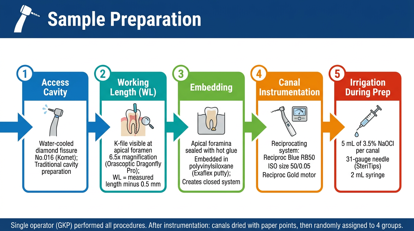
Professional presentation slide, wide 16:9 format, white background, dark blue header bar. Title: "Sample Preparation" with a tooth drill icon. Content in a clean step-by-step horizontal process flow with numbered steps and icons: Step 1 (blue box): "Access Cavity" - Water-cooled diamond fissure No.016 (Komet); Traditional cavity preparation Step 2 (teal box): "Working Length (WL)" - K-file visible at apical foramen; 6.5x magnification (Orascoptic Dragonfly Pro); WL = measured length minus 0.5 mm Step 3 (green box): "Embedding" - Apical foramina sealed with hot glue; Embedded in polyvinylsiloxane (Exaflex putty); Creates closed system Step 4 (orange box): "Canal Instrumentation" - Reciprocating system: Reciproc Blue RB50; ISO size 50/0.05; Reciproc Gold motor Step 5 (red box): "Irrigation During Prep" - 5 mL of 3.5% NaOCl per canal; 31-gauge needle (SteriTips); 2 mL syringe Bottom note bar: "Single operator (GKP) performed all procedures. After instrumentation: canals dried with paper points, then randomly assigned to 4 groups." Arrows connecting each step left to right. Clean modern academic design, readable fonts, well spaced.
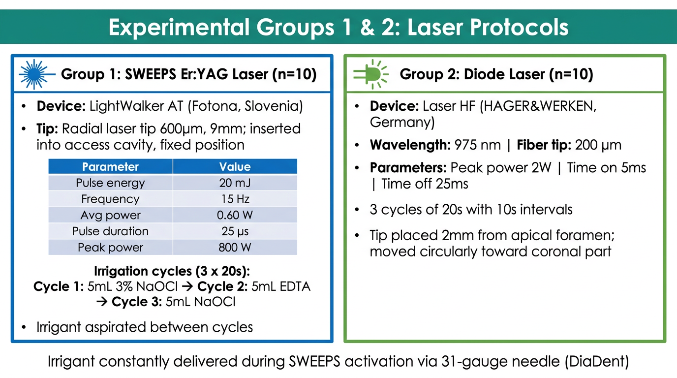
Professional presentation slide, wide 16:9 format, white background, teal green header bar. Title: "Experimental Groups 1 & 2: Laser Protocols". Two clearly separated columns side by side. LEFT COLUMN - blue border, header "Group 1: SWEEPS Er:YAG Laser (n=10)", laser beam icon: - Device: LightWalker AT (Fotona, Slovenia) - Tip: Radial laser tip 600µm, 9mm; inserted into access cavity, fixed position - Parameters table: Pulse energy 20 mJ | Frequency 15 Hz | Avg power 0.60 W | Pulse duration 25 µs | Peak power 800 W - Irrigation cycles (3 x 20s): Cycle 1: 5mL 3% NaOCl → Cycle 2: 5mL EDTA → Cycle 3: 5mL NaOCl - Irrigant aspirated between cycles RIGHT COLUMN - green border, header "Group 2: Diode Laser (n=10)", diode icon: - Device: Laser HF (HAGER&WERKEN, Germany) - Wavelength: 975 nm | Fiber tip: 200 µm - Parameters: Peak power 2W | Time on 5ms | Time off 25ms - 3 cycles of 20s with 10s intervals - Tip placed 2mm from apical foramen; moved circularly toward coronal part Bottom note: "Irrigant constantly delivered during SWEEPS activation via 31-gauge needle (DiaDent)" Clean modern academic design, clear readable fonts.
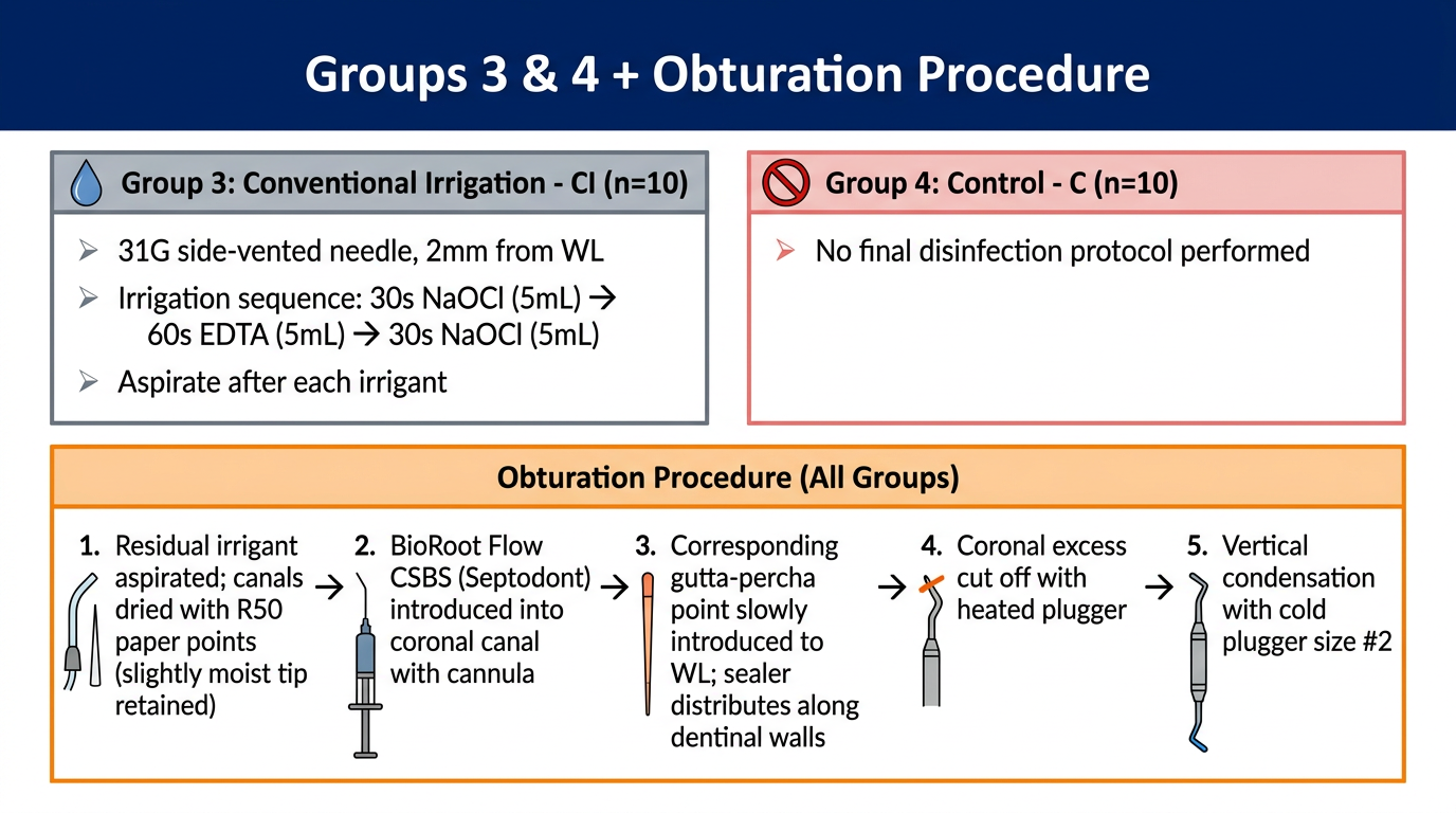
Professional presentation slide, wide 16:9 format, white background, dark navy header bar. Title: "Groups 3 & 4 + Obturation Procedure". TOP HALF - two side-by-side columns: LEFT column - grey border, header "Group 3: Conventional Irrigation - CI (n=10)", water drop icon: - 31G side-vented needle, 2mm from WL - Irrigation sequence: 30s NaOCl (5mL) → 60s EDTA (5mL) → 30s NaOCl (5mL) - Aspirate after each irrigant RIGHT column - light red border, header "Group 4: Control - C (n=10)", no-symbol icon: - No final disinfection protocol performed BOTTOM HALF - full width section, header "Obturation Procedure (All Groups)", orange accent: Step-by-step numbered list with icons: 1. Residual irrigant aspirated; canals dried with R50 paper points (slightly moist tip retained) 2. BioRoot Flow CSBS (Septodont) introduced into coronal canal with cannula 3. Corresponding gutta-percha point slowly introduced to WL; sealer distributes along dentinal walls 4. Coronal excess cut off with heated plugger 5. Vertical condensation with cold plugger size #2 Clean modern academic design, clear fonts, well-organized layout, arrows between steps.
| Slide | Content |
|---|---|
| 1 | Access cavity prep → WL determination → embedding → instrumentation → irrigation during prep |
| 2 | SWEEPS Er:YAG full parameters & irrigation cycles vs. Diode laser parameters & technique |
| 3 | Conventional irrigation (Group 3) + Control (Group 4) + 5-step obturation with BioRoot Flow |
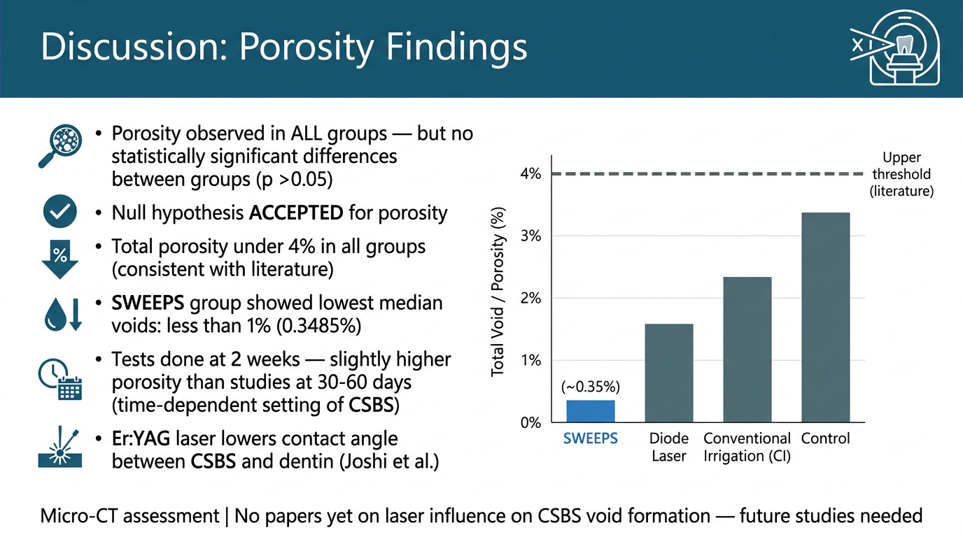
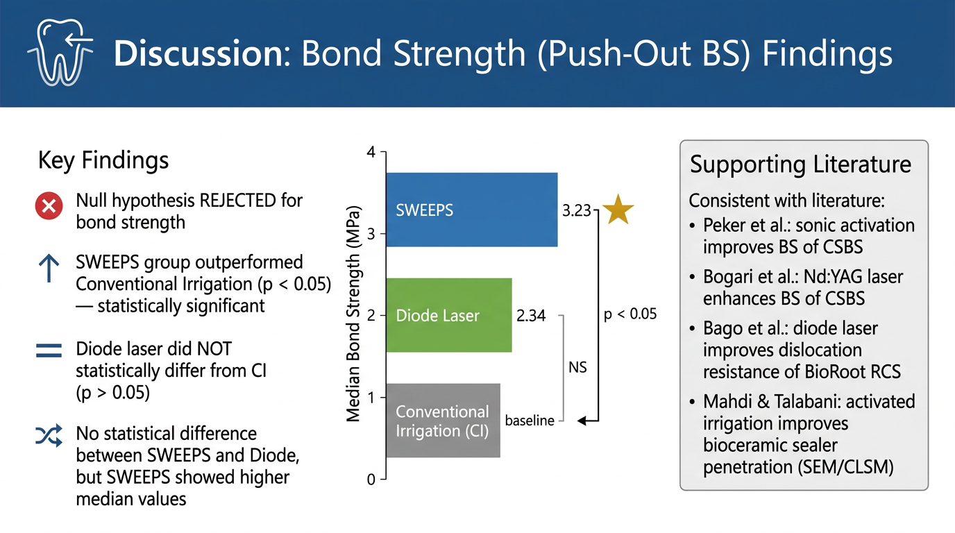
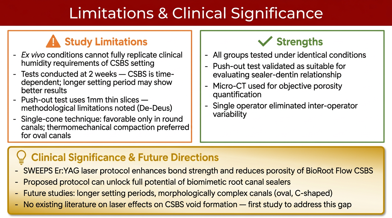
CONVERT THIS INTO IMAGE FOR MY PRESENTATION:The quality of root canal filling is most often estimated according to material ho mogeneity and the BS between the sealer and root canal dentin. Our study evaluated the porosity of a new CSBS, BioRoot Flow, after different laser-assisted protocols using a single-cone obturation technique in round canals. Regarding choosing the filling tech nique to ensure a tight junction between the dentinal walls and sealing material, there are no differences between the single-cone technique and the thermomechanical compaction technique in terms of the marginal gap when the root canal shape is rounded, but favorable results of thermomechanical compaction are noticed when the canal shape is oval, as stated by Zanza et al. [29]. Regarding material homogeneity in our study, porosity was observed in all tested samples, and micro-CT showed no statistical differences (p > 0.05) in the total void formation of BioRoot Flow CSBS after different disinfection protocols, namely two laser-assisted protocols (SWEEPS and diode laser), as well as a CI protocol; thus, the null hypothesis was accepted. In all groups, the total porosity was under 4%, which is https://doi.org/10.3390/bioengineering13060675 Bioengineering 2026, 13, 675 8 of 11 consistent with the results of other studies [30–32]. Inada et al. [32] compared the porosity of CSBS to that of AH plus and found that void formation in all groups was under 5%. A study by Nguyen et al. [30] observed less than 3% total voids when comparing CSBS with silicone bioactive glass-based and epoxy resin-based root canal sealers. The porosity was calculated after 30 and 60 days, which indicates the time dependence of CSBSs. In our study, the tests were done after 2 weeks, which possibly explains why the porosity showed slightly higher values. The porosity in all described studies was assessed by micro-CT. However, in our study, we compared different disinfection protocols in combination with the same CSBS (BioRoot Flow), and only in the SWEEPS group was the median percentage of voids less than 1% (0.3485%). Furthermore, the findings of Joshi et al. [33] showed that irradiation with an Er:YAG laser lowers the contact angle between CSBS and dentin, which could possibly have an effect on the BS and may also support further testing. Although laser-assisted protocols have been tested in various fields in recent years, to our knowledge there are no papers on the influence of laser on CSBS void formation. The benefits of further studies could be clinically important, especially for the treatment of root canals with morphological anomalies. Unlike void formation and porosity, the push-out BS has been analyzed more often in combination with different activated irrigation protocols. The results of our study showed that the SWEEPS laser-assisted protocol was beneficial with regard to the BS of the premixed CSBS (BioRoot Flow), which rejects the null hypothesis. The SWEEPS group outperformed CI (p < 0.05), but the diode laser group did not statistically differ from CI (p >0.05). Although there was no statistical difference between the SWEEPS and diode groups, SWEEPS showed higher median BS values (3.23 MPa) than the diode group (2.34 MPa), which aligns with the results of micro-CT analysis. Peker et al. [25] showed that sonic activation methods improve the BS of CSBS. Regarding laser-activated methods and BS, Bogari et al. [26] reported enhanced BS of CSBS after Nd:YAG laser treatment, and Bago et al. [34] showed that diode laser irradiation improves the dislocation resistance of a CSBS (BioRoot RCS). All results align with the findings of our study and support activation methods for enhancing the BS of bioceramic sealer. Despite the above mentioned results, the beneficial results of existing studies should be further examined due to the lack of information regarding the BS of CSBS after activation methods, especially laser-assisted ones. Although our study used SEM scanning for visualization and descriptive analysis, the results also correspond with other scientific data. Mahdi & Talabani [35] confirmed the bene ficial effect of activated irrigation methods, including diode laser irradiation, on bioceramic sealer penetration using SEM and confocal laser scanning microscopy (CLSM) analysis. One of the limitations of this study relates to the setting procedure of CSBS, which is specific due to its humidity requirements, and clinical conditions that cannot be recreated ex vivo, which may affect the results and, thus, their clinical applicability. Furthermore, considering the trend toward better results in the SWEEPS group and the findings of other studies, future examination should be conducted after a longer setting period [30]. For more than a decade, methodological problems of the push-out test have been pointed out. De-Deus [36] questions the use of 1 mm thin sliced specimens for testing root filling materials with two interfaces (core material and sealer), especially when ranking different materials, but nevertheless makes the point that push-out can contribute to understanding the properties of specific filling materials and their relationship to root dentin, which is exactly what this study aimed for. In addition, research showed that despite these limitations, the push-out test is still a suitable testing tool [37]. Moreover, the aim of this study was to evaluate the effect of different laser-assisted protocols rather than the material itself, and the protocols in all test groups were conducted under the same conditions. https://doi.org/10.3390/bioengineering13060675 Bioengineering 2026, 13, 675 9 of 11 The clinical significance of these results lies in proposing a protocol that can enhance the bonding ability of the premixed CSBSs and decrease their porosity, allowing the full potential of biomimetic root canal sealers to be reached.
Professional presentation slide, wide 16:9 format, white background, dark teal header bar. Title: "Discussion: Porosity Findings" with micro-CT icon. LEFT SIDE - key findings in clean bullet points with icons: - Porosity observed in ALL groups — but no statistically significant differences between groups (p > 0.05) - Null hypothesis ACCEPTED for porosity - Total porosity under 4% in all groups (consistent with literature) - SWEEPS group showed lowest median voids: less than 1% (0.3485%) - Tests done at 2 weeks — slightly higher porosity than studies at 30-60 days (time-dependent setting of CSBS) - Er:YAG laser lowers contact angle between CSBS and dentin (Joshi et al.) RIGHT SIDE - simple bar chart showing 4 bars labeled: SWEEPS (lowest, highlighted in blue ~0.35%), Diode Laser, Conventional Irrigation (CI), Control — all under 4% total void threshold. Dashed horizontal line at 4% labeled "Upper threshold (literature)". Y-axis: "Total Void / Porosity (%)". BOTTOM NOTE: "Micro-CT assessment | No papers yet on laser influence on CSBS void formation — future studies needed" Clean modern academic design, readable fonts.
Professional presentation slide, wide 16:9 format, white background, dark blue header bar. Title: "Discussion: Bond Strength (Push-Out BS) Findings" with bond/adhesion icon. LEFT SIDE - key findings with icons and bullet points: - Null hypothesis REJECTED for bond strength - SWEEPS group outperformed Conventional Irrigation (p < 0.05) — statistically significant - Diode laser did NOT statistically differ from CI (p > 0.05) - No statistical difference between SWEEPS and Diode, but SWEEPS showed higher median values CENTER - clean horizontal comparison bar chart, 3 bars: Bar 1 (blue, tallest): "SWEEPS" = 3.23 MPa — star symbol for best performer Bar 2 (green, medium): "Diode Laser" = 2.34 MPa Bar 3 (grey, shortest): "Conventional Irrigation (CI)" — baseline Y-axis: "Median Bond Strength (MPa)". Bracket showing p<0.05 between SWEEPS and CI. "NS" label between Diode and CI. RIGHT SIDE - supporting literature box: "Consistent with literature: - Peker et al.: sonic activation improves BS of CSBS - Bogari et al.: Nd:YAG laser enhances BS of CSBS - Bago et al.: diode laser improves dislocation resistance of BioRoot RCS - Mahdi & Talabani: activated irrigation improves bioceramic sealer penetration (SEM/CLSM)" Clean modern academic design, clear readable fonts.
Professional presentation slide, wide 16:9 format, white background, dark red header bar. Title: "Limitations & Clinical Significance". TOP HALF - two columns: LEFT column, orange border, header "Study Limitations" with warning icon, bullet points: - Ex vivo conditions cannot fully replicate clinical humidity requirements of CSBS setting - Tests conducted at 2 weeks — CSBS is time-dependent; longer setting period may show better results - Push-out test uses 1mm thin slices — methodological limitations noted (De-Deus) - Single-cone technique: favorable only in round canals; thermomechanical compaction preferred for oval canals RIGHT column, green border, header "Strengths" with checkmark icon, bullet points: - All groups tested under identical conditions - Push-out test validated as suitable for evaluating sealer-dentin relationship - Micro-CT used for objective porosity quantification - Single operator eliminated inter-operator variability BOTTOM HALF - full width, gold/amber accent box, header "Clinical Significance & Future Directions" with lightbulb icon: - SWEEPS Er:YAG laser protocol enhances bond strength and reduces porosity of BioRoot Flow CSBS - Proposed protocol can unlock full potential of biomimetic root canal sealers - Future studies: longer setting periods, morphologically complex canals (oval, C-shaped) - No existing literature on laser effects on CSBS void formation — first study to address this gap Clean modern academic design, readable fonts, well-spaced layout.
| Slide | Focus | Key Takeaway |
|---|---|---|
| 1 | Porosity | All groups under 4%; SWEEPS lowest at 0.35%; null hypothesis accepted |
| 2 | Bond Strength | SWEEPS (3.23 MPa) beats CI (p<0.05); diode laser not significant vs CI; null hypothesis rejected |
| 3 | Limitations & Significance | Ex vivo limitations, push-out test caveats, and clinical case for SWEEPS protocol |