
Mode of transport Diffusion and facilitied diffusion Detailed examination to.students With images
Reading File
Now let me search the medical library for comprehensive content on diffusion and facilitated diffusion.
Finding Sources
Finding Sources
Excellent results. Now let me read the main physiology sources - Costanzo and Guyton - which are the best textbooks for this topic. I'll read them in parallel.
Reading File
Reading File
Good. Let me now read the key diffusion sections from Costanzo (earlier offset) and Guyton's facilitated diffusion section, and also fetch relevant images.
Reading File
Reading File
Reading File
Now let me get the Guyton facilitated diffusion section and fetch key images.
Reading File
Loading Image
Loading Image
Now let me fetch the two critical diagrams - the facilitated diffusion rate comparison graph and the carrier protein mechanism diagram.
Loading Image
Loading Image
Excellent! I now have all the key textbook content and images. Let me also quickly search for Harper's content on GLUT transporters and get one more image showing all transport types.
Reading File
Finding Sources
Let me fetch the ping-pong model image and also get the Guyton section on simple diffusion (earlier section).
Loading Image
Reading File
I now have all the content and images needed for a comprehensive student examination guide. Let me compose the full detailed answer.
Modes of Transport: Diffusion and Facilitated Diffusion
A Detailed Examination Guide for Students
Overview: Classification of Membrane Transport
All membrane transport mechanisms are classified based on two key questions:
- Does it require metabolic energy?
- Does it involve a protein carrier?
| Transport Type | Energy Required | Carrier Protein |
|---|---|---|
| Simple Diffusion | No | No |
| Facilitated Diffusion | No | Yes (channel or carrier) |
| Primary Active Transport | Yes (ATP directly) | Yes |
| Secondary Active Transport | Yes (ATP indirectly) | Yes |
- Costanzo Physiology 7th Ed, p. 12
PART 1: SIMPLE DIFFUSION
Definition
Simple diffusion is the net movement of a substance from an area of higher concentration to an area of lower concentration due to random thermal (kinetic) motion of molecules - without any carrier protein.
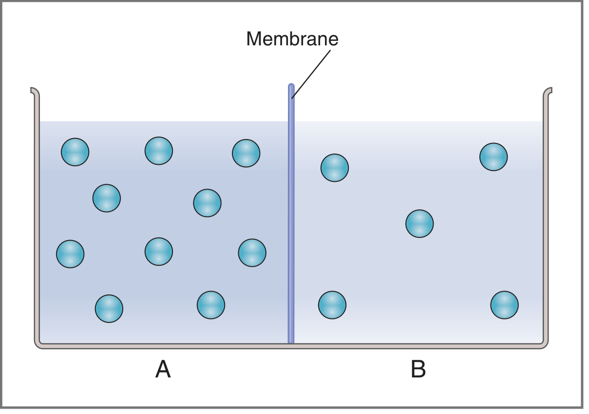
Fig. 1.5 - Simple diffusion. Solution A has a higher concentration; net diffusion moves toward Solution B until equilibrium. (Costanzo Physiology)
Two Subtypes of Simple Diffusion
A. Diffusion Through the Lipid Bilayer (Lipid-Soluble Substances)
Lipid-soluble (hydrophobic) substances dissolve directly in the membrane lipid and diffuse across without any protein help.
Examples: O₂, CO₂, N₂, alcohols, steroid hormones, lipid-soluble drugs
"The lipid solubilities of oxygen, nitrogen, carbon dioxide, and alcohols are high, and all these substances can dissolve directly in the lipid bilayer and diffuse through the cell membrane... Especially large amounts of oxygen can be transported in this way; therefore, oxygen can be delivered to the interior of the cell almost as though the cell membrane did not exist."
- Guyton & Hall Medical Physiology, p. 65
B. Diffusion Through Protein Pores/Channels (Lipid-Insoluble Substances)
Small, water-soluble, lipid-insoluble molecules pass through channel proteins (pores) that span the membrane.
Examples: Water (through aquaporins), small ions through ion channels
Aquaporins are a key example - they form narrow pores permitting water to pass in single file, but exclude ions entirely.
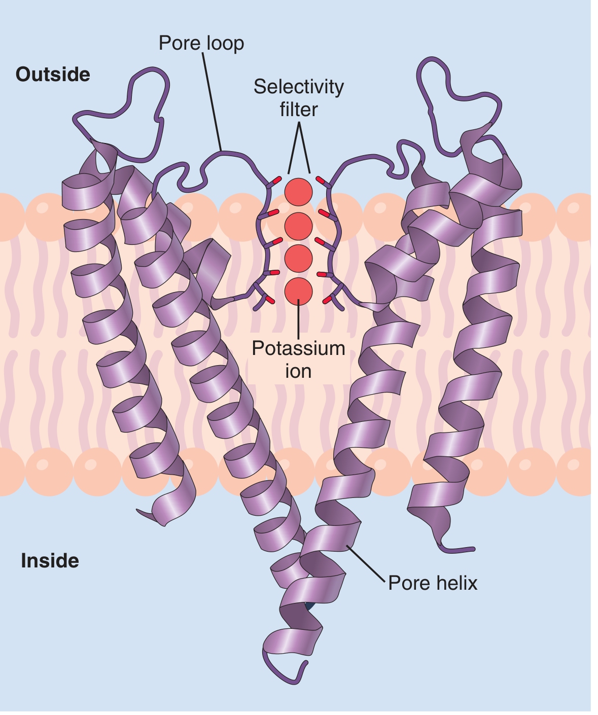
Fig. 4.4 - Potassium channel with selectivity filter. K⁺ ions pass through; Na⁺ ions are excluded due to spacing of carbonyl oxygens. (Guyton & Hall)
Fick's Law of Diffusion
Net diffusion (flux, J) depends on:
J = PA(C_A - C_B)
Where:
- J = Net rate of diffusion (mmol/s)
- P = Permeability (cm/s) - includes partition coefficient, diffusion coefficient, membrane thickness
- A = Surface area (cm²)
- C_A - C_B = Concentration gradient
The full set of determinants of diffusion rate:
| Factor | Effect on Diffusion Rate |
|---|---|
| Concentration gradient (↑) | Rate increases proportionally |
| Molecular weight (↑) | Rate decreases (heavier = slower) |
| Temperature (↑) | Rate increases |
| Membrane thickness (↑) | Rate decreases |
| Surface area (↑) | Rate increases |
| Partition coefficient (↑) | Rate increases (more lipid soluble) |
Diffusion of Electrolytes (Ions)
For charged particles, the driving force is not just concentration gradient but the electrochemical potential gradient (combining concentration + electrical forces). The Nernst equation describes the equilibrium potential for an ion.
PART 2: FACILITATED DIFFUSION
Definition
Facilitated diffusion is transport of a substance down its electrochemical gradient (from high to low concentration), but requiring a membrane carrier protein. No metabolic energy is used.
"Like simple diffusion, facilitated diffusion occurs down an electrochemical potential gradient; thus it requires no input of metabolic energy. Unlike simple diffusion, however, facilitated diffusion uses a membrane carrier and exhibits all the characteristics of carrier-mediated transport: saturation, stereospecificity, and competition."
- Costanzo Physiology 7th Ed, p. 15
Two Subtypes of Facilitated Diffusion
A. Via Channel Proteins
Ion channels allow specific ions to flow down their electrochemical gradients. Channels can be:
- Voltage-gated - opened by changes in membrane potential (e.g., Na⁺ channels in action potentials)
- Ligand-gated - opened by binding of a specific chemical (e.g., ACh receptor)
- Always open (leak channels)
B. Via Carrier (Transporter) Proteins
The substrate binds to a specific site on the carrier protein, which then undergoes a conformational change to release the substrate on the other side.
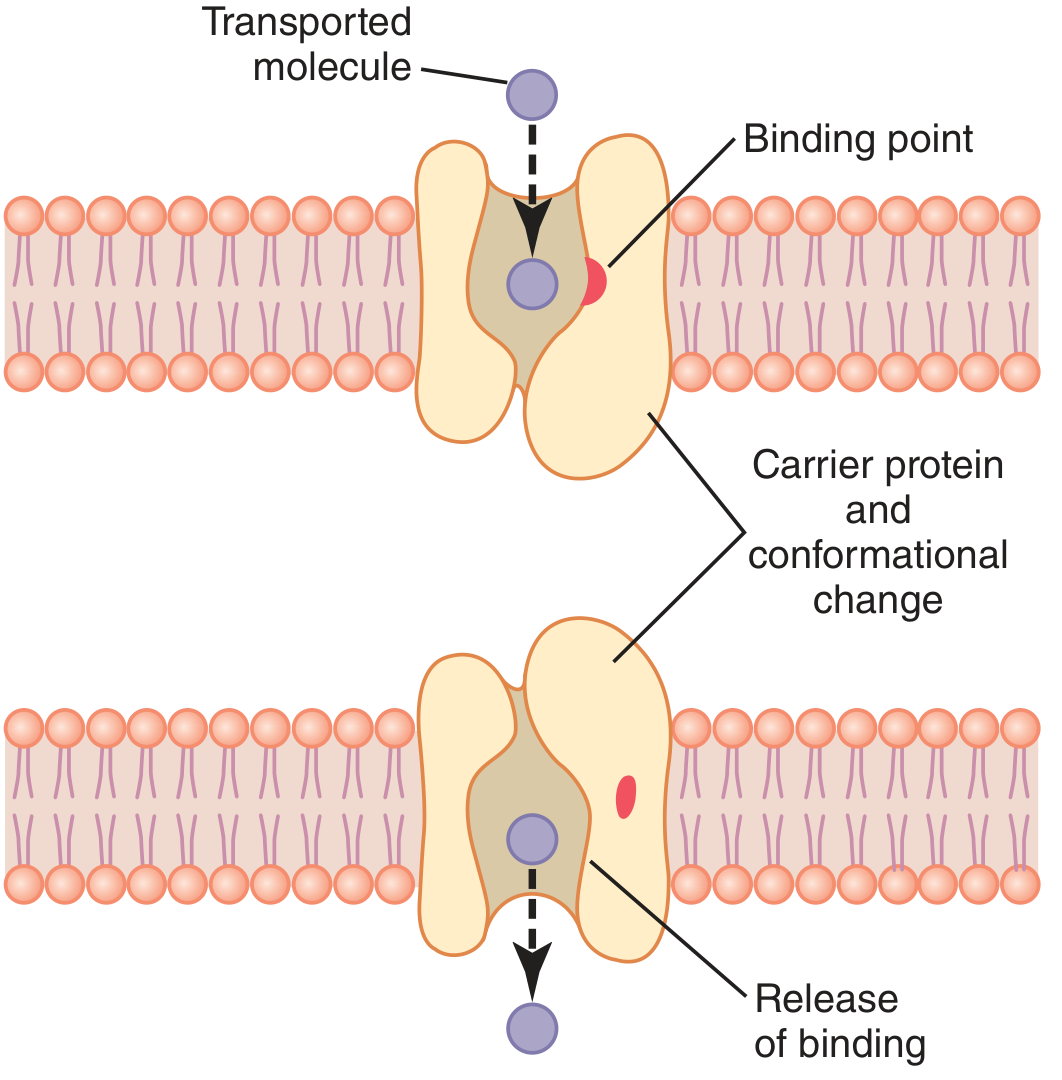
Fig. 4.8 - Mechanism of facilitated diffusion via carrier protein. The molecule enters, binds, conformational change occurs, then it is released on the opposite side. (Guyton & Hall)
The "Ping-Pong" Model (Harper's Biochemistry)
Harper's Illustrated Biochemistry describes facilitated diffusion using a "ping-pong" model:
- "Ping" state: Carrier is exposed to high concentration side - solute binds to specific sites
- "Pong" state: Conformational change exposes carrier to low concentration side - solute is released
- The empty carrier then reverts to the "ping" state to restart the cycle
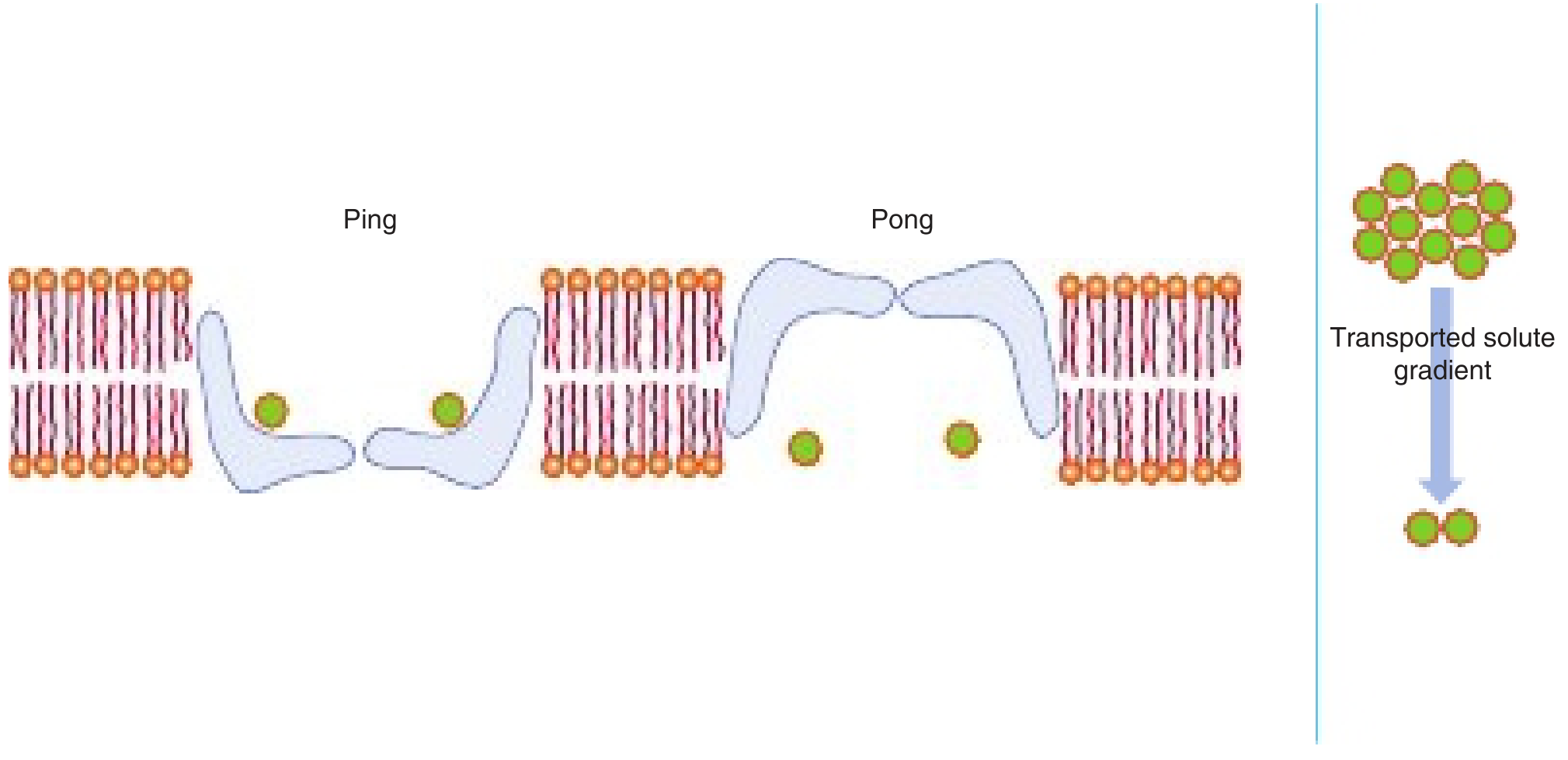
Fig. 40.14 - The "ping-pong" model of facilitated diffusion showing the carrier protein undergoing conformational changes. (Harper's Illustrated Biochemistry 32nd Ed)
The rate of facilitated diffusion is controlled by:
- Concentration gradient across the membrane
- Amount of carrier available (key rate-limiting step)
- Affinity of the solute-carrier interaction
- Speed of the conformational change for both loaded and unloaded carrier
Kinetics: The Critical Difference from Simple Diffusion
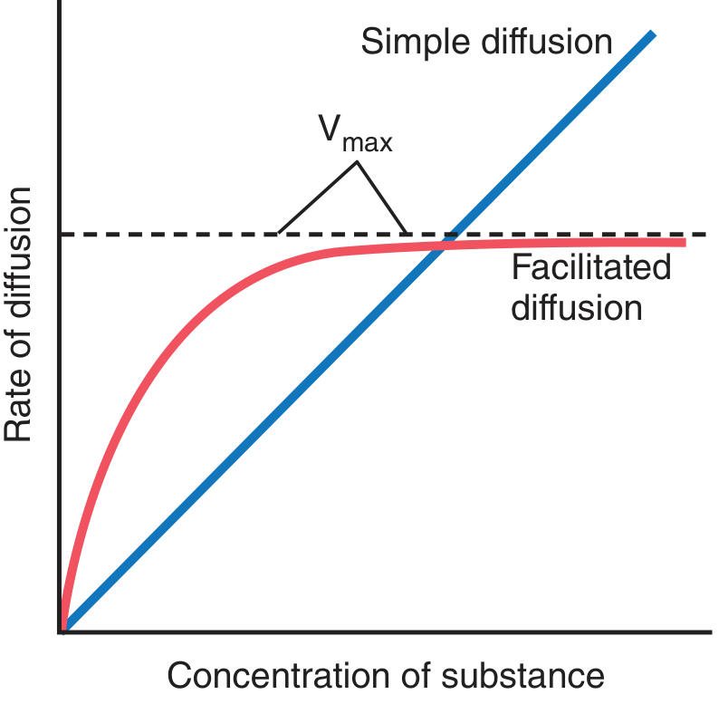
Fig. 4.7 - Comparison of simple vs. facilitated diffusion rates. Facilitated diffusion has a maximum rate (Vmax) due to carrier saturation; simple diffusion increases indefinitely with concentration. (Guyton & Hall)
Key point: At LOW concentrations, facilitated diffusion is FASTER than simple diffusion (due to carrier). At HIGH concentrations, facilitated diffusion plateaus (carriers become saturated) while simple diffusion continues to increase linearly.
Three Hallmark Properties of Carrier-Mediated Transport (All apply to Facilitated Diffusion)
1. Saturation
- Carriers have a finite number of binding sites
- At low concentration: rate increases steeply
- At high concentration: all sites occupied - rate plateaus at Transport Maximum (Tm), analogous to Vmax in enzyme kinetics
- Clinical example: Glucose reabsorption in renal proximal tubule - when plasma glucose exceeds renal Tm (~180-200 mg/dL), glucose spills into urine (glucosuria in diabetes)
2. Stereospecificity
- Carrier binding sites are stereospecific - they recognize molecular shape
- Example: GLUT4 transports D-glucose but NOT L-glucose
- Simple diffusion, having no carrier, does NOT distinguish stereoisomers
3. Competition
- Chemically related solutes can compete for the same binding site
- They may be transported themselves (D-galactose competes with D-glucose and is also transported)
- Or they may block without being transported (phlorizin blocks glucose transport by occupying its site)
Key Clinical Examples of Facilitated Diffusion
| Transporter | Substance Transported | Location | Clinical Relevance |
|---|---|---|---|
| GLUT1 | Glucose | Red blood cells, brain (blood-brain barrier) | Baseline glucose entry |
| GLUT2 | Glucose, fructose | Liver, pancreatic β-cells, intestine | High-Km sensor in β-cells |
| GLUT3 | Glucose | Neurons | High-affinity neuronal glucose uptake |
| GLUT4 | Glucose | Skeletal muscle, adipose, heart | Insulin-stimulated - key to type 2 diabetes |
| GLUT5 | Fructose | Small intestine, sperm | Fructose absorption |
| Aquaporins | Water | All cells | AQP2 regulated by ADH |
"One of these, glucose transporter 4 (GLUT4), can increase the rate of facilitated diffusion of glucose as much as 10- to 20-fold in insulin-sensitive tissues such as skeletal muscle, adipose, and heart. Insulin stimulates translocation of GLUT4 from the cell cytosol to the plasma membrane, thus increasing cell membrane density of the transporter and increasing the maximum rate (Vmax) of glucose diffusion into these cells."
- Guyton & Hall Medical Physiology, p. 68
PART 3: COMPARISON TABLE - Simple vs. Facilitated Diffusion
| Feature | Simple Diffusion | Facilitated Diffusion |
|---|---|---|
| Energy required | No | No |
| Carrier protein | No | Yes |
| Direction of transport | Down concentration gradient | Down concentration gradient |
| Rate vs. concentration | Linear (no limit) | Saturable (plateaus at Vmax/Tm) |
| Saturation | No | Yes |
| Stereospecificity | No | Yes |
| Competition | No | Yes |
| Inhibitable | Not specifically | Yes (competitive inhibitors) |
| Hormonal regulation | No | Yes (e.g., insulin upregulates GLUT4) |
| Examples | O₂, CO₂, steroid hormones, ethanol | Glucose (GLUT), amino acids, water (aquaporins), ions (ion channels) |
PART 4: HIGH-YIELD EXAM POINTS
- Both simple and facilitated diffusion are passive - NO ATP required
- Both move substances down their electrochemical gradient (from high to low)
- Facilitated diffusion is faster than simple diffusion at low concentrations but plateaus at high concentrations
- The Tm (transport maximum) concept applies ONLY to carrier-mediated transport (facilitated and active)
- Insulin increases GLUT4 insertion into the membrane - raising Vmax of glucose transport (not affecting Km)
- Phlorizin is a classic competitive inhibitor of glucose transport (used experimentally)
- Aquaporins are technically a form of facilitated diffusion (channel protein) - ADH regulates AQP2 insertion in collecting duct
- Voltage-gated Na⁺ channels (facilitated diffusion) drive the action potential - tetrodotoxin (TTX) blocks these channels
- Simple diffusion through lipid bilayer follows the partition coefficient - higher lipid solubility = faster diffusion
- Fick's Law formula: J = PA(C_A - C_B)
PART 5: CLINICAL CONNECTIONS
- Diabetes mellitus: Impaired insulin signaling → less GLUT4 on muscle/fat cell membranes → impaired glucose uptake (Guyton & Hall)
- Volatile anesthetics (halothane, sevoflurane): Highly lipid-soluble - cross blood-brain barrier by simple diffusion through lipid bilayer
- Renal glucosuria (Familial): Defective GLUT2 or SGLT2 → glucose spills into urine despite normal blood glucose
- Bartter syndrome / Gitelman syndrome: Defective ion channels/transporters in kidney tubules
Sources:
- Costanzo Physiology, 7th Ed, pp. 12-16
- Guyton and Hall Textbook of Medical Physiology, 14th Ed, pp. 64-68
- Harper's Illustrated Biochemistry, 32nd Ed, pp. 488-489
This is a shared conversation. Sign in to Orris to start your own chat.