
Why teeth are chawky in poisoning
chalky teeth poisoning fluorosis forensic toxicology mechanism
dental fluorosis chalky mottled teeth fluoride poisoning
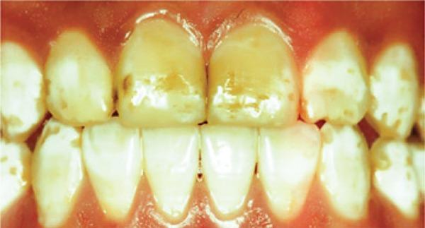
This intraoral clinical photograph displays the anterior dentition of a patient with severe dental fluorosis. The visual presentation is characterized by widespread enamel hypoplasia and significant intrinsic discoloration. The maxillary and mandibular teeth exhibit a loss of normal translucency, appearing opaque or chalky-white with prominent mottled patterns. Focal areas of moderate to dark brown extrinsic staining are visible, particularly on the labial surfaces of the central and lateral incisors. The enamel surface integrity is compromised, showing evidence of pitting, irregularities, and a matte texture rather than a healthy luster. The distribution is generalized across the visible arcade but most pronounced on the maxillary central incisors. This image serves as an educational example of advanced enamel defects caused by excessive fluoride ingestion during tooth development, categorized under the Dean's Index as severe, often requiring invasive restorative treatment.
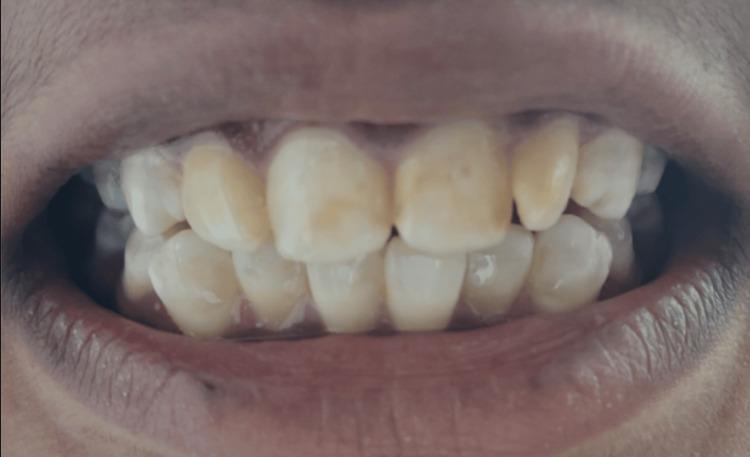
This intraoral clinical photograph displays the permanent dentition of a patient presenting with moderate dental fluorosis. The visual evidence shows a generalized, bilateral distribution of enamel hypomineralization across the maxillary and mandibular anterior teeth. Key findings include diffuse, chalky white opacities interspersed with prominent yellowish-brownish extrinsic staining, particularly localized on the labial surfaces and incisal thirds of the central and lateral incisors. The enamel lacks normal translucency, appearing opaque and mottled. While there is significant color alteration and surface irregularity, there is an absence of discrete pitting or large-scale structural loss of the enamel surface, characteristic of the moderate stage on the Dean's Fluorosis Index. This image serves as an educational example of how chronic fluoride ingestion during odontogenesis manifests as aesthetic dental defects and surface porosity.
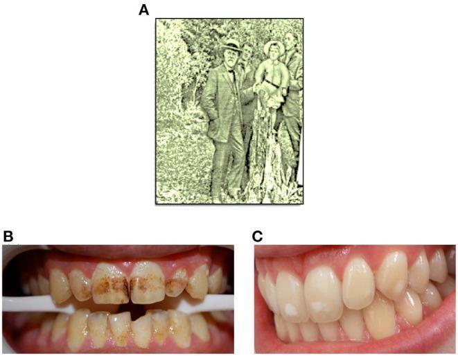
A multi-panel figure illustrating the history and clinical presentation of dental fluorosis. Panel A is a historical photograph from 1909 depicting Dr. G.V. Black in Colorado Springs. Panels B and C are clinical intraoral photographs showing different severities of enamel hypomineralization, historically termed 'mottled enamel.' Panel B demonstrates a severe case with 'Colorado brown stain,' characterized by prominent, intrinsic dark brown to tan discolorations and surface irregularities distributed across the labial surfaces and incisal edges of both maxillary and mandibular anterior teeth. Panel C shows a milder form of fluorosis, presenting as localized opaque white spots and diffuse cloud-like areas (mottling) on otherwise smooth and glossy enamel, indicating altered translucency without significant pitting or dark staining. This figure serves as an educational comparison between severe extrinsic-like staining and mild enamel opacities associated with chronic fluoride exposure during tooth development.
Why Are Teeth "Chalky" in Poisoning?
The Poison: Fluoride (Fluorine)
Mechanism: Why Does Fluoride Make Teeth Chalky?
-
Ameloblast toxicity: Excess fluoride directly damages ameloblasts - the cells that lay down enamel. This disrupts the normal secretory and maturation stages of enamel formation.
-
Disrupted calcium signaling: Fluoride impairs calcium handling within ameloblasts. Since enamel is heavily calcified (hydroxyapatite crystals), any disruption to calcium metabolism leads to hypomineralization - the enamel crystals form poorly and incompletely.
-
Subsurface porosity: Instead of a dense, translucent, well-mineralized enamel, the enamel becomes porous and hypomineralized, especially in the subsurface layer. This porosity scatters light rather than transmitting it, which is what gives the tooth its chalky white, opaque, dull appearance instead of the normal glossy translucency.
-
Progressive severity: As fluoride dose or duration increases:
- Mild: Small paper-white opaque spots (<25% of surface)
- Moderate: Mottled patches up to 50% of surface, brown staining
- Severe: Entire tooth becomes chalky white, pits develop, enamel fractures after eruption, brown discoloration
"Chalky white or yellowish brown areas of discolouration, striations and mottling on enamel, are found in fluorosis (excessive fluoride intake)." - Parikh's Textbook of Medical Jurisprudence, Forensic Medicine and Toxicology
Key Forensic Points
| Feature | Detail |
|---|---|
| Poison | Fluoride (chronic ingestion) |
| Critical window | Only during tooth development (pre-eruption) - adult exposure does NOT cause dental fluorosis |
| Teeth affected | Permanent teeth (those forming during the period of excess fluoride exposure) |
| Appearance | Chalky white, mottled, opaque, dull - can progress to pitting and brown staining |
| Other poisons affecting teeth/gums | Copper - green gum line; Mercury/Lead - blue-black line on gums |
Visual Examples
Other Poisons - Teeth/Gum Changes
- Lead and Mercury - blue-black line (Burton's line) on the gums
- Copper - green pigmentation of gums
- Phosphorus (chronic) - "phossy jaw" with osteonecrosis
- Tetracycline (not a poison, but a drug) - grey/brown intrinsic tooth staining when taken during tooth development