
Saq for bph from harsh mohan
benign prostatic hyperplasia histology nodules BPH gross microscopy
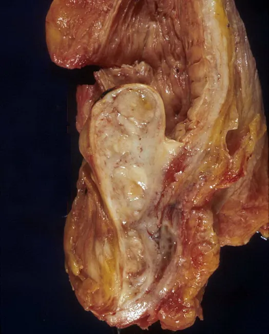
Gross pathology photograph of an autopsy prostatic specimen demonstrating benign prostatic hyperplasia with median lobe enlargement projecting into the bladder neck. The tissue appears tan‑brown and nodular, with a prominent midline lobe extending anteriorly toward the bladder outlet and creating a ball‑valve type obstruction at the bladder neck. The surrounding capsule and adjacent prostatic zones show variable, fibromuscular hyperplasia consistent with nodular hyperplasia rather than malignant invasion. This gross appearance reproduces the classic mechanism of lower urinary tract symptoms in aging men: progressive prostatic enlargement compresses the urethral channel, especially at the bladder outlet, producing obstructive and irritative voiding disorders. The autopsy context confirms a non‑malignant process, though histology would be required to definitively exclude focal carcinoma. Notable features include bilateral symmetry of nodular growth and conspicuous midline protrusion, which can be associated with reduced urinary flow, incomplete emptying, and increased postvoid residuals. Clinically, these findings underpin diagnostic considerations such as benign prostatic hyperplasia and differential diagnoses including prostatic adenocarcinoma or prostatitis; radiologic or endoscopic correlation may help stage obstruction. Educationally, this image serves as a reference for urology trainees and pathology learners to understand gross anatomy, angioarchitecture, and the anatomic basis of bladder outlet obstruction in BPH.
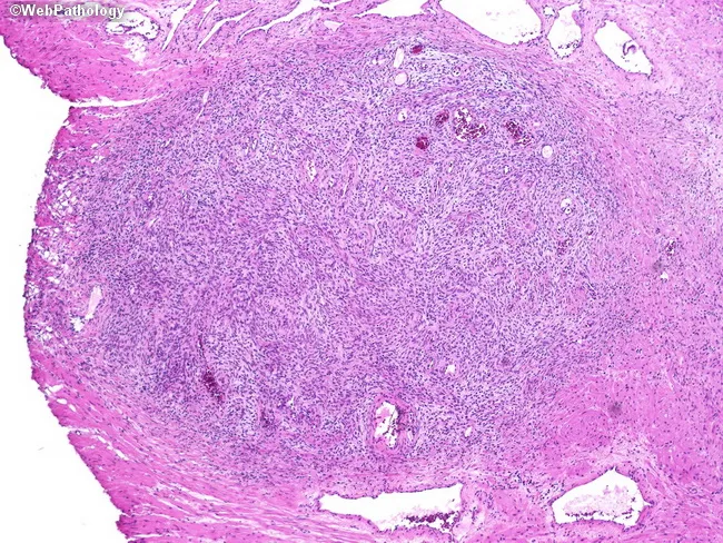
This is a histopathology slide of prostatic tissue obtained from transurethral resection of the prostate (TURP). Imaging modality: light microscopy; staining: hematoxylin and eosin. The specimen demonstrates nodular stromal hyperplasia composed of irregularly sized acini intimately admixed with dense fibromuscular stroma. The nodules display papillary infolding and variable gland size, a hallmark of nodular prostatic hyperplasia (benign prostatic hyperplasia, BPH). The glandular epithelium is typically cuboidal to columnar without significant cytologic atypia; stromal cells are fibromuscular and nonmalignant. Overall architecture is nodular with coarse borders and occasional dilated acini, consistent with benign prostatic tissue arranged in nodules rather than a uniform adenocarcinoma. No malignant features are identified: mitotic activity is not increased, and there is no prominent necrosis or invasion. In TURP specimens, the diagnostic focus rests on obstructive voiding symptoms and clinical presentation rather than morphologic abnormalities. The findings correlate with lower urinary tract symptoms due to prostatic enlargement and bladder outlet obstruction; they support a benign etiology for LUTS and guide management toward medical therapy or TURP as required. Important differentials include prostatic adenocarcinoma, prostatitis, and atrophy; however, absence of invasion or significant atypia favors benign nodular hyperplasia. Correlation with clinical history and imaging enhances diagnostic confidence overall.
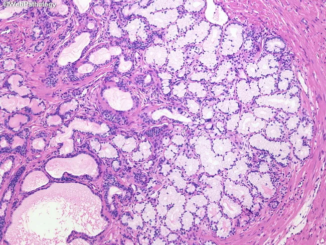
This histopathology image shows prostate tissue with mucinous differentiation within a benign prostatic hyperplasia (BPH) nodule. Prepared as a formalin-fixed, paraffin-embedded section and visualized by brightfield light microscopy, the slide is stained with Hematoxylin and Eosin (H&E). The right half of the field demonstrates extensive mucinous metaplasia in which the normal glandular epithelium is replaced by mucin-secreting cells. The involved glands appear lighter pink due to abundant mucin within the lumen and cytoplasm, producing a pale, glassy appearance compared with adjacent non-mucinous glands. Glandular architecture remains benign without overt invasion or high-grade cytologic atypia. The mucin pools are intra- and peri-luminal, sometimes compressing surrounding stroma but preserving overall prostatic acinar arrangement. This entity is considered an incidental, benign variant, with no malignant potential, and must be distinguished from mucinous (colloid) adenocarcinoma, which would demonstrate infiltrative growth and mucin lakes with malignant epithelial cells. The image highlights a spectrum of prostatic mucinous differentiation, emphasizing that mucinous metaplasia can occur within BPH nodules and often lacks clinical consequence. Clinically, recognition avoids misdiagnosis of prostatic mucinous carcinoma; pathologists should correlate with PSA, imaging, and clinical history for accurate interpretation. Clinical relevance emphasized.
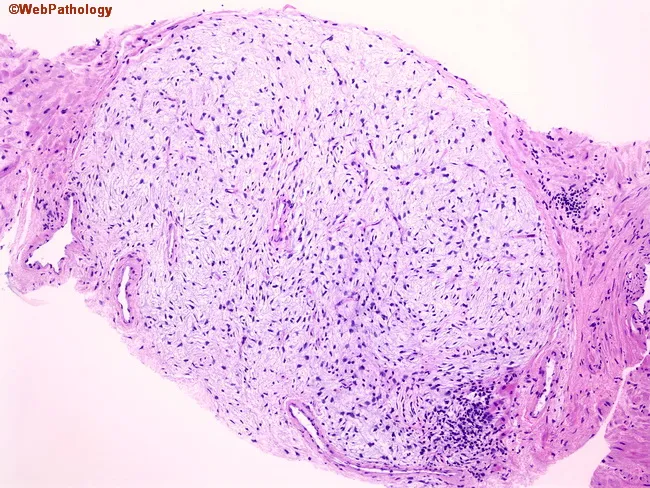
Light microscopy image of prostatic tissue stained with Hematoxylin and Eosin (H&E). The slide displays a well-circumscribed nodule composed of loose, fibromuscular stroma with sparse cellularity, representing an early stromal component of benign prostatic hyperplasia (BPH). Epithelial elements are scant within the nodule, with little glandular proliferation, and the surrounding prostate parenchyma shows a conventional fibromuscular stroma adjacent to residual glands. The stromal cells are spindle-shaped with elongated nuclei and pale cytoplasm embedded in a myxoid to fibrous extracellular matrix; mitotic activity is absent or minimal. There is no cytologic atypia, necrosis, or significant inflammatory infiltrate. This pure stromal nodularity represents one of the earliest histologic changes in BPH and can be encountered incidentally on prostate needle biopsies. Importantly, the diagnosis of prostatic hyperplasia (or nodular hyperplasia) should not be rendered on needle core biopsy; rather, specimens should be signed out as benign prostatic tissue with descriptive comment on the nodular stroma. Clinically, such findings may correlate with lower urinary tract symptoms in the patient, but histology alone cannot confirm severity. The differential considerations include stromal-rich lesions or scar tissue; malignant prostatic neoplasia is not suggested by these features. This pattern emphasizes strict sign-out criteria.
Short Answer Question: Benign Prostatic Hyperplasia (BPH)
Definition
Incidence / Epidemiology
- Most common benign prostatic disease in men over 50 years
- Frequency rises progressively with age
- Reaches ~90% by the 8th decade of life
- Does NOT predispose to carcinoma (important distinction)
Pathogenesis
- Testosterone is converted to DHT in prostatic stromal cells by the enzyme 5α-reductase type 2
- DHT is 10 times more potent than testosterone
- DHT binds nuclear androgen receptors → stimulates growth factors → increases stromal cell proliferation and decreases epithelial cell apoptosis
- With aging, testosterone levels fall but estrogen levels remain stable (or rise due to peripheral conversion); estrogens act synergistically with DHT via estrogen receptors on both stromal and epithelial cells
Morphology
Gross
- Prostate weight increases 3-5 fold (normal ~20 g → 60-100 g or more)
- Predominantly affects the transition zone (inner periurethral zone)
- Compresses the prostatic urethra to a slit-like orifice
- Cut section shows hyperplastic nodules - variable in color and consistency
- Nodules may be solid or contain cystic spaces (dilated glands)
Microscopic
- Hyperplastic nodules composed of variable proportions of glandular elements and fibromuscular stroma
- Glands lined by:
- Tall columnar epithelial cells (inner layer)
- Peripheral flattened basal cell layer (this double-layer distinguishes BPH from carcinoma, which lacks basal cells)
- Glandular lumina often contain corpora amylacea (laminated, eosinophilic, proteinaceous concretions)
- No cytologic atypia, no invasion
Clinical Features
- Mechanical obstruction - enlarging nodules compress the urethra
- Dynamic obstruction - increased α1-adrenergic tone causes smooth muscle contraction
- Difficulty initiating urination (hesitancy)
- Weak/interrupted urinary stream
- Incomplete bladder emptying → residual urine
- Overflow dribbling
- Urinary frequency
- Nocturia
- Dysuria (painful micturition)
- Residual urine → culture medium for bacteria → recurrent UTIs, cystitis, pyelonephritis
- Bladder hypertrophy and distension
- Complete urinary obstruction → hydronephrosis and renal damage
Diagnosis
- Digital Rectal Examination (DRE): smooth, rubbery, symmetrically enlarged prostate
- PSA may be mildly elevated (but not as high as in carcinoma)
- Ultrasound/TRUS: measures prostate volume
- Uroflowmetry: reduced peak flow rate
- Post-void residual urine measurement
Treatment
| Category | Drug/Procedure | Mechanism |
|---|---|---|
| Medical | α1-adrenergic blockers (e.g., tamsulosin, prazosin) | Relax prostatic smooth muscle → reduce dynamic obstruction |
| Medical | 5α-reductase inhibitors (e.g., finasteride, dutasteride) | Block DHT synthesis → reduce gland size over time |
| Surgical | TURP (Transurethral Resection of Prostate) | Gold standard for symptomatic cases refractory to medical therapy |
| Others | HIFU, laser therapy, radiofrequency ablation, thermotherapy | Reserved for selected cases |
Key Distinctions: BPH vs Prostatic Carcinoma
| Feature | BPH | Carcinoma |
|---|---|---|
| Zone | Transition (inner) | Peripheral (outer) |
| Basal cells | Present | Absent |
| Capsule invasion | No | Yes |
| PSA | Mildly raised | Markedly raised |
| Malignant potential | None | N/A |
| DRE | Smooth, rubbery | Hard, nodular, irregular |