
phagocytosis and clearance of offending agent
Reading File
Phagocytosis is the process by which specialized cells (mainly neutrophils and macrophages) engulf and destroy pathogens, dead cells, and foreign particles. Let me pull the detailed textbook content on this.
Searching sources...
Finding Sources
Finding Sources
Reading File
Reading File
Reading File
Reading File
Loading Image
Reading File
Reading File
Loading Image
Here is a comprehensive, textbook-sourced account of phagocytosis and clearance of the offending agent.
Phagocytosis and Clearance of the Offending Agent
Phagocytosis is the cellular ingestion of microbes, dead cells, and foreign particles. It is a central event in acute inflammation and the primary mechanism by which the body eliminates pathogens. The two major phagocytes are neutrophils and macrophages. Neutrophils are rapid first responders that live only 1-2 days in tissues; macrophages arrive later but persist for days to months (or years for tissue-resident forms) and are more powerful phagocytes.
Step 1: Recognition and Attachment
For phagocytosis to begin, the leukocyte must first recognize and bind the target. This occurs via several surface receptors:
- Mannose receptor - a lectin that binds terminal mannose and fucose residues found on microbial glycoproteins/glycolipids. Because mammalian glycoproteins are modified to contain terminal sialic acid, this receptor specifically recognizes microbes, not host cells.
- Scavenger receptors - bind a variety of microbes and also modified LDL particles.
- MAC-1 (CD11b/CD18) - macrophage integrins that also bind microbes.
The efficiency of phagocytosis is dramatically increased by opsonization - coating the microbe with molecules called opsonins. The major opsonins are:
- IgG antibodies - the antibody molecule also engages the complement cascade
- C3b - a breakdown product of complement; recognized by specific receptors on leukocytes
- Plasma lectins - including mannose-binding lectin and collectins
Antibodies adhere to bacterial membranes and combine with C3b of the complement cascade. C3b molecules then attach to receptors on the phagocyte membrane, initiating engulfment. This selection of a pathogen for phagocytosis is called opsonization.
Surface properties also matter: smooth surfaces resist phagocytosis; rough surfaces or surfaces lacking protective protein coats promote it. Most natural host cells have protective protein coats that repel phagocytes, while dead tissues and foreign particles typically lack these coats.
Step 2: Engulfment (Phagosome Formation)
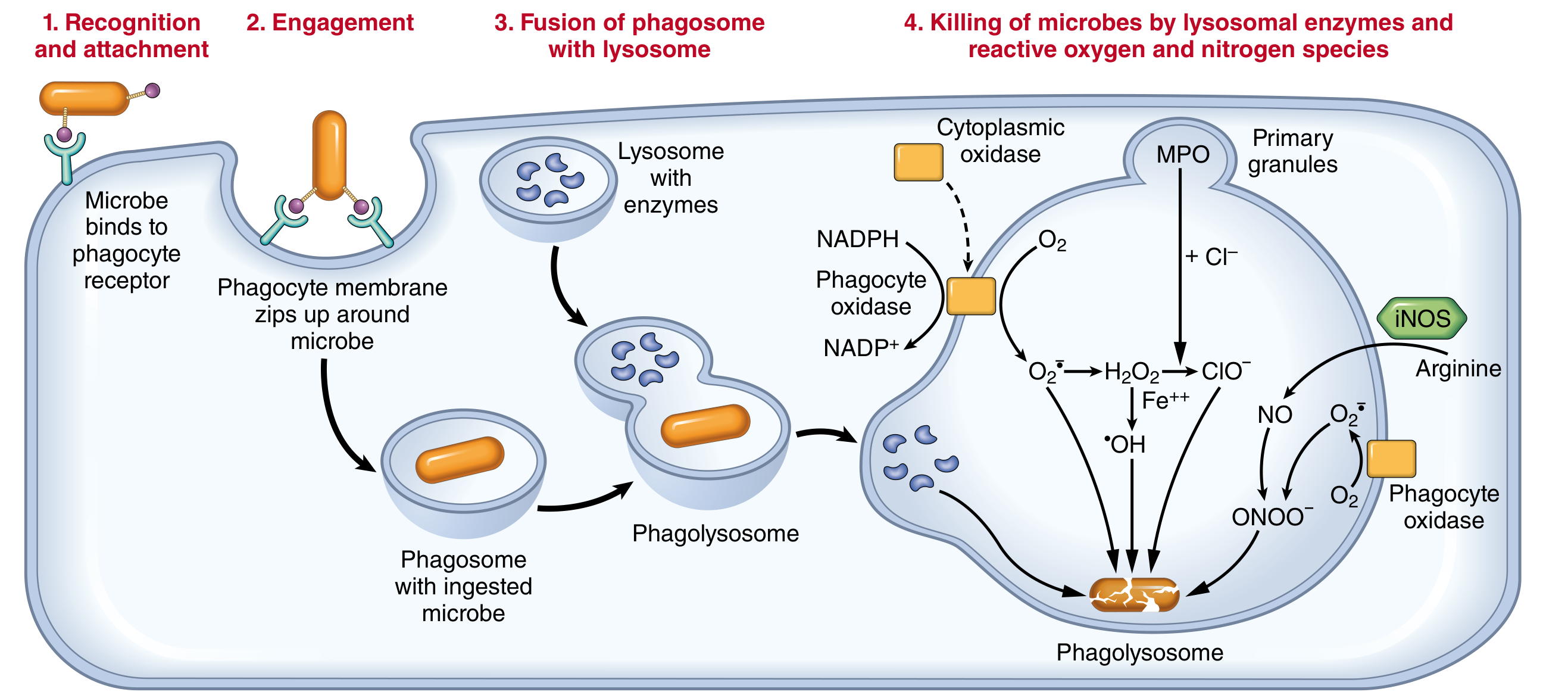
After receptor binding, the phagocyte extends pseudopodia that flow around the particle in all directions. The pseudopodia meet on the far side and fuse, creating an enclosed chamber. This chamber invaginates and breaks free from the outer cell membrane, forming a membrane-bound intracellular vesicle called the phagosome.
This process is complex and involves:
- Integration of multiple receptor-initiated signals
- Membrane remodeling
- Cytoskeletal changes, particularly polymerization of actin filaments
A single neutrophil can typically phagocytize 3-20 bacteria before becoming inactivated and dying. Macrophages (activated by the immune system) are far more powerful - they can phagocytize up to 100 bacteria, can engulf much larger particles (even whole RBCs or malarial parasites), and can survive and function for many more months after digesting particles.
Step 3: Phagolysosome Formation and Intracellular Killing
The phagosome fuses with a lysosomal granule to form the phagolysosome, which discharges lysosomal contents onto the ingested particle. Killing is accomplished by three main mechanisms:
A. Reactive Oxygen Species (ROS) - Respiratory Burst
- NADPH oxidase (phagocyte oxidase) assembles in the phagosomal membrane and oxidizes NADPH, converting O₂ to superoxide anion (O₂⁻)
- In neutrophils, this is accompanied by rapid O₂ consumption - the respiratory burst
- O₂⁻ is converted to hydrogen peroxide (H₂O₂) mostly by spontaneous dismutation
- H₂O₂ alone is a weak microbicide; however, the azurophilic granules of neutrophils contain myeloperoxidase (MPO), which, in the presence of Cl⁻, converts H₂O₂ to hypochlorite (ClO⁻) - the active ingredient in household bleach
- The H₂O₂-MPO-halide system is the most potent bactericidal system in neutrophils
- H₂O₂ can also react with Fe²⁺ (via Fenton reaction) to generate hydroxyl radical (•OH), another potent ROS
- ROS are produced within the phagolysosome to avoid damaging the host phagocyte
B. Reactive Nitrogen Species (RNS)
- Macrophages express inducible nitric oxide synthase (iNOS), which converts arginine to nitric oxide (NO)
- NO reacts with O₂⁻ to form peroxynitrite (ONOO⁻), a highly reactive microbicidal molecule
- Neutrophils produce little or no NO
C. Lysosomal Enzymes
- Lysosomal granules contain a variety of hydrolytic enzymes (proteases, lipases, nucleases) that degrade ingested material
- Neutrophils can also release granule contents into the extracellular space during phagocytosis, contributing to tissue injury if unchecked
- Examples: elastase, cathepsins, lysozyme, defensins (antimicrobial peptides), lactoferrin (sequesters iron from bacteria)
Additional Mechanism: Neutrophil Extracellular Traps (NETs)
NETs are extracellular fibrillar networks that concentrate antimicrobial substances and trap microbes, preventing systemic spread.
- Triggered by bacteria, fungi, inflammatory mediators (chemokines, cytokines, complement proteins, ROS)
- Consist of nuclear chromatin meshwork with bound granule proteins (antimicrobial peptides, MPO, elastase)
- NET formation begins with ROS-dependent activation of an arginine deaminase that converts arginines to citrulline, causing chromatin decondensation
- MPO and elastase enter the nucleus, cause further decondensation, and eventually the nuclear envelope ruptures and chromatin is released
- The neutrophil loses its nucleus and dies (NETosis)
- Particularly effective against large pathogens (e.g., fungi) that are too large to phagocytize
- In sepsis, NETs appear in the bloodstream
- The nuclear chromatin released in NETs (histones + DNA) is a proposed source of autoantigens in systemic lupus erythematosus
Comparison: Neutrophils vs. Macrophages
| Feature | Neutrophils | Macrophages |
|---|---|---|
| Origin | Bone marrow HSCs | Bone marrow HSCs (recruited); yolk sac/fetal liver (tissue-resident) |
| Lifespan in tissues | 1-2 days | Days-weeks (recruited); years (tissue-resident) |
| Response speed | Rapid, short-lived | Slower, prolonged, dependent on gene transcription |
| ROS production | Prominent (respiratory burst) | Less prominent |
| Nitric oxide | Low or none | Produced via transcriptional activation of iNOS |
| Degranulation | Major response | Not prominent |
| Cytokine production | Low or none | Major activity |
| NET formation | Yes | No |
| Capacity | 3-20 bacteria | Up to 100 bacteria; engulfs larger particles |
Summary
The sequence of phagocytosis and clearance is:
- Leukocyte activation by microbial products and cytokines
- Recognition of microbe via mannose receptors, scavenger receptors, or opsonin receptors (IgG, C3b)
- Engulfment - pseudopod extension, phagosome formation
- Phagolysosome formation - fusion with lysosomes
- Killing - by ROS (respiratory burst, MPO-H₂O₂-halide), RNS (NO, peroxynitrite), and lysosomal enzymes
- Exocytosis of digested debris (microbial fragments released by exocytosis)
As the injurious agent is eliminated and anti-inflammatory mechanisms become dominant, inflammation resolves.
Sources: Robbins, Cotran & Kumar - Pathologic Basis of Disease, pp. 89-92; Robbins & Kumar Basic Pathology, p. 31; Guyton and Hall Textbook of Medical Physiology, p. 453
This is a shared conversation. Sign in to Orris to start your own chat.