
patchy hairloss in young male
Reading File
Patchy hair loss in a young male has several common causes - alopecia areata (autoimmune, smooth bald patches), tinea capitis (fungal, scaly patches common in children), and less commonly secondary syphilis or traction alopecia. The pattern, scalp condition, and associated features guide the diagnosis.
Searching sources...
Finding Sources
Finding Sources
Searching PubMed
alopecia areata diagnosis treatment
Reading File
Finding Sources
Finding Sources
Reading File
Finding Sources
Finding Sources
Finding Sources
Reading File
Finding Sources
Loading Image
Here is a structured clinical overview of patchy hair loss in a young male, drawn from Fitzpatrick's Dermatology, Andrews' Diseases of the Skin, Goldman-Cecil Medicine, and Harrison's.
Patchy Hair Loss in a Young Male - Differential Diagnosis & Approach
1. Alopecia Areata (Most Common Cause)
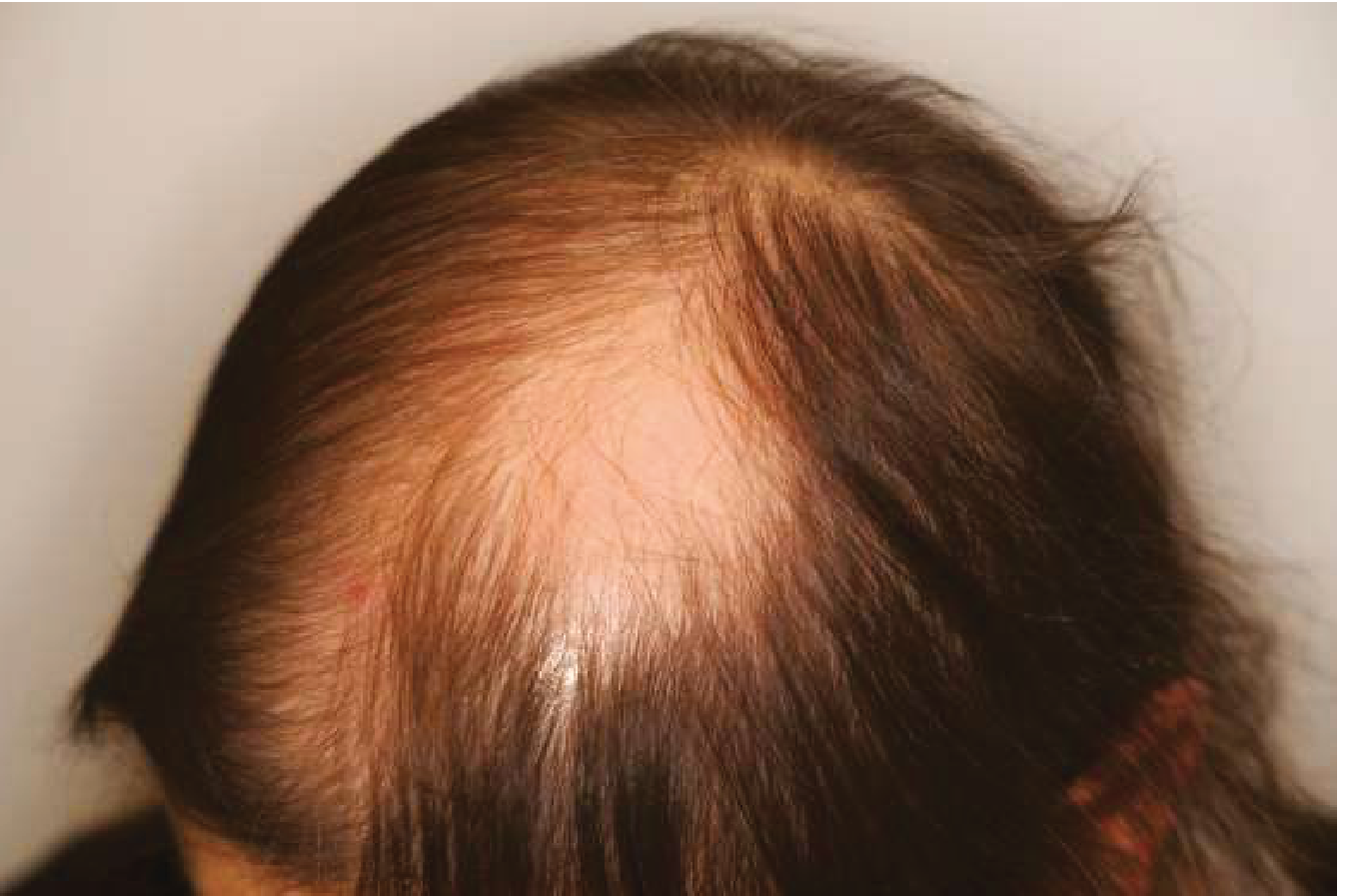
Alopecia areata: the alopecic area is devoid of hairs and the scalp shows no inflammatory changes, with diffuse thinning around the patch.
Pathophysiology: Autoimmune - oligoclonal autoreactive CD8+ T lymphocytes target follicular melanocytes in the hair bulb, collapsing the normal immune privilege of the anagen follicle. Early phase mediated by IL-2, IFN-γ, and TNF-α. - Andrews' Diseases of the Skin, p. 868
Clinical features:
- Rapid, complete loss of hair in round or oval patches, typically 1-5 cm in diameter
- Smooth scalp with no scarring, no scaling, no inflammation
- Pathognomonic: "exclamation point" (exclamation mark) hairs at the patch periphery - 3 mm broken hairs tapered at the base, with a pigmented tip, indicating active disease progression
- Eyebrows, eyelashes, and beard may also be involved
- Nail pitting in ~10% (fine uniform pits, trachyonychia)
- Severe forms: alopecia totalis (entire scalp), alopecia universalis (all body hair)
- Ophiasis pattern: band-like loss along temporal/occipital margins - poor prognosis
Dermoscopy: Diffuse round or polycyclic perifollicular yellow dots - Andrews', p. 868
Associations: Atopic dermatitis, thyroid disease (most common), vitiligo, Down syndrome, SLE, myasthenia gravis, diabetes mellitus
Prognosis: 34-50% recover within 1 year; 15-25% progress to totalis/universalis - Goldman-Cecil Medicine
2. Tinea Capitis (Fungal - especially in prepubertal males)
- Dermatophytosis of the scalp (most often Trichophyton tonsurans in endemic areas)
- Asymptomatic patchy hair loss with scaling, mild erythema, crusting
- "Black dot" pattern: broken hair shafts at scalp level
- More common in children; postpubertal males are relatively resistant due to sebum composition
- Wood's lamp fluorescence (green) with Microsporum species; T. tonsurans does NOT fluoresce
- Treatment: oral griseofulvin or terbinafine (topical antifungals are insufficient for scalp infection)
3. Secondary Syphilis ("Moth-Eaten" Alopecia)
- Irregularly distributed, poorly circumscribed patchy alopecia giving a "moth-eaten" appearance
- Occurs in ~5% of secondary syphilis cases
- Key: look for other secondary syphilis features - diffuse maculopapular rash (including palms/soles), condylomata lata, mucous patches, lymphadenopathy (especially epitrochlear), constitutional symptoms
- Must not be missed - always test RPR/VDRL + TPHA in any young sexually active male with patchy alopecia
- Andrews', p. 150; Harrison's p. 2075
4. Trichotillomania
- Compulsive hair-pulling disorder, more common in children and adolescents
- Patches are irregular, scalp is NOT completely bald - shows broken hairs of various lengths
- Frontal, parietal, and occipital scalp most affected
- Patient may deny the habit; parents may be resistant to accept diagnosis
- Psychiatric referral indicated in adults - Goldman-Cecil Medicine
5. Other Causes to Consider
| Cause | Key Feature |
|---|---|
| Androgenetic alopecia | Gradual bitemporal recession + vertex thinning, not truly "patchy" |
| Traction alopecia | Tight hairstyles, hairline recession |
| Alopecia neoplastica | Metastatic disease to scalp |
| Discoid lupus erythematosus | Scarring, dyspigmentation, follicular plugging |
| Congenital triangular alopecia | Fixed, non-progressive temporal patch |
Diagnostic Approach
History: Onset speed, family history of AA or autoimmune disease, STI risk, hair-pulling behavior, medications, stress
Examination:
- Smooth vs. scaly scalp?
- Scarring vs. non-scarring?
- Exclamation point hairs at margins?
- Nail changes?
- Other mucocutaneous signs?
Investigations to consider:
- KOH scraping and fungal culture if scaling present (r/o tinea capitis)
- RPR/VDRL in sexually active males (r/o syphilis)
- TSH, FT4 if AA confirmed (associated thyroid disease)
- Dermoscopy - yellow dots = AA; black dots = tinea capitis
- Scalp biopsy if diagnosis uncertain
Treatment of Alopecia Areata (most likely diagnosis)
| Severity | Treatment |
|---|---|
| Localized patches | Intralesional triamcinolone 2-10 mg/mL (treatment of choice for cosmetically visible patches) |
| Mild-moderate | High-potency topical corticosteroids, topical minoxidil |
| Rapidly progressive / widespread | Pulsed oral methylprednisolone 500 mg/day x 3 days/month |
| Severe / refractory | Oral JAK inhibitors - baricitinib (FDA-approved), ruxolitinib, tofacitinib, riticitinib |
| Chronic refractory | Contact immunotherapy (DPCP, SADBE), PUVA, anthralin |
Note: Natural history is highly variable - many localized patches regrow spontaneously within weeks to months. Treatment response does not change long-term prognosis. - Andrews' Diseases of the Skin, p. 869
Recent evidence (2024-2025): A network meta-analysis (PMID 39042154) confirmed that oral JAK inhibitors outperform systemic steroids and contact immunotherapy for severe alopecia areata. Baricitinib is currently FDA-approved for severe AA. A further 2025 network meta-analysis (PMID 40231941) corroborates JAK inhibitor superiority across monotherapy options.
This is a shared conversation. Sign in to Orris to start your own chat.