BASIC RADIATION:
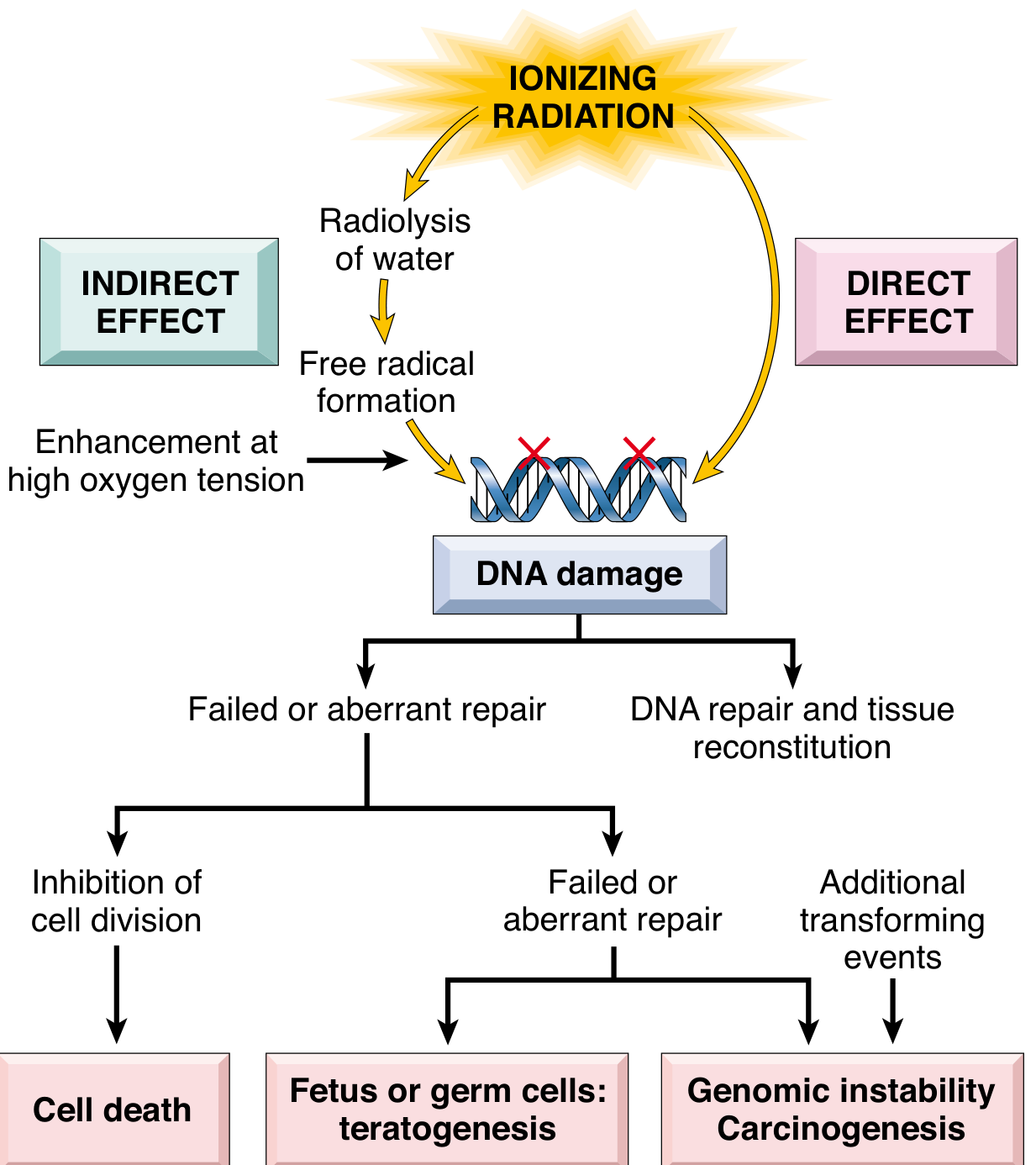
LONG QUESTION: Discuss in detail about the effect of ionizing radiation on Biologic molecules. Write a note on
Osteoradionecrosis. (2018)
Describe the measures of protection from radiation hazards. (16)
7. Define electromagnetic radiation? Describe structure and function of x ray tube. (12)
Biological effects of radiation? (14,23),
NS. Describe x-ray tube with diagram? (13)
6. Discuss in detail about the parts of X-ray tube with diagram. Write in short about production of
x-ray. (19)
What are the protection measures for the patient and the operator against the adverse effect of
radiation? (12)
What is x-ray? How x-ray is produced? What are the radiation hazards? (11)
Describe in details about the hazards of radiation. Describe in detail about various methods of
radiation protection. (20s)
10. Draw a labeled diagram of x-ray head and explain in detail about the working mechanism of x-ray
tube head. (20)
11. Discuss the methods of radiation safety and protection of operator, patient and public. Write a short
note on Acute radiation syndrome. (20)
SHORT NOTE:
Radiation Mucositis. (2018)
Characteristics Radiation. (2018)
Osteoradionecrosis. (16,13)
TLD Badge. (16)
Properties of x-ray. (12,13)
Photoelectric effect. (12,14,16)
Collimation (20s,17,12,14,22)
Breaking Radiation. (15,23)
9. Compton scatters. (12)
10.- Thermo luminescent dose meters. (12)
11. Radiation monitoring badge. (12)
Filtration. 9,)
N.'13. Radiolysis of water 19
14. Write in brief about dosimetry. (20)
15. Production of X-ray. (22)
16 Radiation effects on oral cavity. (22)
EQUIPMENT, FILM AND PROCESSING BAŞICS:
LONG QUESTION:
1. What are the types of Intraoral films? Their size, advantage, disadvantage & uses. (2017)
2 Describe the procedure of processing of radiograph and enumerate the reasons of fatty radiograph.
(15)
2. 4. Describe in detail the process of film development. (14)
Define image sharpness and resolution? Describe the principles and methods for minimizing loss of
image clarity. (07)
5. Factors controlling x-ray beam. (07)
Discuss in detail about processing of radiograph and formation of latent image. (22)
Brought To You By Dr. RADC TMCP UNIT Page 1
Scanned with OKEN Scanner
ORAL RADIOLOGY & ORAL MEDICINE
Final Prof. B.D.S.
Brought To You By Dr. RADC TMCP UNIT
SHORT NOTE:
. Developing & Fixing Solution. (2018)
2. Dark room. (19,17,14).
S. Film fog. (13)
Developer solution. (20s,12)
5. X-ray film. (20s,12)
рб. Composition of OPA film. (14).
Factors affecting contrast of
radiograph. (06)
8. Fixer solution. (19,16,08)
9. Different size of intraoral film. (08)
10. Intensitying screen.
(20s,17,08,09,16,21)
t. Film speed. (06)
Nt2. Fixer solution in radiology. (20)
13. Classification of X-ray films(2023)
TECHNIQUE BASICS:
LONG QUESTION:
PU 3. Write in detail about Sialography & its significance in various diseasés of Salivary glands? (2017)
Briefly describe the principle, advantages, disadvantages, indication and contraindication of Cone
Beam Computed Tomography imaging. (15)
Describe the technique of the Occlusal radiograph to detect a sialolith in the submandibular duct.
Y Describe the principle of OPG along with its uses, advantages, disadvantages, indication and
contraindication. (16,13)
What is the principle of panoramic imaging? Write in detail about the indications, limitations of
panoramic imaging and the faults in OPG images.'20s
Describe the technique of Sialography? What is its indication? (12,09) ~
Write in detail about sialography and its significance in various diseases of the salivary gland. 19 %
Déscribe in detail about different conventional and advanced radiography for TMJ disorder. (07)
Describe in detail about the principles, indication, contraindication, advantages and disadvantages of
orthopantomography. (21)
f. Describe techniques of intraral periapical radiograph in detail with merits and demerits. (22)
11. Describe in detail about different conventional and advanced radiography in TM Joint. (2023)
SHORT NOTE, PNS Radiography. (2018,22)
Ghost Image. (2018)
SLOB Technique. (2017)
Faulty Radiographs. (2017,2019,2020s)
Causes of Dark Radiograph. (16)
6. Lateral oblique radiograph. (16)
Artifacts. (15)
Tomography. (12) /
Grids and Grid Ratio. (15,23)
Common errors in radjography. (12)
Xi 'Causes of light radiograph. (15)
Grid. (2018,13,14)
XI. Bisecting angle technique. (14)
Occlusal radiograph. (17,14)
Diagnostic importance of
Orthopantomograph. (15,22)
Ciezensky's rule of isometry. (13)
What is the basic principle of OPA
technique? (06)
18. Principles of taking ideal radiograph.
(07,23)
Reverse town's projection of skull.
(05,07,21,23)
20. RVG. (06)
Brought To You By Dr. RADC MCP UNIT
Page 2
Scanned with OKEN Scanner
ORAL RADIOLOGY & ORAL MEDICINE
Final Prof. B.D.S.
Brought To You By Dr. RADC TMCP UNIT
21. Bite Wing Radiography. (15)
22. Ultrasound assay in orofacial disorder.
(03)
CBCT in maxillofacial region. (19)
Object localization (19,22)
Importance of CCT. (20s)
(26 Technique of IOPA radiograph. (20s)
Principles and applications of CBCT (20)
28. Grids (20)
Transcranial view (20)
30. Diagnostic importance of sialography.
(22)
P 31. Ghost image in OPG (2023)
NORMAL ANATOMY AND FILM MOUNTING BASICS:
LONG QUESTION:
1. Write in detail about normal anatomical landmarks of maxilla and mandible with diagram. (2018)
• SHORT NOTE:
2. 3. 4. 1. Lamina Dura. (13)
View for maxillary sinus. (12)
Normal anatomical landmarks in mandible as seen in radiograph. (09)
Common radiopaque lesions of the mandible. 08)
RADIOGRAPHIC INTERPRETATION BASICS:
1. Radiological features of Dentigerous Cyst. (2018)
2. Radiographic features of Radicular Cyst. (2017)
3. Pericoronal radiolucencies. (13)
4. Radiographic features of Osteomyelitis. (15)
5. Define Hamartoma. Enumerate hamartomas affecting the oral and dental tissues. Describe in brief
about the clinical features, radiological features and treatment of hemangioma affecting the oral
cavity. (21)
6. Principles of formulation of radiographic diagnosis 21)
7. Radiographic features of adenomatoid odontogenic tumor(2023)