Here are complete answers for all questions in Veterinary Biochemistry context:
QUESTION 5 - Definitions (1×11 = 11 marks)
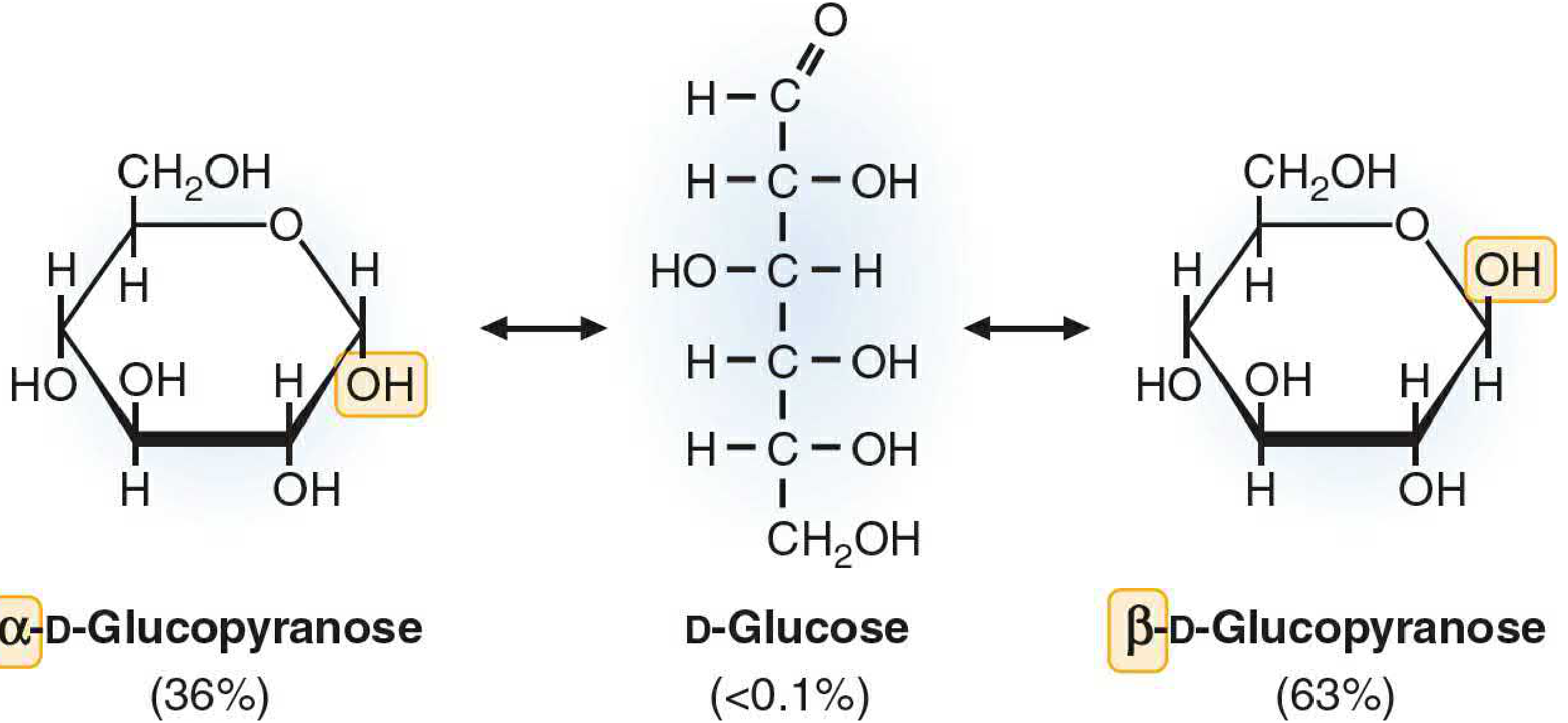
a) Mutarotation
(Covered in detail in previous session - brief recap)
Mutarotation is the spontaneous interconversion between the α and β anomeric forms of a sugar in solution, passing through the open-chain (aldehyde/ketone) intermediate.
- At equilibrium in glucose: ~36% α + ~64% β, with specific rotation = +52.5°
- Catalyzed by mutarotase in cells
- Vet relevance: Glucose oxidase-based analyzers measure only β-glucose; mutarotase is added to commercial kits for accurate results in veterinary diagnostics
b) Glycosidic Bond
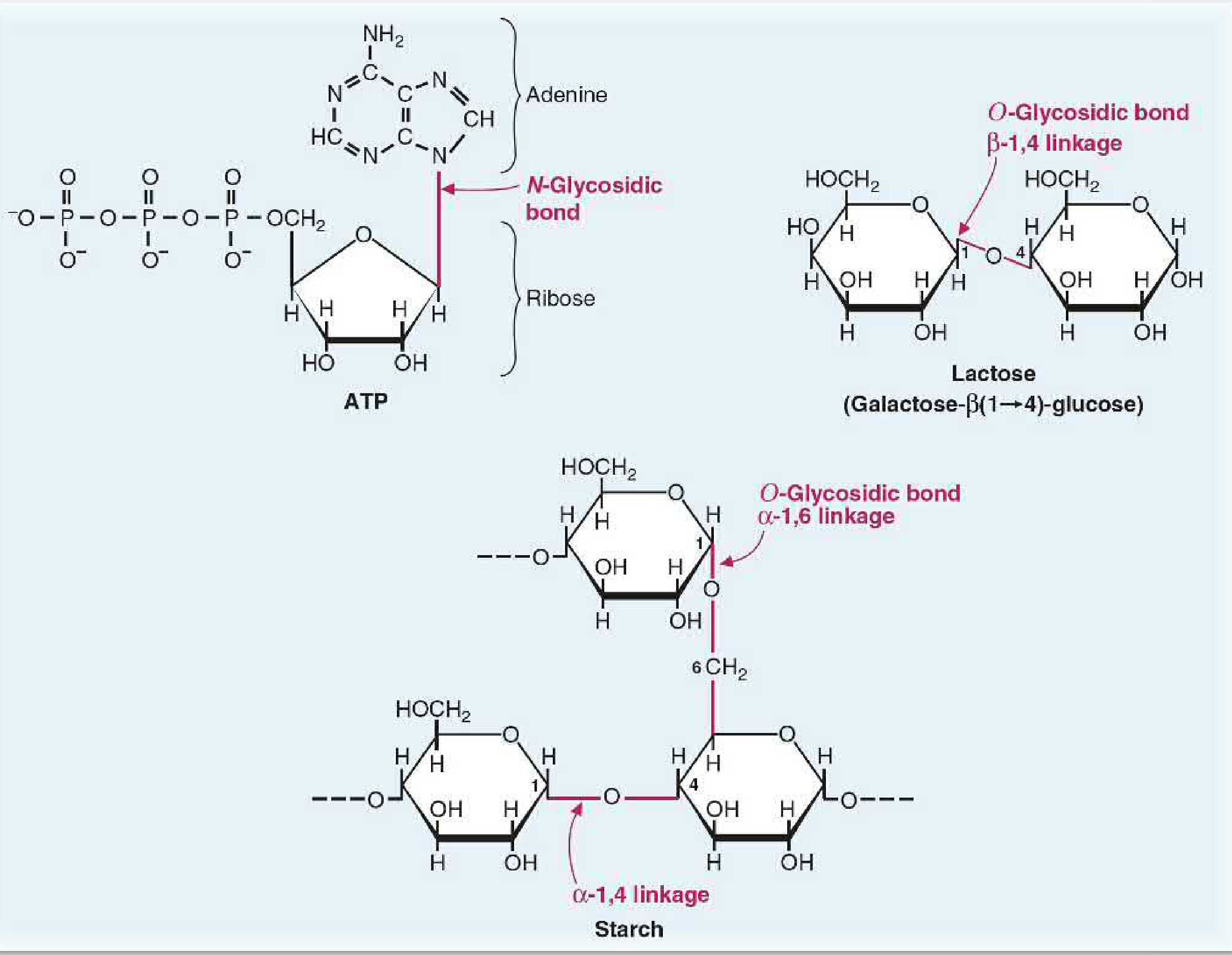
A glycosidic bond is a covalent bond formed between the anomeric carbon (C1) of a monosaccharide and an -OH (O-glycosidic) or -NH (N-glycosidic) group of another molecule.
Mechanism: The -OH group on the anomeric carbon reacts with the -OH or -NH group of another compound → water is eliminated → glycosidic bond forms, fixing the sugar in either the α or β configuration (preventing further mutarotation).
Types:
| Type | Bond | Example |
|---|
| O-glycosidic (α) | Anomeric -OH to another -OH | Starch (α-1,4 bonds), glycogen (α-1,4 and α-1,6 bonds) |
| O-glycosidic (β) | Anomeric -OH to another -OH | Lactose β(1→4), cellulose β(1→4) |
| N-glycosidic | Anomeric -OH to -NH | Nucleosides (base attached to ribose), glycoproteins |
N- and O-glycosidic bonds (Basic Medical Biochemistry, 6e)
Veterinary relevance:
- Ruminants can digest cellulose (β-1,4 glycosidic bonds) via rumen microbial cellulases - monogastrics cannot
- Lactase deficiency in neonatal animals = inability to hydrolyze β(1→4) bond of lactose → lactose intolerance, diarrhea
- Glycogen phosphorylase cleaves α-1,4 bonds in glycogen for energy mobilization in animals
c) Lipoproteins
(Covered in detail - brief recap)
Lipoproteins are macromolecular complexes of lipids and apolipoproteins that transport water-insoluble lipids (triglycerides, cholesterol, phospholipids) through aqueous blood plasma. Classified by density: Chylomicrons → VLDL → IDL → LDL → HDL (increasing density, decreasing size).
Vet note: In cats and dogs, HDL is the predominant lipoprotein. Hypothyroid dogs develop hyperlipidemia (elevated LDL/VLDL).
d) Albumin-Globulin (A:G) Ratio
The A:G ratio is the ratio of serum albumin concentration to serum globulin concentration:
A:G ratio = Albumin (g/dL) / Globulin (g/dL)
Globulin = Total protein - Albumin
Normal values in animals:
| Species | Albumin (g/dL) | Globulin (g/dL) | A:G Ratio |
|---|
| Dog | 2.6-4.0 | 1.6-3.6 | ~1.0-1.5 |
| Cat | 2.3-3.9 | 2.3-5.3 | ~0.8-1.5 |
| Horse | 2.6-3.7 | 2.6-4.0 | ~0.7-1.5 |
| Cattle | 3.0-3.8 | 2.7-4.6 | ~0.8-1.4 |
Clinical interpretation:
| A:G Ratio | Interpretation | Cause |
|---|
| Low (<0.5) | Hypoalbuminemia and/or hyperglobulinemia | Chronic liver failure, protein-losing nephropathy (PLN), PLE, FIP in cats (very low ratio), chronic inflammation |
| High (>2.5) | Hyperglobulinemia absent, or hyperglobulinemia without hypoalbuminemia | Dehydration |
| Reversed ratio | Albumin < Globulin | FIP (cats) - hallmark finding: marked hyperglobulinemia + hypoalbuminemia |
Vet relevance: A reversed or markedly decreased A:G ratio (<0.4) in cats is strongly suggestive of Feline Infectious Peritonitis (FIP) - caused by feline coronavirus.
e) Hyperinsulinism
Hyperinsulinism is a condition of excess insulin relative to the blood glucose level, resulting in hypoglycemia (low blood glucose).
Causes in animals:
| Type | Cause | Species |
|---|
| Insulinoma (functional beta-cell tumor) | Autonomous insulin secretion from pancreatic islet tumor | Dogs most common; also cats, ferrets |
| Insulin overdose | Iatrogenic - too much insulin given to diabetic animal | Dogs, cats |
| Nesidioblastosis | Beta-cell hyperplasia | Rare; neonates |
| Post-prandial reactive hypoglycemia | Excessive insulin response to carbohydrate meal | Rare in animals |
| Neonatal hypoglycemia | Limited gluconeogenesis + high insulin sensitivity | Piglets, foals, puppies |
| Juvenile hypoglycemia | Inadequate glycogen stores + high metabolic demand | Toy breed puppies |
Biochemical consequences:
- Blood glucose <60 mg/dL (dogs) → neurological signs
- Neuroglucopenia → weakness, seizures, collapse
- Counter-regulatory hormones activated: glucagon, epinephrine, cortisol, GH
Diagnosis: Whipple's triad:
- Clinical signs of hypoglycemia
- Blood glucose <60 mg/dL during episode
- Relief of signs with glucose administration
Amended insulin:glucose ratio (AIGR) = [Serum insulin (µIU/mL) × 100] / [Glucose (mg/dL) - 30] > 30 suggests insulinoma in dogs.
f) Dysproteinemia
Dysproteinemia is any abnormality in the type, quantity, or distribution of serum proteins (albumin or globulins).
Classification:
1. Hypoproteinemia (decreased total protein):
- Hypoalbuminemia: liver failure (reduced synthesis), PLN, PLE, malnutrition
- Hypoglobulinemia: failure of passive transfer (neonates), immunodeficiency
- Vet example: Failure of passive transfer (FPT) in foals/calves = insufficient colostral IgG absorption → low IgG → immunodeficiency → septicemia
2. Hyperproteinemia (increased total protein):
- Dehydration (relative increase)
- Hypergammaglobulinemia: chronic infection, FIP, multiple myeloma, ehrlichiosis in dogs
- Monoclonal gammopathy: single clone of plasma cells producing one immunoglobulin → M-spike on electrophoresis = multiple myeloma, lymphoma, ehrlichiosis
3. Paraproteinemia:
- Abnormal immunoglobulin produced by malignant plasma cells
- Vet example: Bence-Jones proteins (free light chains) in urine of dogs with multiple myeloma → damage renal tubules → cast nephropathy
4. Altered electrophoretic patterns:
- Serum protein electrophoresis (SPE) separates proteins into: Albumin, α1, α2, β, γ-globulins
- Polyclonal gammopathy: broad γ-peak = chronic infection, ehrlichiosis, FIP
- Monoclonal gammopathy: sharp M-spike = myeloma
Vet relevance: Serum protein electrophoresis is routinely used in horses (equine infectious anemia, strangles) and dogs (myeloma, Leishmania, Ehrlichia).
g) Emulsification
Emulsification is the process of dispersing large fat globules into smaller, stable droplets (emulsion) in an aqueous medium by bile salts (bile acids), facilitating lipid digestion and absorption.
Mechanism:
- Bile salts (e.g., sodium taurocholate, sodium glycocholate) are amphipathic - have both hydrophilic and hydrophobic regions
- They coat fat globules → reduce surface tension → break large globules into tiny droplets (~1 µm)
- Creates a large surface area for pancreatic lipase to act upon
- Also form micelles with fatty acids and monoglycerides for absorption
Bile salts are synthesized from cholesterol in the liver, conjugated with taurine or glycine, and excreted into bile.
Veterinary relevance:
- Cats: taurine conjugation only (unlike dogs that use both glycine and taurine) → taurine deficiency impairs bile salt synthesis + causes dilated cardiomyopathy
- Bile duct obstruction (pancreatitis in dogs, cholangiohepatitis in cats) → failure of emulsification → steatorrhea (fat malabsorption, greasy stool)
- Ruminants: bile delivered continuously (no gallbladder) vs. dogs/cats/horses (have gallbladder); pigs/horses also lack bile storage in some circumstances
h) Peptide Bond
A peptide bond is a covalent amide bond formed between the α-carboxyl group (-COOH) of one amino acid and the α-amino group (-NH₂) of the next amino acid, with the elimination of water (condensation reaction):
-CO-NH- (peptide linkage)
Properties of the peptide bond:
- Has partial double-bond character (resonance) → rigid, planar structure
- Trans configuration is preferred (side chains on opposite sides of the plane)
- NOT freely rotatable (unlike the Cα-N and Cα-CO bonds which can rotate, defining backbone conformation)
- Hydrolyzed by: proteases (trypsin, chymotrypsin, pepsin), strong acid/base, heat
Nomenclature:
- 2 amino acids = dipeptide
- 3-10 amino acids = oligopeptide
-
10 amino acids = polypeptide
-
100 amino acids (generally) = protein
Veterinary relevance:
- Trypsin and chymotrypsin (pancreatic proteases) hydrolyze peptide bonds during digestion in all animals
- Exocrine Pancreatic Insufficiency (EPI) in German Shepherds → insufficient protease → undigested protein in feces (polyphagia + weight loss)
- Proline and hydroxyproline in collagen cause unique kinking of the polypeptide - critical for tendon and ligament strength in horses
i) pKa Value
(Covered in detail in previous session - recap)
pKa = -log₁₀Ka = pH at which 50% of the acid is dissociated (equal concentrations of HA and A⁻).
- In amino acids: each ionizable group has its own pKa (pKa1 for α-COOH, pKa2 for α-NH₃⁺, pKaR for side chain)
- At the isoelectric point (pI) = (pKa1 + pKa2)/2 → net charge on amino acid = 0
Veterinary relevance: Critical for buffer systems in blood (bicarbonate buffer pKa = 6.1), rumen (pKa of volatile fatty acids ~4.8), and urine acidification in cats (struvite uroliths form at alkaline pH).
j) Oxidative Stress
Oxidative stress is a state of imbalance between the production of reactive oxygen species (ROS) and the capacity of the cellular antioxidant defense system to neutralize them, resulting in oxidative damage to cells.
ROS include: Superoxide radical (O₂·⁻), hydrogen peroxide (H₂O₂), hydroxyl radical (·OH), singlet oxygen
Sources of ROS:
- Mitochondrial electron transport chain (normal metabolism)
- Inflammatory cells (NADPH oxidase in neutrophils - respiratory burst)
- CYP450 reactions (xenobiotic metabolism)
- Ischemia-reperfusion injury
Antioxidant defenses:
| Defense | Mechanism |
|---|
| Superoxide dismutase (SOD) | O₂·⁻ → H₂O₂ |
| Catalase | H₂O₂ → H₂O + O₂ |
| Glutathione peroxidase (GPx) | H₂O₂ + GSH → H₂O + GSSG |
| Vitamin E (α-tocopherol) | Lipid peroxidation chain breaker |
| Vitamin C (ascorbate) | Aqueous phase antioxidant |
| Selenium | Cofactor of GPx |
Damage caused by ROS:
- Lipid peroxidation of cell membranes
- Protein oxidation and carbonylation
- DNA strand breaks, base oxidation (8-OHdG)
Veterinary relevance:
- Vitamin E/Selenium deficiency in ruminants → white muscle disease (nutritional myodegeneration) - oxidative damage to cardiac and skeletal muscle
- Heinz body hemolytic anemia in cats (onion, garlic, acetaminophen) → oxidation of hemoglobin
- Equine rhabdomyolysis - oxidative damage to muscle
- Aflatoxin → oxidative liver damage in poultry and cattle
- Selenium supplementation in livestock (selenium-deficient soils in many regions) prevents white muscle disease and retained placenta in cattle
k) Pregnancy Toxaemia
(Covered in detail in previous session - recap)
Pregnancy toxaemia is a metabolic disorder of late gestation in small ruminants (ewes, does) and cattle characterized by severe negative energy balance, hypoglycemia (<40 mg/dL), hyperketonemia (BHBA >3 mmol/L), ketonuria, and hepatic lipidosis, occurring primarily in animals carrying multiple fetuses.
Pathogenesis: Fetuses' energy demand > dietary supply → NEFA mobilization → excess ketogenesis → ketoacidosis + CNS glucose deficiency → neurological signs → death.
Vet relevance: Also called twin lamb disease in ewes; fatty liver-ketosis syndrome in dairy cows. Treatment: IV/oral glucose (propylene glycol), glucocorticoids, vitamin B12, ensure adequate feed in late gestation.
QUESTION 6 - Classification of Amino Acids Based on R-Group Solubility in Water (5 marks)
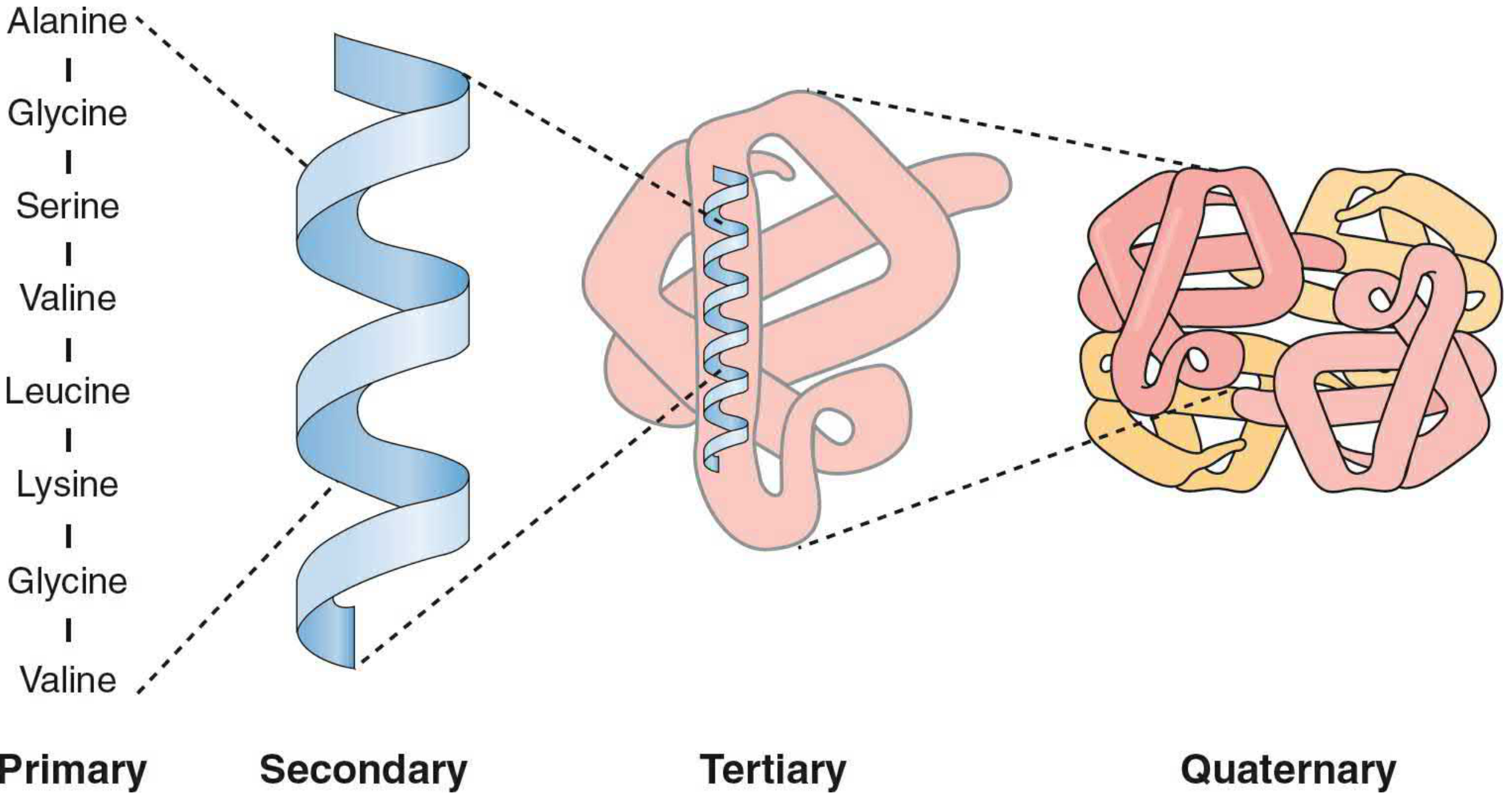
Amino acids are the building blocks of proteins. All have the same basic structure: a central α-carbon bonded to an amino group (-NH₂), a carboxyl group (-COOH), a hydrogen, and a variable side chain (R group) that determines their properties.
The most biochemically important classification is by polarity of the R group in water (solubility of R group):
CLASS 1: Nonpolar, Hydrophobic Amino Acids (R groups are insoluble in water)
These have aliphatic or aromatic hydrocarbon side chains that are hydrophobic - they avoid water and tend to cluster in the interior of folded proteins.
Members (9 total):
| Amino Acid | Symbol | R Group |
|---|
| Glycine (Gly, G) | Smallest; R = -H | Simplest; found in tight turns, collagen (every 3rd residue) |
| Alanine (Ala, A) | -CH₃ | Most abundant; α-helix former |
| Valine (Val, V) | Branched: -CH(CH₃)₂ | Branch-chain amino acid (BCAA); major energy source in muscle |
| Leucine (Leu, L) | Branched: -CH₂CH(CH₃)₂ | Most abundant BCAA; ketogenic |
| Isoleucine (Ile, I) | Branched: -CH(CH₃)CH₂CH₃ | Both glucogenic and ketogenic |
| Proline (Pro, P) | Cyclic; -CH₂CH₂CH₂- forms ring with N | Unique: amino group is tertiary; forms kinks in helix; abundant in collagen |
| Phenylalanine (Phe, F) | Benzyl: -CH₂-C₆H₅ | Aromatic; precursor of tyrosine; essential |
| Tryptophan (Trp, W) | Indole ring | Most hydrophobic; precursor of serotonin, niacin; essential |
| Methionine (Met, M) | -CH₂CH₂-S-CH₃ | Contains sulfur; first amino acid in all proteins (start codon AUG); SAM precursor |
Properties and functions:
- These R groups are insoluble in water
- Cluster in hydrophobic core of globular proteins - major force of protein folding
- BCAAs (Val, Leu, Ile): primary fuel for skeletal muscle during exercise and starvation
- Vet relevance: BCAA-enriched diets used in horses for muscle maintenance; proline + hydroxyproline constitute 1/3 of amino acids in collagen (tendons, bones, ligaments)
CLASS 2: Polar, Uncharged (Neutral) Amino Acids (R groups are soluble but neutral at physiological pH)
These have polar but uncharged R groups that can form hydrogen bonds with water and other polar molecules.
Members (6 total):
| Amino Acid | Symbol | R Group |
|---|
| Serine (Ser, S) | -CH₂-OH | Hydroxyl group; phosphorylated by kinases (signaling) |
| Threonine (Thr, T) | -CH(OH)-CH₃ | Hydroxyl group; essential; phosphorylated |
| Cysteine (Cys, C) | -CH₂-SH | Sulfhydryl (-SH) group; forms disulfide bonds (S-S) |
| Tyrosine (Tyr, Y) | -CH₂-C₆H₄-OH | Phenolic -OH; precursor of thyroid hormones, catecholamines, melanin |
| Asparagine (Asn, N) | -CH₂-CO-NH₂ | Amide of aspartate; N-glycosylation site |
| Glutamine (Gln, Q) | -CH₂CH₂-CO-NH₂ | Amide of glutamate; major nitrogen carrier in blood; fuel for intestinal enterocytes |
Properties and functions:
- R groups are soluble in water (form H-bonds with water)
- Located on surface of proteins facing aqueous environment
- Serine and threonine: key sites of phosphorylation in signal transduction cascades
- Cysteine: forms disulfide bonds that stabilize extracellular proteins (immunoglobulins, keratin)
- Vet relevance:
- Cystine urolithiasis in dogs (Newfoundlands, English Bulldogs) - defect in cystine reabsorption → cystine crystals/stones in urine
- Tyrosine deficiency → reduced melanin → coat color changes; precursor of thyroxine (critical for metabolic rate in all animals)
- Glutamine is the most abundant amino acid in blood; critical fuel for rapidly dividing cells (enterocytes, lymphocytes) → glutamine supplementation in critically ill veterinary patients
CLASS 3: Charged Polar Amino Acids - Acidic (Negatively Charged at pH 7.4)
These have R groups with carboxyl (-COOH) that are deprotonated (COO⁻) and therefore negatively charged at physiological pH.
Members (2):
| Amino Acid | Symbol | R Group | pKaR |
|---|
| Aspartate (Asp, D) | -CH₂-COOH → -CH₂-COO⁻ | ~3.7 | Participates in active sites of many enzymes (transaminases) |
| Glutamate (Glu, E) | -CH₂CH₂-COOH → -CH₂CH₂-COO⁻ | ~4.3 | Excitatory neurotransmitter; central to amino acid metabolism |
Properties and functions:
- Highly soluble in water (charged, interact strongly with water)
- Located on protein surfaces, form ionic bonds (salt bridges) with basic amino acids
- Aspartate and Glutamate are central to transamination reactions (ALT, AST enzymes)
- Glutamate is the major excitatory neurotransmitter in the CNS
- Vet relevance:
- AST (Aspartate aminotransferase) is the primary liver enzyme used in cattle and horses for liver disease diagnosis
- Glutamate participates in urea cycle (glutamate → α-ketoglutarate → enters TCA cycle)
- Glutamate + ammonia → Glutamine (ammonia detoxification)
CLASS 4: Charged Polar Amino Acids - Basic (Positively Charged at pH 7.4)
These have R groups with amino/guanidino groups that are protonated and therefore positively charged at physiological pH.
Members (3):
| Amino Acid | Symbol | R Group | pKaR | Notes |
|---|
| Lysine (Lys, K) | -(CH₂)₄-NH₃⁺ | ~10.5 | Positive; cross-links collagen; histone acetylation | |
| Arginine (Arg, R) | Guanidinium group; very basic | ~12.5 | Most basic; precursor of nitric oxide (NO); urea cycle intermediate | |
| Histidine (His, H) | Imidazole ring; pKaR ~6.0 | ~6.0 | Partially charged at pH 7.4; key catalytic residue in enzyme active sites | |
Properties and functions:
- All are soluble in water (charged)
- Form ionic bonds with negatively charged molecules (DNA, acidic lipids, acidic amino acids)
- Histidine has a pKa near physiological pH → acts as a proton acceptor/donor → key in enzyme catalysis, hemoglobin's Bohr effect, and intracellular buffering
- Arginine: precursor of NO (nitric oxide) via NO synthase → vasodilation; intermediate in urea cycle
- Lysine: essential amino acid; cross-links collagen and elastin via lysyl oxidase → important in connective tissue
- Vet relevance:
- Lysine deficiency in poultry and pig diets → growth retardation (most limiting AA in grain diets); lysine supplementation of swine rations
- Arginine is essential in cats (cannot synthesize ornithine from glutamate) → short arginine-free meal → life-threatening hyperammonemia
- Histidine in hemoglobin: Bohr effect (H⁺ binds to His residues at high CO₂ → promotes O₂ release in tissues)
Summary Table
| Class | Solubility in Water | Key AA | Properties |
|---|
| Nonpolar hydrophobic | Insoluble (avoid water) | Gly, Ala, Val, Leu, Ile, Pro, Phe, Trp, Met | Cluster in protein interior; hydrophobic core |
| Polar, uncharged | Soluble (H-bond with water) | Ser, Thr, Cys, Tyr, Asn, Gln | Form H-bonds; on protein surface; phosphorylation sites |
| Acidic (negatively charged) | Highly soluble | Asp, Glu | COO⁻ at pH 7.4; salt bridges; enzyme active sites |
| Basic (positively charged) | Highly soluble | Lys, Arg, His | NH₃⁺ at pH 7.4; bind nucleic acids; enzyme catalysis |
QUESTION 7 - Lipoproteins and Their Functions (5 marks)
Structure of Lipoproteins
Lipoproteins are spherical macromolecular complexes that transport lipids in blood. Their general structure:
- Outer shell: Phospholipids (monolayer, polar heads outward), free cholesterol, apolipoproteins
- Hydrophobic core: Triglycerides, cholesterol esters, fat-soluble vitamins
Classification by Density and Function
1. Chylomicrons
- Origin: Intestinal enterocytes (from dietary fat)
- Core lipid: Dietary triglycerides (>85%)
- Key apoprotein: ApoB-48, ApoC-II, ApoE
- Function: Transport exogenous (dietary) triglycerides from intestine to peripheral tissues (muscle, adipose) via lymph → blood
- Metabolism: ApoC-II activates lipoprotein lipase (LPL) on capillary endothelium → TG hydrolyzed → FFA released to tissues → chylomicron remnants taken up by liver (via ApoE-receptor)
- Vet note: Postprandial lipemia (milky plasma) in dogs/cats = normal after fatty meal; pathological chylomicronemia in lipoprotein lipase deficiency (cats, miniature Schnauzers)
2. VLDL (Very Low-Density Lipoprotein)
- Origin: Liver hepatocytes
- Core lipid: Endogenous triglycerides (55%)
- Key apoprotein: ApoB-100, ApoC-II, ApoC-III, ApoE
- Function: Transport endogenous triglycerides synthesized in liver to peripheral tissues
- Metabolism: LPL cleaves TG → releases FFA → VLDL becomes IDL, then LDL
- Vet note: Elevated VLDL in bovine fatty liver syndrome; hypothyroidism in dogs; equine metabolic syndrome
3. IDL (Intermediate-Density Lipoprotein)
- Origin: Formed from VLDL after TG removal
- Function: Transient intermediate; taken up by liver (via ApoE) OR converted to LDL
- Short-lived; not usually measured in veterinary diagnostics
4. LDL (Low-Density Lipoprotein) - "Bad Cholesterol"
- Origin: Formed from VLDL/IDL after most TG removed
- Core lipid: Cholesterol esters (50%)
- Key apoprotein: ApoB-100 only
- Function: Deliver cholesterol to peripheral cells via LDL receptor (ApoB-100 binds LDL receptor) → endocytosis → cholesterol released for membrane synthesis, steroid hormone production
- Vet note: In dogs (unlike humans), LDL is NOT the predominant lipoprotein - HDL is. LDL elevated in hypothyroid dogs, chronic kidney disease
5. HDL (High-Density Lipoprotein) - "Good Cholesterol"
- Origin: Liver and intestine; also formed from surface remnants of chylomicrons/VLDL
- Core lipid: Cholesterol esters (largest proportion relative to its size)
- Key apoprotein: ApoA-I, ApoA-II
- Function: Reverse cholesterol transport - removes excess cholesterol from peripheral tissues → transport to liver for excretion as bile acids
- ApoA-I activates LCAT (Lecithin-Cholesterol Acyl Transferase) → esterifies free cholesterol → incorporated into HDL core
- Vet note: HDL is the major plasma lipoprotein in dogs, cats, horses, cattle (unlike humans where LDL predominates). Cats completely lack CETP (cholesteryl ester transfer protein).
6. Lipoprotein(a) [Lp(a)]
- LDL-like particle with an extra apoprotein: Apo(a) linked to ApoB-100 by disulfide bond
- Structurally similar to plasminogen → may inhibit fibrinolysis → thrombotic tendency
- Measured in dogs; significance unclear in veterinary medicine
Functions of Lipoproteins
| Function | Details |
|---|
| TG Transport (exogenous) | Chylomicrons carry dietary fat from intestine to liver and periphery |
| TG Transport (endogenous) | VLDL carries liver-synthesized TG to muscle and adipose |
| Cholesterol delivery | LDL delivers cholesterol to cells for membranes, hormones, bile acids |
| Reverse cholesterol transport | HDL removes cholesterol from peripheral tissues → liver (anti-atherosclerotic) |
| Fat-soluble vitamin transport | Chylomicrons carry vitamins A, D, E, K from intestine |
| Lipid signaling | Arachidonic acid in phospholipids of lipoproteins |
| Enzyme activation | ApoC-II activates LPL; ApoA-I activates LCAT |
| Receptor-mediated uptake | ApoB-100 on LDL → LDL receptor; ApoE on remnants → hepatic uptake |
QUESTION 8 - Watson-Crick DNA Model + DNA vs RNA (5+4 = 9 marks)
(Detailed answer provided in the previous session - presented here in expanded form)
A. Watson-Crick Double Helix Model (1953)
Watson and Crick, utilizing X-ray crystallography data from Rosalind Franklin and Maurice Wilkins, proposed the double helix model of DNA in 1953 (published in Nature).
Features of the B-DNA Double Helix:
1. Two antiparallel polynucleotide strands
- DNA consists of two complementary strands wound around each other
- Strands run antiparallel: one 5'→3', the other 3'→5'
- The backbone (exterior) = alternating deoxyribose + phosphate groups (hydrophilic)
- The bases (interior) face inward (hydrophobic stacking)
2. Complementary Base Pairing (Chargaff's Rules)
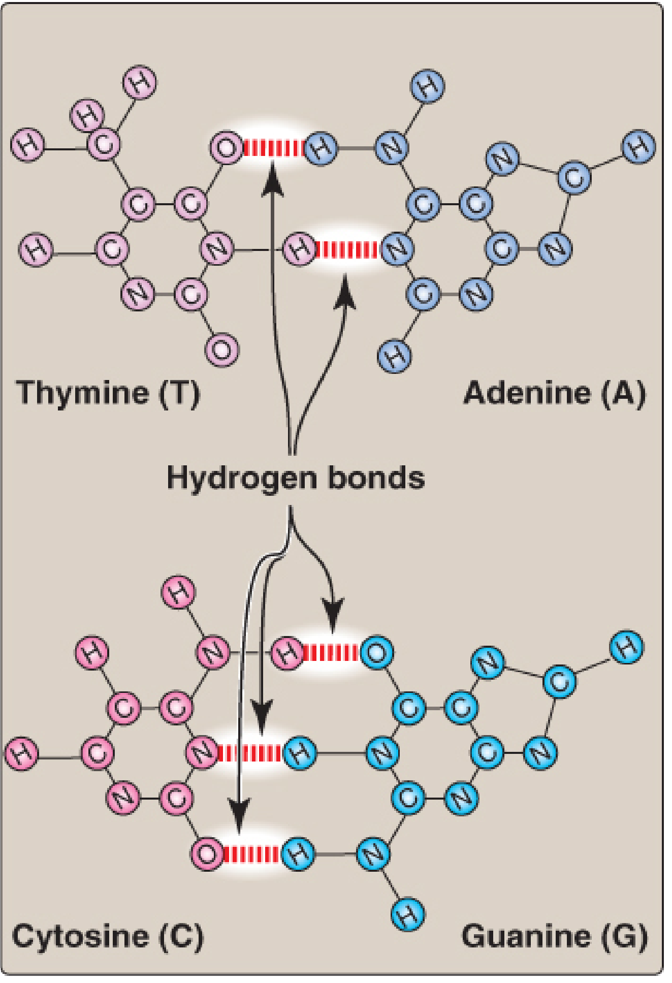
- A=T: Adenine pairs with Thymine via 2 hydrogen bonds
- G≡C: Guanine pairs with Cytosine via 3 hydrogen bonds
- This ensures [A]=[T] and [G]=[C] in any double-stranded DNA (Chargaff's rule)
- G-C pairs = more stable (3 H-bonds); G+C content determines melting temperature (Tm)
Hydrogen bonds between A:T and G:C base pairs (Lippincott Biochemistry, 8e)
3. Structural Parameters of B-DNA:
| Parameter | Value |
|---|
| Diameter | 2 nm (20 Å) |
| Pitch (full turn) | 3.4 nm |
| Base pairs per turn | 10 bp |
| Rise per base pair | 0.34 nm |
| Helix direction | Right-handed |
| Base plane orientation | Perpendicular to helical axis |
4. Major and Minor Grooves
- Unequal wrapping of the two strands creates:
- Major groove (wider, ~22 Å): binding site for regulatory proteins, transcription factors
- Minor groove (narrower, ~12 Å): binding site for some drugs (e.g., berenil in trypanosomiasis treatment in animals)
5. Forms of DNA:
| Form | Helix | bp/turn | Context |
|---|
| B-DNA | Right-handed | 10 | Physiological; described by Watson-Crick |
| A-DNA | Right-handed | 11 | Dehydrated B-DNA; RNA:DNA hybrids |
| Z-DNA | Left-handed | 12 | Alternating purines-pyrimidines; may regulate transcription |
6. Stabilization:
- Hydrogen bonds between bases (hold strands together)
- Base stacking interactions (π-π stacking of aromatic rings above/below each other - the strongest stabilizing force)
- Phosphodiester bonds (covalent, within each strand)
B. Differences Between DNA and RNA
| Feature | DNA | RNA |
|---|
| Full name | Deoxyribonucleic acid | Ribonucleic acid |
| Sugar | 2'-Deoxyribose (no -OH at C2') | Ribose (-OH at C2') |
| Unique base | Thymine (T) - methylated uracil | Uracil (U) - unmethylated |
| Common bases | A, G, C | A, G, C |
| Strandedness | Usually double-stranded | Usually single-stranded |
| Stability | More stable (no 2'-OH) | Less stable (2'-OH → alkaline hydrolysis) |
| Location | Nucleus, mitochondria | Nucleus + cytoplasm + ribosomes |
| Size | Very large (billions of bp) | Smaller |
| Helix type | B-form right-handed | A-form where double-stranded |
| Types | One type | mRNA, tRNA, rRNA, snRNA, miRNA, siRNA |
| Function | Stores genetic information | Protein synthesis (transcription, translation) |
| Replication | Semiconservative (DNA → DNA) | No standard replication (except RNA viruses) |
| Base pairing | A=T (2 H-bonds), G≡C (3 H-bonds) | A=U, G≡C |
Veterinary relevance: RNA viruses (FMDV, BVDV, FIV, Rabies, Canine distemper, Influenza) use RNA as genetic material and require RNA-dependent RNA polymerase. Retroviruses (FeLV, FIV) reverse-transcribe RNA → DNA via reverse transcriptase.
QUESTION 9 - Membrane Transport & Membrane Lipids (5 marks each)
OPTION A: Types of Membrane Transport
(Complete answer given in previous session - summary below with additional vet details)
1. Passive Transport (No energy - moves down gradient)
- Simple diffusion: O₂, CO₂, lipid-soluble molecules directly through bilayer
- Facilitated diffusion: Polar/charged molecules via carrier proteins (GLUT transporters for glucose) or channel proteins (aquaporins, ion channels)
2. Active Transport (Energy required - against gradient)
- Primary active transport: Direct ATP hydrolysis by ATPase pumps (Na⁺/K⁺-ATPase, Ca²⁺-ATPase, H⁺/K⁺-ATPase)
- Secondary active transport: Uses Na⁺ gradient (created by Na⁺/K⁺-ATPase) as energy source - SGLT1/2 (glucose symport), Na⁺/Ca²⁺ exchanger (antiport)
3. Vesicular Transport
- Endocytosis: Pinocytosis, phagocytosis, receptor-mediated endocytosis (colostral IgG in neonatal calves/foals)
- Exocytosis: Insulin secretion, neurotransmitter release
OPTION B: Membrane Lipids and Their Functions
The plasma membrane of all animal cells is a fluid mosaic - a lipid bilayer embedded with proteins. The three main lipid classes are:
1. Phospholipids (Major component - ~50% of membrane lipids)
Structure: Glycerol + 2 fatty acids + phosphate + polar head group (choline, ethanolamine, serine, inositol)
Types:
| Phospholipid | Head Group | Location / Function |
|---|
| Phosphatidylcholine (Lecithin) | Choline | Outer leaflet; most abundant; structural |
| Phosphatidylethanolamine | Ethanolamine | Inner leaflet; curls membrane; cell signaling |
| Phosphatidylserine | Serine | Inner leaflet; when externalized → apoptosis signal, coagulation (platelet activation) |
| Phosphatidylinositol (PI) | Inositol | Inner leaflet; PI → PIP₂ → IP₃ + DAG (second messenger cascade) |
| Sphingomyelin | Phosphocholine (via ceramide) | Outer leaflet; lipid rafts; myelin sheath |
Functions of phospholipids:
- Form the lipid bilayer barrier (hydrophilic heads outward, hydrophobic tails inward)
- Lung surfactant: Dipalmitoylphosphatidylcholine (DPPC) reduces alveolar surface tension - deficiency in neonatal foals/premature lambs causes respiratory distress syndrome
- Platelet activation: Externalized phosphatidylserine on activated platelets binds coagulation factors
- Signal transduction: PIP₂ → IP₃ (releases Ca²⁺) + DAG (activates PKC)
- Emulsification: Bile contains phosphatidylcholine for fat emulsification
2. Cholesterol (25-30% of membrane lipids in animal cells)
Structure: Four-ring sterol nucleus (cyclopentanoperhydrophenanthrene) + hydroxyl group + hydrocarbon tail
Functions:
| Function | Mechanism |
|---|
| Membrane fluidity regulator | At high temp: reduces fluidity (intercalates between fatty acids, restricts motion); at low temp: prevents crystallization (disrupts packing) → cholesterol is the "fluidity buffer" |
| Reduces membrane permeability | Fills gaps between phospholipid tails → reduces ion/water leakage |
| Lipid raft formation | With sphingomyelin, forms ordered microdomains (rafts) that concentrate signaling proteins, receptors |
| Precursor | Bile acids, steroid hormones (cortisol, sex hormones), Vitamin D₃ |
Vet relevance:
- Cell membranes of ruminants adjust cholesterol content seasonally for temperature adaptation
- Cholesterol toxicosis in horses (hyperlipemia): Ponies, donkeys in NEB → massive VLDL release → hyperlipidemia + hepatic lipidosis
3. Glycolipids (5-10% of membrane lipids, outer leaflet only)
Structure: Ceramide + one or more sugar residues (no phosphate)
Types:
| Glycolipid | Sugar | Location |
|---|
| Cerebrosides | Glucose or Galactose | Myelin, brain |
| Gangliosides | Oligosaccharide + sialic acid | Neuron cell surface; abundant in gray matter |
| Globosides | Multiple sugars | RBC membranes (blood group antigens) |
Functions:
| Function | Details |
|---|
| Cell recognition | ABO blood group antigens (glycolipids on RBC surface) - relevant in blood transfusion compatibility (dogs: DEA system, horses: Aa/Qa system) |
| Cell-cell adhesion | Glycocalyx formation - mediates tissue organization |
| Neural signal transduction | Gangliosides modulate receptor tyrosine kinase signaling |
| Pathogen binding | Many bacteria, viruses bind glycolipids for cell entry (e.g., cholera toxin binds GM1 ganglioside) |
| Lipid rafts | Concentrated in lipid raft microdomains |
Vet relevance:
- GM1/GM2 gangliosidosis: Lysosomal storage diseases in dogs (Beagles, Portuguese Water Dogs), cats, cattle - deficiency of ganglioside-degrading enzymes → accumulation in neurons → progressive neurodegeneration
- Feline blood types (A, B, AB): Determined largely by glycolipid antigens on RBCs - critical for blood transfusions (Type B cats have strong anti-A antibodies → neonatal isoerythrolysis in Type A kittens of Type B mothers)
Membrane Asymmetry
The lipid bilayer is asymmetric - different lipids in each leaflet:
- Outer leaflet: Phosphatidylcholine, sphingomyelin, glycolipids, gangliosides
- Inner leaflet: Phosphatidylethanolamine, phosphatidylserine, phosphatidylinositol
Significance: Phosphatidylserine externalization = apoptosis marker + platelet activation signal
QUESTION 10 - Methods of Urine Analysis and Diagnostic Applications (5+5 = 10 marks)
Urinalysis (UA) is one of the most valuable and cost-effective diagnostic tests in veterinary medicine. It involves three components: physical, chemical, and microscopic examination.
I. Physical Examination of Urine
1. Color
| Color | Significance |
|---|
| Yellow (pale to amber) | Normal; intensity varies with concentration |
| Colorless/very pale | Dilute urine; polyuria (DI, CKD, hypoadrenocorticism) |
| Dark yellow/amber | Concentrated; dehydration; bilirubinuria |
| Red/pink | Hematuria (blood), hemoglobinuria (intravascular hemolysis) |
| Brown | Myoglobinuria (rhabdomyolysis); methemoglobinuria |
| Orange | Bilirubinuria; urobilinogen |
| Green/blue | Biliverdin; Pseudomonas UTI (rare) |
| Milky/white | Pyuria, chyluria, lipiduria (cats with hepatic lipidosis) |
Vet note: Red/brown urine differentiating test: Centrifuge → if RBC pellet forms → hematuria; if supernatant remains red → hemoglobin or myoglobin. Ammonium sulfate precipitation distinguishes hemoglobinuria from myoglobinuria.
2. Turbidity (Clarity)
- Normal: clear to slightly cloudy
- Turbid: Pyuria (pus), crystalluria, bacteriuria, lipiduria
- Vet note: Healthy horse urine is normally turbid due to calcium carbonate crystals and mucus (not pathological)
3. Volume (Urine Output)
- Normal urine output: dogs ~20-40 mL/kg/day; cats ~22-30 mL/kg/day
- Polyuria (PU): >50 mL/kg/day → DI, CKD, diabetes, hyperadrenocorticism, hypercalcemia
- Oliguria: <0.5 mL/kg/hr → acute kidney injury, severe dehydration, urethral obstruction
- Anuria: No urine production → severe AKI, complete obstruction
4. Specific Gravity (USG)
- Measures solute concentration (urine-concentrating ability of renal tubules)
- Measured by: Refractometer (most accurate), urinometer, dipstick (less accurate)
| USG | Interpretation | Significance |
|---|
| 1.030-1.060 (dogs), 1.035-1.060 (cats) | Normal concentrated | Adequate renal function, euhydration |
| >1.030 (dogs), >1.035 (cats) | Adequately concentrated | Rules out primary renal failure |
| 1.013-1.029 | Suboptimally concentrated | Partial concentrating defect; early CKD |
| 1.008-1.012 | Isosthenuria | Same as glomerular filtrate; loss of tubular concentrating/diluting ability; severe CKD |
| <1.008 | Hyposthenuria | Tubules actively diluting; intact tubular function; DI or psychogenic polydipsia |
5. Odor
- Ammoniacal: urease-positive bacteria (UTI), alkaline urine, urea degradation
- Fruity/sweet: ketonuria (diabetes, pregnancy toxaemia, starvation)
II. Chemical Examination (Urine Dipstick + Specific Tests)
1. pH
- Normal range: dogs/cats pH 6.0-7.5; pH varies with diet
- Alkaline urine (pH >7.5): UTI with urease bacteria, post-prandial alkaline tide, diet (plant-based), metabolic alkalosis
- Acidic urine (pH <6.0): Protein-rich diet, metabolic acidosis, respiratory acidosis
- Clinical use:
- Struvite uroliths form in alkaline urine (cats, dogs) → treat by acidifying diet
- Urate and oxalate uroliths form in acidic urine
2. Protein (Proteinuria)
- Detected by dipstick (reaction with tetrabromophenol blue - detects albumin primarily)
- Quantified by UPC (urine protein:creatinine ratio) - gold standard
- Normal: UPC <0.2 dogs, <0.4 cats
- Confirmed with SSA (sulfosalicylic acid) precipitation test - detects all proteins including Bence-Jones
- Causes: Glomerulonephritis, amyloidosis, UTI, CKD, post-exercise (physiological)
3. Glucose (Glycosuria)
- Detected by dipstick (glucose oxidase/peroxidase method) - specific for glucose
- Benedict's test: Non-specific reducing sugar test (also detects fructose, galactose, lactose) - useful in ruminants and neonates
- Normal: absent/trace
- Positive → diabetes mellitus, stress hyperglycemia (cats), Fanconi syndrome, renal glycosuria
4. Ketones (Ketonuria)
- Detected by dipstick - Rothera's nitroprusside test (detects acetone + acetoacetate; NOT β-hydroxybutyrate)
- Positive in: Diabetic ketoacidosis (dogs, cats), starvation ketosis, pregnancy toxaemia (ewes, does, dairy cows), bovine ketosis
- Cowside test for ketosis: Keto-Test strips for β-hydroxybutyrate in milk (most sensitive in cattle)
5. Bilirubin (Bilirubinuria)
- Only conjugated (direct) bilirubin appears in urine
- Dogs: Trace bilirubinuria may be normal (especially in concentrated urine of male dogs - kidney tubules conjugate some bilirubin)
- Cats: Any bilirubinuria is significant (low renal threshold)
- Detected by dipstick (diazonium salt reaction)
- Positive → hepatic disease, biliary obstruction, severe hemolysis
6. Urobilinogen
- Colorless product of bilirubin metabolism by intestinal bacteria; partially reabsorbed and excreted in urine
- Increased: Hemolysis, hepatocellular disease
- Absent: Complete biliary obstruction (no bilirubin reaching intestine → no urobilinogen → also absent in urine and stool)
7. Blood (Occult Blood / Hematuria / Hemoglobinuria)
- Dipstick detects heme (in RBCs, free hemoglobin, or myoglobin)
- Must differentiate: Hematuria (intact RBCs) vs. Hemoglobinuria vs. Myoglobinuria
- Hematuria confirmed by microscopy (RBCs in sediment)
- Causes: UTI, urolithiasis, trauma, tumor, coagulopathy, immune-mediated hemolytic anemia
8. Nitrite
- Produced by nitrate-reducing bacteria (Gram-negative)
- Positive → bacteriuria (UTI)
- Less reliable in veterinary species (urine often doesn't sit long enough for bacterial conversion)
9. Leukocyte Esterase
- Detects WBCs (neutrophils); indicates pyuria
- Not reliable in dogs and cats - species difference in WBC enzyme; confirmed by microscopy
10. Urine Creatinine
- Used to calculate UPC ratio and fractional excretion of electrolytes
III. Microscopic Examination of Urine Sediment
Preparation:
- Centrifuge urine (400g × 5 min) → pour off supernatant → resuspend pellet → examine wet mount
A. Cells
| Cell Type | Normal | Significance if Increased |
|---|
| Red Blood Cells (RBC) | 0-5/HPF | Hematuria → UTI, trauma, uroliths, neoplasia, coagulopathy |
| White Blood Cells (WBC/Pus cells) | 0-5/HPF | Pyuria → UTI, pyelonephritis, urethritis, vaginitis (contamination) |
| Epithelial cells | Occasional | Transitional (normal shedding) vs. clumps (neoplasia); renal tubular cells (AKI) |
| Spermatozoa | In intact males post-ejaculation | Normal incidental finding |
B. Casts (Indicate Renal Origin)
Casts = protein cylinders formed in renal tubules (Tamm-Horsfall mucoprotein matrix)
| Cast Type | Significance |
|---|
| Hyaline casts | Protein only; normal in small numbers; fever, dehydration |
| Granular casts (fine) | Degenerated cellular material; tubular stress |
| Granular casts (coarse) | Active renal tubular damage |
| RBC casts | Glomerulonephritis (bleeding into tubule) |
| WBC casts | Pyelonephritis (infection/inflammation in tubule) |
| Waxy casts | Severe, chronic renal disease; end-stage |
| Fatty casts | Lipiduria; feline hepatic lipidosis, hypothyroidism in dogs |
C. Crystals
| Crystal | pH | Significance |
|---|
| Struvite (MgNH₄PO₄) | Alkaline | UTI (urease bacteria), iatrogenic in cats on dry food |
| Calcium oxalate (monohydrate/dihydrate) | Acidic/neutral | Ethylene glycol toxicosis (dogs/cats - rosette crystals), hyperoxaluria |
| Urate (ammonium biurate) | Acidic | Portosystemic shunt, Dalmatians (purine metabolism defect) |
| Cystine | Acidic | Cystinuria - genetic defect (Newfoundlands, English Bulldogs) |
| Bilirubin | - | Bilirubinuria; hepatic disease |
| Tyrosine/Leucine | Acidic | Severe hepatic failure (rarely seen) |
| Calcium carbonate | Alkaline | Normal in horses and rabbits |
D. Bacteria, Fungi, Parasites
- Bacteria: Must distinguish contamination vs. true infection (quantitative culture)
- Candida sp.: Immunocompromised animals, prolonged antibiotic use
- Capillaria plica (Pearsonema plica): Bladder worm in dogs/cats → hematuria
QUESTION 11 - Metabolic Acidosis and Alkalosis + Compensatory Mechanisms (6 marks)
(Detailed answer given in previous session - see Q16 above. Complete answer reproduced here.)
See the detailed answer in the previous session Q16 which covers:
- Metabolic Acidosis: definition, causes (DKA, lactic acidosis, diarrhea in calves, rumen acidosis, uremia), biochemical changes, respiratory and renal compensation
- Metabolic Alkalosis: definition, causes (abomasal displacement in cattle, GDV in dogs, vomiting), biochemical changes, respiratory and renal compensation, paradoxical aciduria
- Summary comparison table
QUESTION 12 - Ketosis and Its Diagnosis (5+1 = 6 marks)
Ketosis
Definition: Ketosis is a metabolic state characterized by elevated concentrations of ketone bodies (acetoacetate, β-hydroxybutyrate/BHBA, and acetone) in blood (ketonemia), urine (ketonuria), and/or milk (ketonemia of milk), arising when fat catabolism exceeds the liver's capacity to oxidize acetyl-CoA via the TCA cycle.
Biochemistry of Ketone Body Formation (Ketogenesis)
Location: Hepatic mitochondria (EXCLUSIVELY - liver cannot use its own ketones)
Pathway:
- Lipolysis in adipose → NEFA released into blood
- NEFA → Liver → undergoes β-oxidation → Acetyl-CoA
- When energy demand is high (starvation, NEB, uncontrolled DM) and oxaloacetate (OAA) is depleted (diverted to gluconeogenesis), Acetyl-CoA cannot enter TCA cycle
- Excess Acetyl-CoA → Ketogenesis:
- 2 Acetyl-CoA → Acetoacetyl-CoA
- Acetoacetyl-CoA + Acetyl-CoA → HMG-CoA (via HMG-CoA synthase)
- HMG-CoA → Acetoacetate (via HMG-CoA lyase)
- Acetoacetate → β-hydroxybutyrate (via BHBA dehydrogenase; reduced by NADH)
- Acetoacetate → Acetone (spontaneous decarboxylation - fruity smell in breath)
The key biochemical principle: "Fat burns in the flame of carbohydrates" - OAA (from glucose/TCA) is needed to accept Acetyl-CoA. When OAA is diverted to gluconeogenesis (starvation) → Acetyl-CoA accumulates → ketogenesis.
Ketone Bodies and Their Fates
Peripheral tissues (muscle, heart, brain, kidney) CAN use ketones:
- Acetoacetate/BHBA → Acetyl-CoA → TCA cycle → ATP
- During prolonged starvation, brain adapts to use ketones as primary fuel (normally uses glucose only)
- Liver CANNOT use its own ketones (lacks succinyl-CoA transferase/thiophorase)
Types of Ketosis in Veterinary Species
1. Bovine Ketosis (Acetonaemia)
Species: Dairy cattle (most common metabolic disease in high-producing cows)
Type I (Underfeeding ketosis - classic):
- Occurs 2-6 weeks post-partum (peak milk production)
- High-producing cows cannot consume enough feed → Negative Energy Balance (NEB)
- Fat mobilization → elevated NEFA → hepatic ketogenesis
- BHBA >1.0 mmol/L = subclinical; >3.0 mmol/L = clinical
Type II (Fatty liver / Hepatic ketosis):
- Overconditioned cows at calving (BCS >3.5)
- Massive fat mobilization post-partum → fatty liver → impaired hepatic function → impaired gluconeogenesis → worsening ketosis
- High NEFA + high BHBA + elevated liver enzymes (AST, GLDH)
Clinical signs: Decreased milk production, anorexia, decreased rumen motility, abnormal dung, neurological form (circling, bellowing, blindness - "nervous ketosis")
2. Pregnancy Toxaemia (Small Ruminants)
- Ewes/does with multiple fetuses in late gestation
- Signs: blindness, teeth grinding, recumbency, coma
- BHBA >3 mmol/L; blood glucose <40 mg/dL
3. Diabetic Ketoacidosis (DKA) - Dogs and Cats
- Severe insulin deficiency + stress hormones (glucagon, cortisol, epinephrine)
- Massive NEFA mobilization + ketogenesis
- Metabolic acidosis (anion gap) + dehydration + electrolyte imbalance
- Life-threatening; requires intensive care
4. Equine Hyperlipaemia/Hyperlipidaemia
- Particularly in ponies, donkeys, and miniature horses under stress/NEB
- Massive fat mobilization → VLDL overload → hypertriglyceridemia + ketosis + hepatic lipidosis
- Triggers: late pregnancy, lactation, starvation, illness
Diagnosis of Ketosis
1. Clinical Diagnosis
- History and clinical signs (milk drop, anorexia, neurological signs)
- Breath smell: fruity/acetone odor (acetone is volatile)
2. Bedside/Cowside Tests
| Test | Detects | Method |
|---|
| Rothera's nitroprusside test (urine/milk) | Acetone + Acetoacetate | Sodium nitroprusside + ammonium sulfate + ammonia → purple color |
| Keto-Test strip (milk) | β-hydroxybutyrate | Colorimetric strip; most practical cowside test |
| Urine dipstick ketone pad | Acetone + Acetoacetate | Semi-quantitative; NOT sensitive for BHBA |
3. Laboratory Tests
| Test | Normal | Ketosis (Clinical) | Severe |
|---|
| Serum/Plasma BHBA | <0.6 mmol/L (cattle) | 1.0-3.0 mmol/L (subclinical); >3.0 (clinical) | >6 mmol/L (DKA in dogs) |
| Blood glucose | 3.0-5.0 mmol/L (cattle) | Low (<2.0 mmol/L) | |
| Serum NEFA | <0.3 mmol/L (cattle) | Markedly elevated | |
| Liver enzymes | Normal | Elevated AST, GLDH (fatty liver) | |
| Urine ketones | Negative | Positive | |
| Milk ketones | Negative | Positive BHBA in milk | |
| Blood pH, HCO₃⁻ | pH 7.35-7.45; HCO₃⁻ 18-24 mEq/L | Decreased (metabolic acidosis in DKA) | |
| HbA1c/Fructosamine | Normal | Elevated in DM-associated ketosis | |
4. Herd Monitoring (Dairy Cattle)
- Routine milk BHBA testing at 5-7 days post-partum identifies subclinical ketosis
- BHBA >0.10 mmol/L in milk (Keto-Test) = subclinical ketosis → intervention
QUESTION 13 - Phase I Metabolism of Xenobiotics (3 marks) OR Carbohydrate Classification (5 marks)
OPTION A: Phase I Metabolism of Xenobiotics
(Detailed answer in previous session Q18 - brief summary below)
Phase I reactions (functionalization reactions) are the first step of xenobiotic biotransformation, converting lipophilic compounds into more polar metabolites by introducing or exposing a functional group (-OH, -NH₂, -SH, -COOH).
Three types of Phase I reactions:
1. Oxidation (Most Important)
- Catalyzed by Cytochrome P-450 (CYP) enzyme system in hepatocyte ER
- Reaction: Substrate + O₂ + NADPH → Oxidized product + H₂O + NADP⁺
- Types: Hydroxylation (aromatic/aliphatic), epoxidation, N-oxidation, S-oxidation, deamination
- Examples in animals:
- Barbiturate hydroxylation → more water-soluble (cats are slow at this)
- Acetaminophen → NAPQI (N-acetyl-p-benzoquinoneimine) via CYP2E1 - toxic intermediate especially in cats
- Carbon tetrachloride → CCl₃· free radical → hepatotoxicity
- Aflatoxin B₁ → epoxide metabolite → carcinogen in cattle, poultry
2. Reduction
- Reduces -NO₂ (nitro) and -N=N- (azo) groups
- Nitrate reduction by rumen bacteria → nitrite → oxidizes hemoglobin → methemoglobin → nitrite poisoning in ruminants (grazing on nitrate-rich forages, preserved silage)
3. Hydrolysis
- Cleaves ester, amide, or glycoside bonds
- Atropine hydrolysis by atropinase (rabbits, goats can eat deadly nightshade safely - they have plasma atropinase)
- Organophosphate hydrolysis by paraoxonase (PON1) - species differences (horses have very low PON1 activity)
Key Enzyme: Cytochrome P-450 (CYP)
- Heme-containing mixed-function oxidases
- Located in ER of hepatocytes (also intestine, lung, skin)
- CYP1A2, CYP2B6, CYP2C, CYP2D6, CYP2E1, CYP3A4 - different substrate specificities
- Species differences: Cats have reduced CYP activity for certain substrates → drug toxicity (aspirin, acetaminophen, phenols, NSAIDs)
OPTION B: Classification of Carbohydrates
Definition: Carbohydrates are polyhydroxy aldehydes or ketones (or compounds that hydrolyze to yield them), with the general formula (CH₂O)n. They are the primary energy source in most animals and serve structural roles in glycoproteins, glycolipids, and the glycocalyx.
Classification of Carbohydrates
I. MONOSACCHARIDES (Simple Sugars - Cannot be hydrolyzed further)
A. By number of carbons:
| Name | Carbons | Examples |
|---|
| Triose | 3 | Glyceraldehyde, DHAP (glycolysis intermediates) |
| Tetrose | 4 | Erythrose |
| Pentose | 5 | Ribose (RNA), Deoxyribose (DNA), Ribulose (Calvin cycle), Xylulose |
| Hexose | 6 | Glucose, Fructose, Galactose, Mannose - most important biologically |
| Heptose | 7 | Sedoheptulose (pentose phosphate pathway) |
B. By functional group:
- Aldoses: Have aldehyde group (-CHO) at C1 → e.g., Glucose, Galactose, Mannose, Ribose
- Ketoses: Have ketone group (C=O) at C2 → e.g., Fructose, Ribulose, Xylulose
Properties:
- Exist in ring forms (pyranose = 6-membered; furanose = 5-membered) in solution
- All are reducing sugars (free anomeric carbon can reduce Cu²⁺ in Benedict's test)
- Show mutarotation and optical activity (D- and L- forms)
Key monosaccharides in animals:
- Glucose (D-glucose): Primary blood sugar; universal fuel; normal blood glucose: dogs 70-120 mg/dL; cats 70-150 mg/dL; cattle 45-75 mg/dL; horses 70-110 mg/dL
- Galactose: From lactose digestion; converted to glucose-1-phosphate (Leloir pathway) in liver
- Fructose: From sucrose; fruit sugar; phosphorylated by fructokinase in liver (bypasses PFK regulation)
- Ribose/Deoxyribose: Pentoses; components of RNA/DNA nucleotides
II. DISACCHARIDES (2 monosaccharides joined by glycosidic bond)
| Disaccharide | Monosaccharides | Bond | Source |
|---|
| Lactose (milk sugar) | Galactose + Glucose | β(1→4) O-glycosidic | Milk of all mammals |
| Sucrose (cane sugar) | Glucose + Fructose | α(1→2) O-glycosidic | Plant-derived |
| Maltose (malt sugar) | Glucose + Glucose | α(1→4) O-glycosidic | Starch digestion product |
| Cellobiose | Glucose + Glucose | β(1→4) O-glycosidic | Cellulose digestion product |
| Trehalose | Glucose + Glucose | α(1→1) O-glycosidic | Insects, fungi |
Veterinary note:
- Lactose intolerance = deficiency of intestinal lactase → undigested lactose → osmotic diarrhea. Common in adult carnivores (cats/dogs lose lactase after weaning)
- Sucrose is not normally present in significant amounts in animal feeds; excess sucrose → equine metabolic disorders
III. OLIGOSACCHARIDES (3-10 monosaccharides)
- Short chains of 3-10 sugar units linked by glycosidic bonds
- Often attached to proteins (glycoproteins) or lipids (glycolipids) via N- or O-glycosidic bonds
- Examples: Raffinose (3 units, in legumes → flatulence in monogastrics), Stachyose (4 units), Human milk oligosaccharides (HMO), Blood group antigens
Vet relevance: N-linked oligosaccharides on immunoglobulins (IgG in bovine colostrum), complement proteins, and cell surface receptors - critical for immune recognition
IV. POLYSACCHARIDES (>10 monosaccharides - most with thousands of units)
A. Homopolysaccharides (one type of monosaccharide)
1. Starch (Storage polysaccharide in plants)
- Two forms: Amylose (linear, α-1,4 bonds) + Amylopectin (branched: α-1,4 + α-1,6 bonds every 24-30 glucose units)
- Digested by amylase (salivary + pancreatic) → maltose → glucose
- Major carbohydrate source in monogastric rations (pigs, poultry, dogs, cats)
- Ruminants: Starch digested predominantly by ruminal microbes (amylolytic bacteria) → VFAs (acetate, propionate, butyrate); excess starch → rumen acidosis
2. Glycogen (Animal storage polysaccharide)
- More branched than starch: α-1,4 bonds + α-1,6 branches every 8-12 glucose units
- Stored in liver (up to 5-6% of liver weight; ~100g in adult dog) and muscle (1-2% of muscle weight)
- Liver glycogen: Maintains blood glucose during fasting (glycogenolysis)
- Muscle glycogen: Provides energy for muscle contraction; CANNOT release glucose to blood (no glucose-6-phosphatase)
- Vet relevance:
- Glycogen storage diseases (GSD): GSD Type I (von Gierke's) - glucose-6-phosphatase deficiency - reported in dogs
- Exertional rhabdomyolysis in horses (Polysaccharide Storage Myopathy/PSSM): Abnormal glycogen accumulation in muscle; treat with low-starch, high-fat diet
- Neonatal hypoglycemia in piglets/puppies: limited glycogen stores → rapid hypoglycemia when not feeding
3. Cellulose (Structural polysaccharide in plants)
- Linear polymer of glucose with β(1→4) glycosidic bonds
- Cannot be digested by any vertebrate enzyme
- Ruminants: Rumen microbes (Fibrobacter succinogenes, Ruminococcus sp.) produce cellulases → digest cellulose → VFAs → energy (up to 70% of ruminant energy needs)
- Horses: Hindgut fermentation (cecum + large colon) of cellulose
- Monogastrics (dogs, cats, pigs): Cannot digest cellulose; acts as dietary fiber → bulks stool, feeds colonic microbiome
4. Chitin
- Polymer of N-acetyl-D-glucosamine with β(1→4) bonds
- Major component of insect exoskeletons, crustacean shells, fungal cell walls
- Digested by chitinase (acid mammalian chitinase in gastric juice of insectivores)
5. Inulin
- Fructose polymer (β-2,1 bonds)
- Found in roots of chicory, Jerusalem artichoke
- Exogenous use: Gold standard for GFR measurement (inulin clearance) in all veterinary species
- Dietary use: Prebiotic fiber in some pet foods
B. Heteropolysaccharides (two or more types of monosaccharides)
1. Glycosaminoglycans (GAGs) / Mucopolysaccharides
- Long unbranched chains of repeating disaccharide units
- One unit = amino sugar (GlcNAc or GalNAc) + uronic acid (GlcUA or IdUA)
- Often sulfated and linked to proteins = proteoglycans
| GAG | Components | Location | Function |
|---|
| Hyaluronic acid | GlcUA + GlcNAc (no sulfate) | Synovial fluid, vitreous humor, ECM | Lubrication, water retention |
| Chondroitin sulfate | GlcUA + GalNAc-4-SO₄ | Cartilage, bone, skin | Structural support, water retention |
| Heparan sulfate | GlcUA + GlcNAc (variably sulfated) | Cell surfaces, basement membranes | Cell signaling, coagulation |
| Heparin | IdUA + GlcNAc (highly sulfated) | Mast cell granules | Anticoagulant (antithrombin III activator) |
| Dermatan sulfate | IdUA + GalNAc-4-SO₄ | Skin, heart valves, tendons | Structural |
| Keratan sulfate | Galactose + GlcNAc | Cornea, cartilage | Structural, corneal transparency |
Vet relevance:
- Hyaluronic acid injections used in equine joint disease (osteoarthritis)
- Chondroitin sulfate + glucosamine supplements widely used for joint disease in dogs, horses
- Mucopolysaccharidoses (MPS): Lysosomal storage diseases in cats (MPS I, VI, VII) and dogs (MPS III, VII) due to enzyme deficiency → GAG accumulation in tissues
2. Glycoproteins
- Short oligosaccharide chains covalently attached to proteins via N- or O-glycosidic bonds
- Examples: Mucins (airway mucus), blood group antigens, collagen, immunoglobulins
- Mucus in respiratory and GI tract is a glycoprotein gel - protects epithelium from pathogens
Sources: Basic Medical Biochemistry (Lieberman & Peet, 6e); Biochemistry, Lippincott Illustrated Reviews (8e); Harper's Illustrated Biochemistry (32e); Robbins & Kumar Pathologic Basis of Disease (10e); Henry's Clinical Diagnosis and Management by Laboratory Methods. All content applied to veterinary species throughout.