
Syringomyeliq in detail
I'll start by reading the medical library skill, then search for comprehensive information on syringomyelia in parallel.
Reading File
Now let me search the library and PubMed in parallel for comprehensive coverage.
Finding Sources
Searching PubMed
syringomyelia pathophysiology treatment
Excellent. Now let me read from the most comprehensive textbook sources in parallel.
Reading File
Reading File
Reading File
Now let me get the Goldman-Cecil and Campbell's Operative Orthopaedics sections plus the Rosen's Emergency Medicine and deeper Campbell sections for full clinical detail.
Reading File
Reading File
Reading File
Excellent data collected. Let me also fetch a key MRI image and search for deeper paragraphs on pathophysiology and the Campbell's SYRINGOMYELIA section (block 18, line 6682).
Loading Image
Reading File
Finding Sources
Let me read the Harrison's and Miller's Review sections for pathophysiology and the complete clinical picture.
Reading File
Reading File
Now let me read the rest of the Harrison's section (treatment details) and fetch the Harrison's MRI image.
Reading File
Loading Image
I now have comprehensive data from multiple authoritative sources. Let me compile the detailed answer.
Syringomyelia - A Detailed Review
Definition and Terminology
Syringomyelia is a condition characterized by the formation of a fluid-filled cavity (syrinx) within the substance of the spinal cord, lined primarily by glial tissue, containing fluid that is similar or identical to CSF. The word "syrinx" derives from the Greek surinx meaning "pipe" or "channel."
- Syringomyelia - the cavity lies within cord parenchyma
- Hydromyelia - dilation of the central spinal canal itself
- Syringohydromyelia - overlapping term used when cerebellar ectopia/Chiari is the cause
- Syringobulbia - extension of the syrinx cranially into the medulla
The two are clinically used interchangeably and the distinction is often not possible without histology.
- Harrison's Principles of Internal Medicine 22E, p. 3611
- Goldman-Cecil Medicine, p. 4047
Epidemiology
-
Symptoms classically appear in late adolescence or early adulthood
-
More than 50% of all cases are associated with Chiari type I malformation
-
90% of patients with syringomyelia have a Chiari I malformation (cerebellar tonsillar herniation)
-
Scoliosis develops in 63-73% of children with syringomyelia and may be the first presenting sign
-
Approximately 10-20% of cases are idiopathic (no identifiable cause)
-
The spinal cord is enlarged in ~80% of cases, normal in size in 10%, and diffusely atrophic in 10%
-
Rosen's Emergency Medicine, p. 2073
-
Grainger & Allison's Diagnostic Radiology, p. 1309
-
Campbell's Operative Orthopaedics 15th Ed 2026, p. 3363
Etiology and Classification
Syringomyelia is categorized by its underlying cause:
1. Communicating Syringomyelia (associated with CSF flow obstruction)
- Chiari I malformation - most common cause (~90%); cerebellar tonsils project below the foramen magnum (typically to C1-C2 level), intermittently obstructing fourth ventricular outflow and foramen magnum CSF flow
- Chiari II malformation - associated with myelomeningocele
- Basilar invagination, other craniocervical junction malformations
2. Non-communicating (Post-injury / Secondary) Syringomyelia
- Post-traumatic - develops months to years after spinal cord injury; one of the most important delayed complications
- Intramedullary tumors (ependymoma, astrocytoma) - cause local necrosis that organizes into cavities
- Spinal arachnoiditis - post-infectious (TB meningitis) or post-inflammatory causes
- Post-myelitis - transverse myelitis, multiple sclerosis
- Tethered cord syndrome
3. Idiopathic
-
~10-20% with no identifiable cause
-
Rosen's Emergency Medicine, p. 2073
-
Goldman-Cecil Medicine, p. 4047
-
Harrison's Principles of Internal Medicine 22E, p. 3611
Pathophysiology
The pathophysiology of syrinx expansion remains controversial. Several theories exist:
Gardner's Hydrodynamic Theory (Piston Theory)
Intermittent obstruction of CSF at the foramen magnum (by Chiari tonsils) acts like a piston, driving CSF pulsations into the central canal with each cardiac cycle, gradually distending it. A patent communication between the fourth ventricle and central canal is required.
Williams' Craniospinal Pressure Dissociation Theory
Valsalva maneuvers or coughing create a venous surge that transiently raises intracranial pressure. CSF flows caudally but the return flow is blocked by the herniated tonsils, creating a pressure gradient that sucks CSF into the cord ("slosh" mechanism). This explains why Chiari patients develop cough headaches and why symptoms worsen with straining.
Oldfield's Propagating Wave Theory
The herniated cerebellar tonsils act as a piston on the spinal subarachnoid space with each systole, generating a propagating pressure wave that forces extracellular fluid and CSF into the cord interstitium and ultimately into the central canal.
Whatever the mechanism, interference with normal CSF flow is the common denominator. The cavity, once formed, propagates due to hydrodynamic forces into normal cord tissue.
- Harrison's Principles of Internal Medicine 22E, p. 3611
- Grainger & Allison's Diagnostic Radiology, p. 1309
Anatomy of the Syrinx
- Most commonly involves the cervical cord, though can extend to the thoracic cord
- Rarely extends cranial to C2 (only ~10% of cysts)
- Small cavities begin at the bases of the posterior columns, often communicating with the central canal
- Larger cavities cause extensive loss of cord substance
- Double cavities or multilocular cavities can occur
- The cord is enlarged in 80% of cases, correlating with internal pressure
The critical anatomical fact explaining the classic symptoms: the syrinx lies centrally, first damaging decussating spinothalamic fibers at the anterior commissure (crossing fibers carrying pain and temperature), while sparing the posterior columns (proprioception, vibration, light touch) in the early stages.
- Grainger & Allison's Diagnostic Radiology, p. 1308-1309
- Bradley and Daroff's Neurology in Clinical Practice, p. 540
Clinical Features
Symptom Onset
Progressive, insidious onset in adolescence or early adulthood. Characteristically sporadic progression with intermittent stable periods. Symptoms can be exacerbated by activities that raise intracranial pressure: coughing, sneezing, straining, Valsalva maneuver.
Classic Presentation: Central Cord Syndrome
1. Dissociated Sensory Loss ("Cape Distribution")
- Loss of pain and temperature in the upper extremities and shoulders ("cape-like" or "shawl-like" distribution), with preservation of light touch, vibration, and proprioception
- The anatomical basis: damage to the crossing fibers of the lateral spinothalamic tract at the anterior commissure, while the dorsal columns are spared
- Often begins asymmetrically - unilateral hand numbness that leads to burns and injuries unnoticed by the patient
- The sensory deficit is said to be "suspended" (not extending to the feet) in early stages
- Facial numbness can occur if the descending tract of the trigeminal nerve (at C2 or above) is damaged
2. Upper Limb Motor Findings
- Segmental lower motor neuron signs in the arms/hands: muscle wasting, weakness, areflexia (from damage to anterior horn cells in the gray matter)
- Atrophy particularly of the intrinsic hand muscles
3. Lower Limb Motor Findings (with cavity enlargement)
- Upper motor neuron signs in the legs: spasticity, hyperreflexia, weakness
- From compression/damage of the descending corticospinal tracts
4. Autonomic Findings
- Horner's syndrome (ptosis, miosis, anhidrosis) - from damage to the ciliospinal center at C8-T1
- Bladder and bowel dysfunction (late)
5. Syringobulbia Features (medullary extension)
- Palatal/vocal cord paralysis
- Dysarthria
- Horizontal or vertical nystagmus
- Episodic dizziness/vertigo
- Tongue weakness with atrophy (CN XII)
- Lower cranial nerve palsies (CN IX-XII)
6. Musculoskeletal Manifestations
-
Scoliosis - very common, especially in children (63-73%); the curve is often left-sided with thoracic kyphosis (>40°) rather than hypokyphosis (unlike idiopathic scoliosis); lack of rotation through the apex; increased cervical lordosis
-
Pes cavus deformity
-
Occipital and upper cervical headaches
-
Charcot (neuropathic) joints - from loss of pain sensation; classically shoulder and cervical joints
-
Rosen's Emergency Medicine, p. 2073-2077
-
Harrison's Principles of Internal Medicine 22E, p. 3611
-
Campbell's Operative Orthopaedics 15th Ed 2026, p. 3363-3385
MRI Appearance
MRI is the gold standard and the only modality in widespread use that adequately images the syrinx.
MRI Findings:
- Well-circumscribed central cord cavity with signal characteristics similar to CSF
- T1-weighted: hypointense (like CSF)
- T2-weighted: hyperintense (like CSF) - a key diagnostic sequence
- Pulsatile cysts may show flow-related signal changes on standard sequences
- Cord may appear enlarged (80% of cases) or atrophic (10%)
- Must image from brain to lumbar spine to assess: full syrinx extent, Chiari malformation, hydrocephalus
Phase-contrast MRI can demonstrate abnormal CSF flow dynamics at the foramen magnum, useful in surgical planning.
A moderate correlation exists between syrinx location and clinical features, but importantly, clinical severity does NOT correlate with syrinx size relative to cord substance.
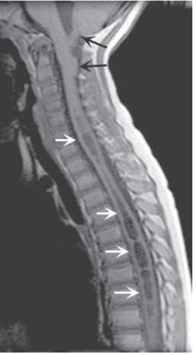
Fig. 453-7 from Harrison's: Sagittal T1 MRI showing cerebellar tonsillar descent (black arrows) and syrinx within the cervical-thoracic cord (white arrows).
- Harrison's Principles of Internal Medicine 22E, p. 3611
- Grainger & Allison's Diagnostic Radiology, p. 1309
Differential Diagnosis
| Condition | Distinguishing Feature |
|---|---|
| Intramedullary spinal cord tumor | Enhances on contrast MRI; clinical course faster |
| Central cord syndrome (traumatic) | Acute onset after hyperextension injury |
| Multiple sclerosis (myelitis) | Demyelinating plaques on MRI; relapsing-remitting |
| Amyotrophic lateral sclerosis | Pure motor, no sensory loss |
| Hematomyelia | Acute onset after trauma; cord hemorrhage on MRI |
| Subacute combined degeneration (B12) | Posterior column dominant; serum B12 low |
| Spinal cord edema | Interstitial (not confluent cavity); associated with contusion/tumor |
- Rosen's Emergency Medicine, p. 2079
- Miller's Review of Orthopaedics, p. 8111
Investigations
- MRI spine + brain - investigation of choice; must include craniocervical junction to identify Chiari
- Phase-contrast MRI - CSF flow dynamics at foramen magnum
- CT myelography - reserved for when MRI is contraindicated; C1-C2 puncture preferred (a subarachnoid web at a lower level acts as a one-way valve that may not be seen with lumbar contrast)
- EMG/NCS - to assess extent of anterior horn cell damage
- Somatosensory evoked potentials - baseline and monitoring
Treatment
Treatment is primarily surgical. The goal is to arrest neurological deterioration; some improvement occurs but complete recovery is uncommon.
Indications for Surgery
- Progressive neurological deficit
- Large symptomatic syrinx
- Acute neurological deterioration (~5% of Chiari-associated cases require urgent decompression)
Asymptomatic/Stable Cases
- Serial clinical and MRI monitoring without surgery
Surgical Options
1. Foramen Magnum Decompression (First-line for Chiari-associated)
- Suboccipital craniectomy + upper cervical laminectomy (C1 ± C2) + duraplasty (dural graft)
- Restores fourth ventricular outflow and normal CSF circulation
- Most effective approach: 70-80% of cases achieve syrinx collapse
- If the curve/syrinx continues to progress, further intervention may be needed
- Note: direct syrinx drainage in addition to decompression has NOT been shown to add benefit and carries more complications
2. Syrinx Shunting
- For large syrinx not associated with Chiari, or residual/recurrent syrinx after decompression
- Types: syringosubarachnoid, syringopleural, or syringoperitoneal shunting
- The three approaches (foramen magnum decompression, syringo-subarachnoid shunting, lumboperitoneal shunting) are reported to be equally effective in achieving syrinx collapse in 70-80% of cases
- Important caveat: clinical outcome and syrinx extension on interval imaging bear NO reliable relationship to whether the cavity remains collapsed on MRI
3. Tumor Resection
- When syrinx is secondary to intramedullary tumor (ependymoma, astrocytoma), resection of the tumor is the primary treatment
4. Management of Hydrocephalus
- Shunting of hydrocephalus, when present, generally precedes correction of the syrinx
5. Scoliosis Management
-
Drainage of the cyst followed by observation is recommended initially; 70% reduction in syrinx size can lead to curve stabilization
-
If curve continues to progress after posterior fossa decompression, scoliosis surgery may be indicated
-
Caution: instrumentation/fusion in a patient with untreated syringomyelia carries risk of paraplegia and cyst rupture; neurosurgical consultation before orthopedic surgery is mandatory
-
Harrison's Principles of Internal Medicine 22E, p. 3611-3612
-
Grainger & Allison's Diagnostic Radiology, p. 1309
-
Campbell's Operative Orthopaedics 15th Ed 2026, p. 3387-3393
-
Rosen's Emergency Medicine, p. 2083
Prognosis
- Natural history is variable; many patients have prolonged periods of stability with sporadic progression
- Surgery can stabilize the deficit; some patients improve
- Neurological improvement is more likely with early intervention before significant cord damage
- Clinical severity does not correlate reliably with syrinx size on MRI
- Even with successful syrinx collapse post-operatively, clinical improvement may not occur
Summary Table
| Feature | Detail |
|---|---|
| Definition | CSF-filled cavity within spinal cord |
| Most common cause | Chiari I malformation (~90%) |
| Classic sensory loss | Dissociated (pain/temp lost, proprioception/vibration spared) |
| Distribution | Cape distribution over shoulders/upper arms |
| Motor signs (UL) | LMN: atrophy, weakness, areflexia |
| Motor signs (LL) | UMN: spasticity, hyperreflexia |
| Worsening triggers | Cough, sneeze, Valsalva |
| Key imaging | MRI (T2-hypointense central cavity) |
| First-line treatment | Foramen magnum decompression (if Chiari) |
| Success rate (syrinx collapse) | 70-80% with surgery |
Recent literature note: A 2025 review (PMID 40364175) comprehensively covers orthopedic manifestations of syringomyelia (scoliosis, Charcot joints, cavus foot), and a 2024 review (PMID 39452020) discusses evolving understanding of Chiari pathophysiology and surgical approaches, consistent with the textbook content above.
This is a shared conversation. Sign in to Orris to start your own chat.