
Explain Hodgkins lymphoma and non hodgkins lymphoma . Their relation in ent head and neck . Their complications , clinical features , differentiations , types, appearance , pathophysiology ,etiology and management , treatment and their regime in both respectively and in last their comparison . In short everything and focusing more over ent
Reading File
Finding Sources
Finding Sources
Finding Sources
Reading File
Reading File
Reading File
Searching PubMed
"Hodgkin lymphoma" AND "head and neck"
Hodgkin Lymphoma (HL) vs Non-Hodgkin Lymphoma (NHL) — Focus on ENT/Head & Neck
1. What they are
Both are malignant clonal proliferations of lymphocytes arising in lymph nodes or lymphoid tissue.
- Hodgkin lymphoma: a B-cell derived malignancy defined by the presence of Reed-Sternberg (RS) cells (large binucleate/multinucleate cells with prominent "owl-eye" nucleoli) sitting in a background of reactive inflammatory cells (lymphocytes, eosinophils, plasma cells, histiocytes). Only 1-2% of the tumor mass is actually malignant cells - most of the mass is the reactive infiltrate.
- Non-Hodgkin lymphoma: a heterogeneous group (>60 subtypes) of B-cell, T-cell, or NK-cell neoplasms without RS cells, composed predominantly of the malignant clone itself.
2. Relevance to ENT / Head and Neck
Lymphoma is the second most common malignancy of the head and neck after squamous cell carcinoma, and the neck/Waldeyer ring is a frequent presenting site for both diseases (Cummings Otolaryngology, ch. 116).
- HL: presents almost exclusively as cervical/supraclavicular lymphadenopathy; classic HL spreads in a contiguous nodal fashion and rarely involves Waldeyer ring, tonsils, sinuses, or preauricular/occipital nodes. Extranodal H&N presentation of HL is rare.
- NHL: extranodal head and neck involvement is common (15-20% of oropharyngeal tumors in some series are NHL, mostly B-cell, MALT-type - Bailey & Love). NHL commonly involves Waldeyer ring (tonsil, base of tongue, nasopharynx), salivary glands, thyroid, nasal cavity/paranasal sinuses (extranodal NK/T-cell lymphoma, nasal type - classically associated with EBV), and preauricular/occipital nodes - sites HL rarely touches.
This nodal-distribution pattern is a key clinical differentiator ENT surgeons use at the bedside.
3. Pathophysiology
- HL: RS cells are derived from germinal-center B cells that have lost normal B-cell gene expression; EBV infects the malignant clone in a large proportion of cases (especially mixed cellularity subtype). RS cells secrete cytokines (IL-5, TNF, TGF-β) that recruit the massive reactive infiltrate responsible for systemic "B symptoms."
- NHL: results from genetic alterations (translocations, e.g., t(14;18) BCL2 in follicular lymphoma, t(8;14) MYC in Burkitt, t(11;14) cyclin D1 in mantle cell) causing uncontrolled clonal lymphocyte proliferation with loss of apoptosis/differentiation arrest. Chronic antigenic stimulation (H. pylori for gastric MALT, EBV for NK/T-cell nasal lymphoma, HIV) also drives many subtypes.
4. Etiology / Risk factors
HL (Cummings, ch.116): EBV infection (prior infectious mononucleosis raises risk ~3x), HIV/chronic immunosuppression (~20x risk), genetic predisposition (first-degree relatives, twin concordance, HLA linkage), tobacco use. Childhood viral illnesses (measles, mumps, rubella) show a negative association; breastfeeding may be protective.
NHL: immunosuppression (post-transplant, HIV/AIDS-related NHL), autoimmune disease, EBV (Burkitt, NK/T-cell nasal type, PTLD), H. pylori (MALT lymphoma), HTLV-1 (adult T-cell leukemia/lymphoma), radiation/chemotherapy exposure, certain occupational chemical exposures.
5. Types / Classification (WHO)
Hodgkin Lymphoma
- Nodular lymphocyte-predominant HL (NLPHL) - ~5%, usually solitary node (cervical/axillary/inguinal), mediastinum spared, non-contiguous spread
- Classical HL (~95%): Nodular sclerosis (most common, especially young women, mediastinal), Mixed cellularity (strongest EBV association), Lymphocyte-rich, Lymphocyte-depleted (rare, elderly/HIV, aggressive)
Non-Hodgkin Lymphoma (relevant to ENT)
- B-cell: Diffuse large B-cell lymphoma (DLBCL, most common NHL overall, aggressive), Follicular lymphoma (indolent), Chronic lymphocytic leukemia/small lymphocytic lymphoma (SLL, indolent), MALT lymphoma (salivary gland, thyroid), Mantle cell, Burkitt lymphoma (highly aggressive, jaw/facial masses in endemic form)
- T/NK-cell: Extranodal NK/T-cell lymphoma, nasal type (classically midline destructive nasal/palatal lesion, EBV-driven - important ENT entity), Angioimmunoblastic T-cell lymphoma, Anaplastic large-cell lymphoma
- AIDS-related NHL: aggressive, often extranodal (H&N, CNS, GI)
- Post-transplant lymphoproliferative disorder (PTLD)
6. Clinical Features
| Feature | Hodgkin Lymphoma | Non-Hodgkin Lymphoma |
|---|---|---|
| Typical age | Bimodal: young adults (15-35) and >55 | Any age, median older; some subtypes in children (Burkitt) |
| Nodal pattern | Painless, contiguous spread; cervical/supraclavicular; bulky, matted nodes | Non-contiguous, unpredictable spread; multiple sites |
| Extranodal H&N disease | Rare | Common (Waldeyer ring, sinuses, salivary glands, thyroid, orbit) |
| B symptoms (fever, night sweats, weight loss) | 30-40% in stage III/IV, <10% in early stage | Variable, more common in aggressive subtypes |
| Pruritus | Classic feature (~25%) | Uncommon |
| Alcohol-induced nodal pain | Rare but classic for HL | Not typical |
| Mediastinal involvement | >85% (often asymptomatic despite bulk) | Variable |
| Course | Predictable, orderly progression | Erratic; indolent subtypes wax/wane (up to 20% spontaneous regression in follicular lymphoma), aggressive subtypes progress rapidly |
ENT-specific presentations: neck mass/painless cervical adenopathy (both), tonsillar asymmetry or enlargement (NHL >> HL), nasal obstruction/epistaxis/palatal ulceration with midfacial destruction (NK/T-cell nasal lymphoma), parotid/salivary gland swelling (MALT NHL, often in Sjögren's patients), thyroid mass (thyroid MALT/DLBCL, often on background of Hashimoto thyroiditis), sore throat/dysphagia/muffled voice from Waldeyer ring involvement, hoarseness or airway compromise from bulky nodal disease.
7. Appearance
- Gross/clinical: firm, rubbery, painless, mobile (early) or matted/fixed (advanced) lymph nodes; NK/T-cell nasal lymphoma shows necrotic, ulcerative, midline destructive palatal/nasal lesions.
- Histology: HL shows scattered RS cells (large, binucleate, "owl-eye" nucleoli, CD30+/CD15+) in a mixed inflammatory background, often with fibrous bands (nodular sclerosis). NHL shows a monomorphic sheet of malignant lymphocytes replacing normal nodal architecture (loss of follicular pattern), immunophenotype-specific markers (CD20 for B-cell, CD3 for T-cell).
- Imaging: CT/PET show bulky, sometimes matted nodal conglomerates (HL) or variable single/multiple nodal and extranodal masses (NHL); PET-CT is central to staging and response assessment in both.
8. Complications
HL: airway/SVC compression from mediastinal bulk, secondary malignancies (leukemia, solid tumors) from treatment, treatment-related cardiotoxicity (anthracyclines) and pulmonary fibrosis (bleomycin), hypothyroidism/xerostomia after neck/mediastinal radiotherapy, infertility, immune dysregulation (increased infection risk).
NHL: local destruction (nasal septal/palatal perforation in NK/T-cell type), airway obstruction from Waldeyer ring bulk, tumor lysis syndrome (especially Burkitt, DLBCL - rapid cell turnover), CNS involvement/spread, bone marrow infiltration/cytopenias, hypercalcemia, secondary infections from immunosuppression, transformation of indolent to aggressive subtype (e.g., follicular to DLBCL).
9. Diagnostic Differentiation Workup
Both require excisional or core biopsy (not FNA alone) of an accessible node - ENT surgeons frequently provide this via neck node or tonsillectomy/adenoidectomy specimens. Workup includes immunohistochemistry (CD15/CD30 for RS cells vs CD20/CD3/CD10/BCL2/BCL6/Ki-67 panels for NHL subtyping), flow cytometry, EBV studies, bone marrow biopsy, CT chest/abdomen/pelvis or PET-CT, and Ann Arbor staging (I-IV, with A/B for absence/presence of systemic symptoms) for both diseases.
10. Management / Treatment Regimens
Hodgkin Lymphoma
- Early stage (I-II, favorable): ABVD chemotherapy (Adriamycin/doxorubicin, Bleomycin, Vinblastine, Dacarbazine), 2-4 cycles + involved-site radiotherapy (ISRT)
- Advanced stage (III-IV): ABVD x 6 cycles, or escalated BEACOPP in high-risk/bulky disease
- Relapsed/refractory: high-dose chemotherapy + autologous stem cell transplant; brentuximab vedotin (anti-CD30 antibody-drug conjugate); checkpoint inhibitors (nivolumab, pembrolizumab) for relapsed classical HL
- NLPHL: often managed with radiotherapy alone if localized, or rituximab (CD20+) based regimens
Non-Hodgkin Lymphoma (subtype-driven)
- Aggressive B-cell (DLBCL): R-CHOP (Rituximab, Cyclophosphamide, doxorubicin/Hydroxydaunorubicin, Vincristine/Oncovin, Prednisone), typically 6 cycles +/- radiotherapy for bulky/localized nodal disease
- Indolent (follicular, SLL): watch-and-wait if asymptomatic; rituximab-based chemoimmunotherapy (R-CHOP, R-Bendamustine) when treatment indicated; radiotherapy alone for truly localized disease
- Burkitt lymphoma: intensive short-cycle regimens (e.g., CODOX-M/IVAC, hyper-CVAD) plus tumor lysis prophylaxis (aggressive hydration, rasburicase/allopurinol)
- Extranodal NK/T-cell lymphoma, nasal type: concurrent radiotherapy + chemotherapy (asparaginase-based regimens, e.g., SMILE), since it is relatively radioresistant to standard CHOP but radiosensitive at higher doses
- MALT lymphoma (salivary/thyroid): localized radiotherapy is often curative; H. pylori eradication for gastric variants
- Relapsed/refractory NHL: autologous/allogeneic stem cell transplant, CAR-T cell therapy (axicabtagene, tisagenlecleucel) for relapsed DLBCL, bispecific antibodies
Per Bailey & Love: radiotherapy is treatment of choice for localized H&N NHL, while systemic chemotherapy is required for widespread/disseminated NHL; ENT surgery's main role in lymphoma is diagnostic (biopsy), airway management, and managing local complications rather than definitive oncologic resection (lymphoma is not surgically excised as primary treatment).
11. Overall Comparison Summary
| Parameter | Hodgkin Lymphoma | Non-Hodgkin Lymphoma |
|---|---|---|
| Cell of origin | B-cell (RS cells) with reactive background | B, T, or NK cell - the malignant clone itself |
| Spread pattern | Orderly, contiguous | Unpredictable, often multicentric |
| Extranodal H&N disease | Rare | Common (Waldeyer ring, sinuses, salivary glands, thyroid) |
| Age | Bimodal (young + elderly) | Any age, generally older median |
| B symptoms | Classic, correlates with stage | Variable |
| Prognosis | Generally excellent (>80-90% cure in early stage) | Highly variable - indolent subtypes prolonged survival without cure; aggressive subtypes potentially curable, high-grade untreated fatal in months |
| First-line regimen | ABVD +/- radiotherapy | R-CHOP (or subtype-specific) |
| EBV association | Strong (especially mixed cellularity) | Present in specific subtypes (Burkitt, NK/T-cell nasal, PTLD) |
| Surgical role in ENT | Diagnostic biopsy only | Diagnostic biopsy; occasional debulking for airway relief |
A recent systematic review (PMID 38862882, 2024) on pediatric/adolescent B-cell lymphoma presentation and a population-modeling study on HIV-attributable HL incidence (PMID 40879655) reinforce that immunosuppression (HIV) remains a major modifiable risk driver for both HL and aggressive NHL - consistent with the etiology described above; no findings there contradict the standard management approach outlined.
If you want, I can go deeper into any single piece of this - for example a focused breakdown of extranodal NK/T-cell nasal lymphoma (a uniquely ENT-relevant entity), staging systems (Ann Arbor/Lugano), or detailed chemotherapy toxicity profiles.
Deep dive into extranodal NK/T-cell nasal lymphoma: pathology, imaging, and surgical management
Finding Sources
Finding Sources
Searching Images
extranodal NK/T-cell lymphoma nasal type histology angiocentric necrosis
Searching Images
nasal NK/T-cell lymphoma CT MRI midline destructive lesion palate
Searching PubMed
"extranodal NK/T-cell lymphoma" nasal type treatment
Reading File
Reading File
Reading File
Searching Images
nasal lymphoma CT scan midline destructive lesion palate perforation
Reading Article
PMID: 33458595
Extranodal NK/T-Cell Lymphoma, Nasal Type — Deep Dive
Overview
Extranodal NK/T-cell lymphoma, nasal type (ENKTL) is a rare, aggressive, almost universally EBV-driven non-Hodgkin lymphoma arising from NK cells (or, less commonly, cytotoxic T cells). It was historically called "lethal midline granuloma" or angiocentric lymphoma before its lymphomatous nature was recognized. It is one of the few lymphomas an ENT surgeon will personally diagnose and manage the local complications of, since the nasal cavity/nasopharynx is the classic primary site.
Epidemiology: Markedly more common in East Asia and Central/South America; rare in Europe/North America, where it accounts for only 0.2-0.4% of all NHL (van Doesum et al., HemaSphere 2021, PMID 33458595). Affects young-to-middle-aged adults with a male predominance.
Pathophysiology / Pathogenesis
- Nearly 100% EBV-associated - the tumor cells harbor clonal episomal EBV, most often the LMP1-deletion variant strain.
- Malignant cells express cytotoxic granule proteins (perforin, granzyme B, TIA-1), consistent with an NK/cytotoxic-T origin.
- The defining biological feature is angiocentricity and angioinvasion: tumor cells infiltrate and destroy vessel walls, causing thrombosis and ischemic necrosis. This vascular destruction, not direct tumor bulk alone, drives the characteristic tissue destruction and explains why biopsies are so often non-diagnostic (necrotic tissue predominates over viable tumor).
- T-cell receptor (TCR) genes are typically in germline configuration (true NK lineage) but can be clonally rearranged in the rarer cytotoxic T-cell-derived cases.
- Circulating plasma EBV DNA correlates with tumor burden and is used as a biomarker for disease monitoring and prognosis.
Pathology
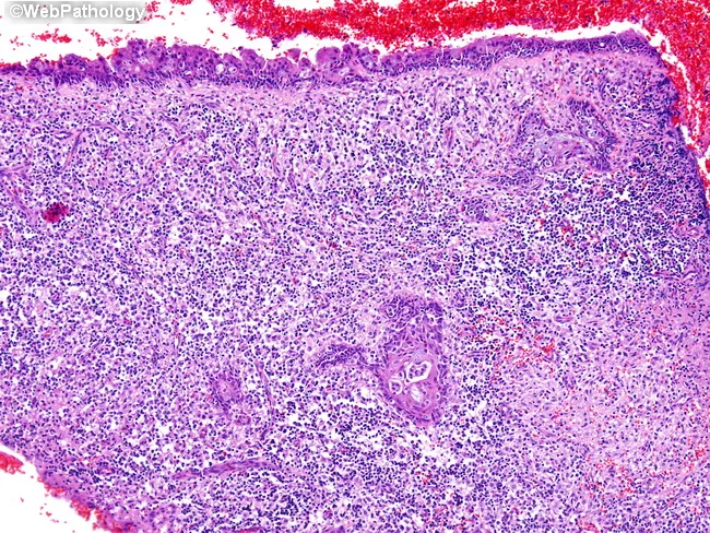
Histology: Dense infiltrate involving the mucosa/submucosa (or dermis/subcutis when skin is involved - the second most common site). Cell size ranges from small to large, but most cases show medium-sized cells with irregular/oval nuclei, moderately dense chromatin, and pale cytoplasm. Prominent angiocentric and angiodestructive growth with extensive necrosis is the histologic hallmark, often admixed with a reactive inflammatory background (small lymphocytes, histiocytes, plasma cells, eosinophils) that can mimic an infectious or inflammatory process. Overlying mucosa frequently shows squamous metaplasia or pseudoepitheliomatous hyperplasia, which can be mistaken for squamous cell carcinoma on limited sampling.
Immunophenotype: CD2+, cytoplasmic CD3+ (surface CD3 negative — a key diagnostic point), CD56+, and cytotoxic markers (TIA-1, granzyme B, perforin) positive. EBV positivity by EBER in-situ hybridization is required for diagnosis — CD56+/EBV+ lymphocytes are essentially never seen in normal or inflammatory nasal mucosa, making this combination highly specific.
Diagnostic pitfall: Because of extensive necrosis, a single biopsy is frequently non-diagnostic. Multiple, deep biopsies (often repeated) are usually needed to capture viable tumor tissue away from the necrotic center.
Clinical Features
- Nasal obstruction, epistaxis, foul-smelling discharge
- Progressive midfacial destructive ulceration - palatal perforation, septal perforation, facial swelling/erythema
- Can extend to paranasal sinuses, orbit, nasopharynx, skin
- Clinical course may be complicated by a hemophagocytic syndrome with pancytopenia and fever - an aggressive, sometimes fatal complication
- Systemic B symptoms and disseminated disease occur in advanced stages
Imaging
Both CT and MRI are used, and PET-CT is now standard for staging (per van Doesum et al. 2021):
- CT: best for delineating bone destruction - septal/palatal erosion, sinus wall destruction, extent of midfacial bony involvement. Shows a soft-tissue infiltrative mass centered on the nasal cavity with variable degrees of osteolysis.
- MRI: better for soft-tissue extent - defines invasion into orbit, pterygopalatine fossa, skull base, and perineural spread; lesions are typically iso- to hypointense on T1, variable T2 signal, with heterogeneous enhancement due to necrosis.
- PET-CT: essential for accurate staging (differentiating localized stage I/IIE from disseminated stage III/IV disease) and for post-treatment response assessment.
- MRI of the nasopharynx specifically is recommended at diagnosis given the propensity for deep extension along this route.
- Plasma EBV DNA PCR is a required adjunct at diagnosis and during follow-up, complementing imaging in disease monitoring.
Staging and Workup
Ann Arbor/Lugano staging is used, but risk-adapted models specific to ENKTL are applied. Key risk factors defining "high-risk" stage I/II disease: stage II (vs. I), age >60, elevated LDH, ECOG performance status ≥2, and primary tumor invasion beyond the nasal cavity. Workup includes bone marrow biopsy, full-body PET-CT, MRI nasopharynx, and plasma EBV DNA quantification.
Surgical Management (ENT-specific role)
Surgery is not curative in ENKTL — this is fundamentally a radiosensitive/chemosensitive systemic disease, and aggressive resection has no survival benefit and risks unnecessary morbidity. The ENT surgeon's role is therefore mainly:
- Diagnostic: endoscopic nasal/nasopharyngeal biopsy — often requiring multiple deep biopsies (image-guided or repeated endoscopic sampling) because of the necrotic tumor core, plus biopsy of any skin lesions if present.
- Staging endoscopy: assessing extent of mucosal/bony involvement for radiotherapy planning and to define resectable necrotic/infected tissue.
- Debridement of necrotic tissue: endoscopic debridement can be needed to control secondary infection, foul discharge, or crusting from the necrotic ulcerated mass, and to relieve nasal obstruction — this is palliative/supportive, not oncologic.
- Management of complications: control of epistaxis (which can be significant given the angioinvasive, vessel-destructive biology), airway management if there is significant obstruction, and treatment of secondary sinus infection.
- Reconstruction after treatment-related tissue loss: a growing surgical literature (e.g., Ueda et al., Cureus 2025, PMID 41561241) addresses external nasal reconstruction in patients left with saddle-nose deformity, septal/palatal perforation, or significant soft-tissue and cartilage loss after the disease process and/or radiotherapy — using local flaps, cartilage grafts, or free flaps once the disease is in remission. This is reconstructive/rehabilitative surgery performed after oncologic treatment, not primary treatment.
- There is no role for radical surgical resection or wide local excision as primary curative therapy — attempting this delays definitive chemoradiotherapy and does not improve outcomes, unlike squamous cell carcinoma of the sinonasal tract.
Definitive (Non-Surgical) Management — for context
- Stage I/II (localized): Radiotherapy is the backbone (minimum dose 50-56 Gy). Low-risk stage I can sometimes be treated with RT alone; high-risk stage I/II (per criteria above) requires combined chemoradiotherapy (concurrent platinum-based or sequential regimens).
- Chemotherapy regimen of choice: non-P-glycoprotein (non-MDR)-based regimens, since ENKTL cells frequently over-express multidrug-resistance proteins that confer resistance to anthracycline-based regimens like CHOP. L-asparaginase-based regimens are preferred:
- SMILE regimen (dexamethasone, methotrexate, ifosfamide, L-asparaginase, etoposide) — response rates 80%+ in trials
- Stage III/IV (disseminated): chemotherapy (e.g., 3 cycles) followed by autologous hematopoietic stem cell transplant; allogeneic transplant reserved for relapsed/refractory disease.
- Monitoring: serial plasma EBV DNA levels track tumor burden and relapse risk during and after treatment.
Prognosis
Historically poor with RT alone (high relapse rates despite good initial response), improved substantially with asparaginase-based chemoradiotherapy protocols. Advanced-stage and hemophagocytic-syndrome-complicated disease carry a markedly worse prognosis. Early, accurate diagnosis (which depends heavily on adequate ENT biopsy technique given the necrosis-diagnosis pitfall) is a major determinant of outcome.
This is a shared conversation. Sign in to Orris to start your own chat.