
Frostnip and frostbite
I'll look this up in the medical library for you.
Reading File
Now let me search the library and PubMed in parallel.
Finding Sources
Searching PubMed
frostbite frostnip treatment
Good hits. Now let me read the most comprehensive sources in parallel - Fitzpatrick's (the gold standard dermatology text), Rosen's Emergency Medicine, and the Washington Manual.
Reading File
Reading File
Reading File
Good content. Now let me read more of Fitzpatrick's (treatment section) and Rosen's (management details), plus fetch the clinical images.
Reading File
Reading File
Now let me get the treatment/management details from Rosen's and Fitzpatrick's, and fetch the key clinical images.
Reading File
Reading File
Loading Image
Loading Image
Let me fetch the deep frostbite and gangrene images, and get the Rosen's management details.
Loading Image
Loading Image
Reading File
Now let me get the management/treatment section from Rosen's.
Reading File
I now have excellent, comprehensive content. Let me compile the full answer.
Frostnip and Frostbite
Overview
These are freezing cold injuries caused by exposure to cold air, liquids, or metal. They exist on a spectrum, from frostnip (fully reversible, no tissue destruction) to deep frostbite (irreversible tissue death, potential amputation). Historically, frostbite caused over 1 million casualties in World Wars I & II and the Korean War.
- Fitzpatrick's Dermatology, Vol. 1-2, p. 1697
- Rosen's Emergency Medicine, p. 2775
Frostnip
Frostnip is the only form of cold injury safely treated in the field with first aid alone.
Pathophysiology: Superficial vasoconstriction without true tissue freezing. No ice crystal formation, no cell death.
Clinical features:
- Intense cold sensation progressing to numbness, then pain
- Erythema on cheeks, ears, nose, fingers, toes
- No edema, no blisters (blebs)
- Skin remains soft and pliable
- Damage is fully reversible
Treatment:
- Remove from cold exposure immediately
- Rewarm the affected area (e.g. warm hands placed over the area, breath warming)
- No specific medical intervention needed; heals completely
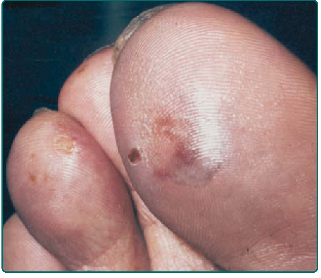
Frostbite
Frostbite involves actual freezing of tissue with ice crystal formation and potential permanent damage.
Pathophysiology - The Freezing Injury Cascade
The injury unfolds in sequential phases (Rosen's, p. 2776):
| Phase | Events |
|---|---|
| Pre-freeze | Superficial cooling, increased blood viscosity, microvascular constriction, endothelial plasma leakage |
| Freeze-thaw | Extracellular ice crystal formation → water shifts across cell membranes → intracellular dehydration, hyperosmolality → cell membrane denaturation and collapse |
| Vascular stasis | Vasospasticity, stasis coagulation, AV shunting, endothelial damage, prostanoid release, interstitial hypertension |
| Late ischemia | Necrosis, mummification, demarcation, slough |
Key mediators: Blister fluid (especially clear blebs) is rich in prostaglandin F2α and thromboxane A2, causing vasoconstriction, platelet aggregation, and leukocyte adhesion - perpetuating progressive tissue loss even after warming.
The acral structures (fingers, toes, ears, nose, penis) are at greatest risk because they contain arteriovenous anastomoses that shut down during cold stress (the "life-versus-limb" mechanism).
Classification
Modern classification (Cauchy/Wilderness Medicine) is based on the extent of the initial lesion after rapid rewarming and day-2 bone scan findings:
| Grade | Initial Lesion | Blisters (Day 2) | Prognosis |
|---|---|---|---|
| 1 | None after rewarming | None | No amputation, no sequelae |
| 2 | Distal phalanx | Clear blisters | Tissue amputation; nail sequelae |
| 3 | Mid/proximal phalanx | Hemorrhagic blisters on digit | Bone amputation of digit; functional sequelae |
| 4 | Carpal/tarsal | Hemorrhagic blisters over carpal/tarsal | Limb amputation ± sepsis |
Rosen's Emergency Medicine, p. 2779
Traditional degree-based classification (still widely used):
- 1st degree (frostnip): Anesthesia, erythema - fully reversible
- 2nd degree (superficial frostbite): Superficial vesiculation with clear/milky blebs, edema, erythema; subcutaneous tissue soft and resilient
- 3rd degree (deep frostbite): Hemorrhagic blebs, deeper involvement into subcutaneous tissue/muscle
- 4th degree: Extends to tendon and bone; leads to gangrene
Clinical Presentation
Superficial frostbite (2nd degree):
- Waxy, firm skin, but deeper tissue remains soft
- Pain subsides to a sensation of warmth - this is a warning sign of severe involvement
- Clear blebs form 24-36 hours after thawing
- Erythema and edema present
Deep frostbite (3rd/4th degree):
- Skin becomes white or bluish-white, totally numb and anesthetic
- Tissue hard and indurated; joints immobile; muscles may be paralyzed
- Large blisters form 1-2 days after rewarming
- Eschar forms → dry gangrene → autoamputation weeks later along the line of demarcation
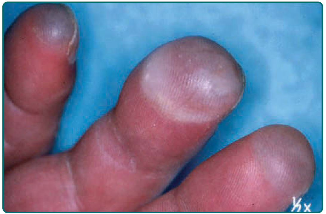
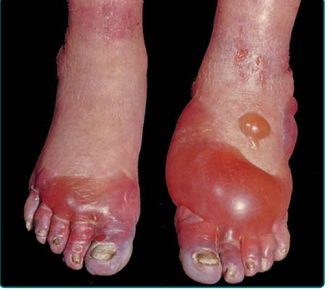
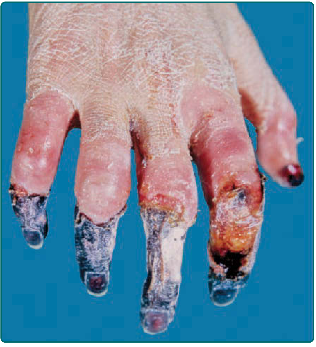
Prognostic Signs
| Good Signs | Poor Signs |
|---|---|
| Large clear blebs extending to digit tips | Hard, white, cold, insensitive skin |
| Rapid return of sensation | Cold, cyanotic skin without blebs after rewarming |
| Rapid return of warmth | Dark hemorrhagic blebs |
| Pink skin after rewarming | Early mummification |
| Rapid capillary refill | Freeze-thaw-refreeze injury |
| Constitutional signs (fever, tachycardia) |
Fitzpatrick's, Table 98-2
Predisposing Factors
- Physiologic: Dehydration, overexertion, prior cold injury, hypoxia, lack of acclimatization
- Mechanical: Constricting or wet clothing, tight boots, immobility
- Psychological: Intoxication, mental status changes, fatigue, hunger
- Environmental: Low ambient temperature, wind chill, high altitude, humidity
- Cardiovascular: Diabetes, PVD, Raynaud syndrome, atherosclerosis, sickle cell disease, anemia
Rosen's Emergency Medicine, Box 128.5
Management
Pre-hospital
- Remove wet/constricting clothing; insulate and immobilize affected area
- Do NOT massage - increases tissue loss
- Do NOT rub with snow
- Keep away from dry heat (forced air, open fire)
- Critical rule: Do not rewarm in the field if there is any risk of refreezing - the freeze-thaw-refreeze cycle is catastrophically destructive
Hospital - Rapid Rewarming (cornerstone of treatment)
- Immerse in water bath at 37-39°C (98.6-102.2°F) for 15-30 minutes until the most distal parts flush pink
- Hexachlorophene or povidone-iodine can be added to the bath
- Premature termination of thawing is a common error
- Reperfusion is painful - parenteral analgesia is often required
- Elevate the affected part
- Leave blisters intact - do not debride
- Check for and treat systemic hypothermia first
Adjunctive Therapies
- Topical aloe vera - inhibits thromboxane synthetase, reduces vasoconstriction
- Oral ibuprofen - inhibits COX, counters prostanoid-mediated injury
- Thrombolysis - may salvage severely frostbitten tissue if given within 24 hours of thawing (tPA)
- Vasodilators, hyperbaric oxygen in selected cases
- Tetanus toxoid for open wounds
- Sympathectomy / intra-arterial reserpine - controversial, some benefit reported
Imaging for Surgical Planning
- Triple-phase bone scan, MRI, and MR angiography can predict tissue viability and accelerate surgical decision-making. The historical aphorism "frostbite in January, amputate in July" has given way to earlier imaging-guided intervention.
- Surgical debridement is generally best delayed 1-3 months after demarcation.
Fitzpatrick's, p. 1698-1699; Washington Manual, p. 989; Rosen's, p. 2779
Sequelae
Long-term complications of frostbite include:
- Permanent cold hypersensitivity (most common)
- Hyperhidrosis
- Neuropathic pain, paresthesias, complex regional pain syndrome
- Frostbite arthritis (resembling osteoarthritis) - weeks to years later
- Epiphyseal plate damage in children - shortened digits, joint deviation, dystrophic nails
- Ossification of the ear cartilage
- Squamous cell carcinoma (rare, heel, 20-30 years later)
Fitzpatrick's, p. 1699
Quick Comparison: Frostnip vs Frostbite
| Feature | Frostnip | Superficial Frostbite | Deep Frostbite |
|---|---|---|---|
| Tissue freezing | No | Yes (superficial) | Yes (deep) |
| Cell death | None | Minimal-moderate | Yes |
| Blisters | Absent | Clear blebs | Hemorrhagic blebs |
| Skin appearance | Red, warm | Waxy | White/blue, hard |
| Deeper tissue | Soft | Soft | Hard, indurated |
| Reversibility | Complete | Partial | Incomplete to none |
| Field treatment | Yes | No - hospital needed | No - hospital needed |
| Sequelae | None | Possible | Likely |
Can you give me an animated video on pathophysiology of frostbite
Searching the Web
frostbite pathophysiology animated video YouTube
I can't create an animated video myself, but here are the best YouTube videos on frostbite pathophysiology:
Most relevant - dedicated pathophysiology video (graphic content warning):
Dr. Nabil Ebraheim's educational animated overview of frostbite:
Nursing-focused walkthrough covering pathophysiology and management:
The first link ("The pathophysiology of frostbite") is the most directly relevant to what you're looking for. Dr. Ebraheim's video is a good animated alternative if you want a clean, concise educational format without graphic clinical imagery.
I'm not able to generate animated videos - that's outside what I can produce. If you'd like, I can create a static illustrated diagram of the frostbite pathophysiology cascade, or a detailed written summary formatted for study/teaching purposes.
This is a shared conversation. Sign in to Orris to start your own chat.