
Fontanelle
Reading File
Searching sources...
Finding Sources
Finding Sources
Reading File
Reading File
Reading File
Loading Image
Finding Sources
Here is a complete overview of fontanelles from the medical library:
Fontanelles
Fontanelles are membranous gaps (fibrous soft spots) at the junctions of the cranial bones in the newborn skull, where ossification is incomplete. They allow the skull bones to overlap during childbirth (moulding) and permit continued brain growth postnatally.
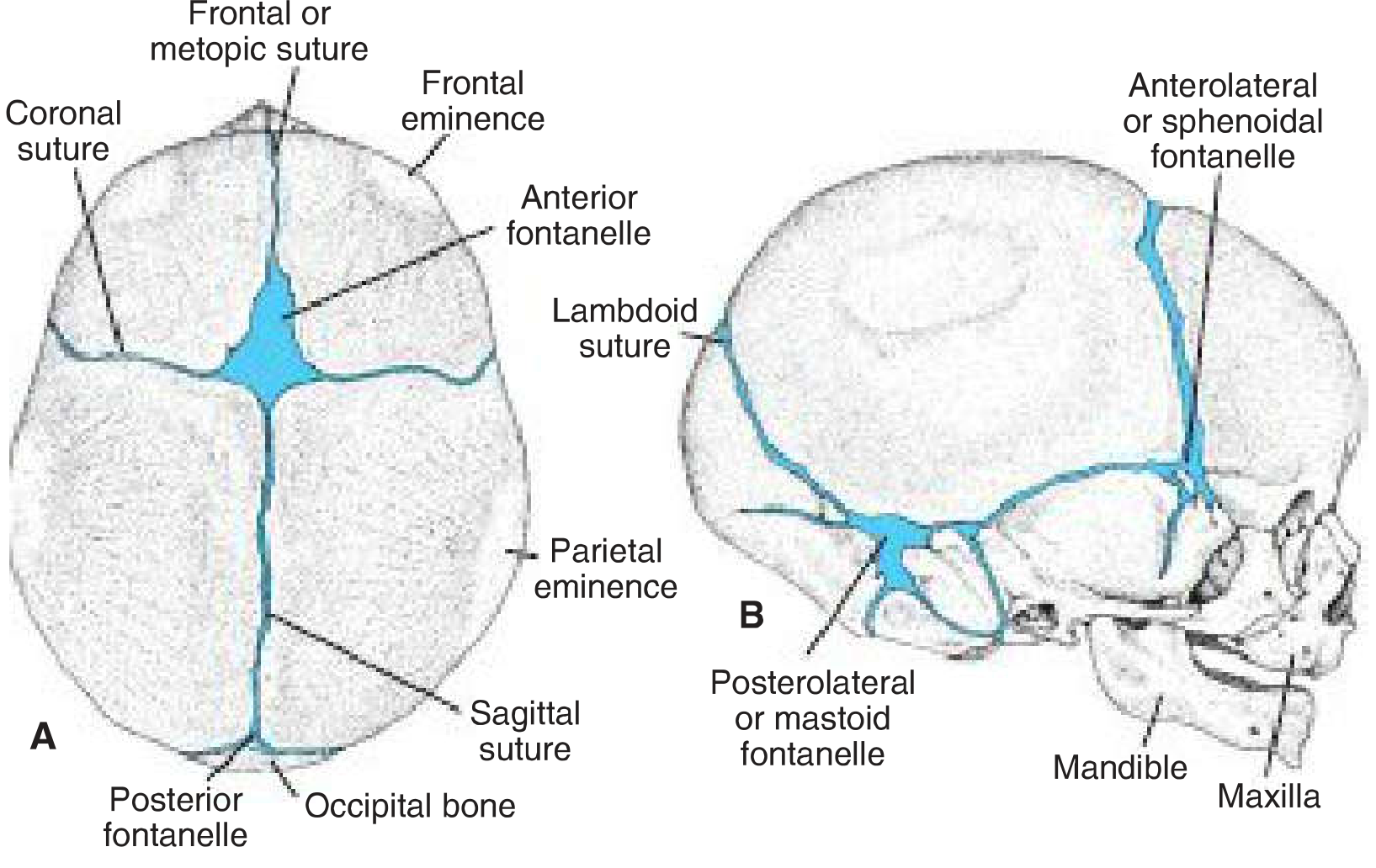
Skull of a newborn seen from above (A) and right side (B), showing the anterior and posterior fontanelles and associated sutures - Langman's Medical Embryology
Types of Fontanelles
| Fontanelle | Location | Shape | Closure |
|---|---|---|---|
| Anterior (bregmatic) | Junction of frontal, sagittal, and coronal sutures | Diamond / cross (+) | 18 months (range 1.5-2 years) |
| Posterior (lambdoid) | Junction of sagittal and lambdoid sutures | Triangular / Y-shape | 1-3 months after birth |
| Anterolateral (sphenoidal) | At the pterion (junction of frontal, parietal, sphenoid, temporal) | - | ~3 months |
| Posterolateral (mastoid) | At the asterion (junction of parietal, temporal, occipital) | - | 6-18 months |
The anterior fontanelle is always the largest and most clinically significant. The posterior fontanelle is smaller and closes earliest.
Clinical Significance of the Anterior Fontanelle
In the first years of life, palpation of the anterior fontanelle gives important clinical information about:
- Normal ossification - whether skull development is proceeding normally
- Intracranial pressure
- Bulging fontanelle - suggests raised intracranial pressure (e.g., meningitis, hydrocephalus, intracranial hemorrhage)
- Sunken fontanelle - seen in hypovolaemia/dehydration (one of the clinical signs of dehydration in infants under 1 year)
"A bulging fontanelle may suggest raised intracranial pressure, whereas a sunken fontanelle is seen in hypovolaemia." - Bailey and Love's Short Practice of Surgery, 28th Ed.
Forensic & Age Estimation Relevance
From PC Dikshit's Textbook of Forensic Medicine & Toxicology, fontanelle closure timing is used to estimate age at death in infants/neonates:
- Lateral and occipital fontanelle: closes at 2 months after birth
- Posterior fontanelle: closes at 6-8 months after birth
- Anterior fontanelle: closes at 1.5-2 years after birth
- Metopic suture: closes at 2-4 years (may extend to 6 years)
Nasal Fontanelles (ENT Context)
In otolaryngology/sinus anatomy, the term "fontanelle" also refers to membranous areas in the lateral nasal wall (nasal fontanelles), found in the region between the uncinate process and the inferior turbinate:
- Anterior nasal fontanelle - anterior to the uncinate bone
- Posterior nasal fontanelle - posterior to the uncinate bone
Accessory ostia of the maxillary sinus (found in 20-25% of patients) occur in perforated fontanelles, typically the posterior nasal fontanelle, and may indicate prior sinus disease. This is relevant in functional endoscopic sinus surgery (FESS).
Obstetric Context (Fetal Head Assessment)
During labour and delivery, fontanelles are used to assess fetal head position:
- The anterior fontanelle (cross/diamond shape) and posterior fontanelle (Y/triangular shape) are felt on vaginal examination to determine fetal presentation
- The posterior fontanelle pointing anteriorly indicates occiput anterior (OA) position (normal)
- An occiput posterior (OP) position is associated with a palpable anterior fontanelle anteriorly
Sources: Langman's Medical Embryology; Bailey and Love's Short Practice of Surgery 28th Ed.; PC Dikshit Textbook of Forensic Medicine and Toxicology; Cummings Otolaryngology; KJ Lee's Essential Otolaryngology; Tintinalli's Emergency Medicine
This is a shared conversation. Sign in to Orris to start your own chat.