

sorosis
onychomycosis nail fungal infection subungual hyperkeratosis
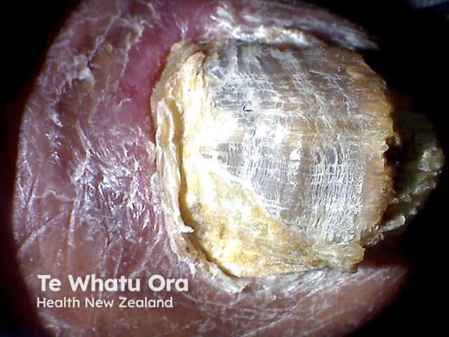
This is a dermatology clinical photograph of a toenail demonstrating total onychomycosis with complete dystrophy of the nail plate. The image captures a dorsal, macro view of the nail unit showing an extremely thickened, opaque, yellow-white to brownish nail plate with surface cracking and irregular layering. Subungual hyperkeratosis is evident with overlying rim of discolored keratin and distal lifting of the plate from the nail bed. Surrounding epidermis appears mildly inflamed or reactive. The appearance is characteristic of advanced fungal involvement (onychomycosis), typically caused by dermatophytes (e.g., Trichophyton spp.), non-dermatophyte molds, or Candida species; however, etiological confirmation requires mycological culture or microscopy. The image illustrates a severe, long-standing infection often associated with secondary bacterial colonization, discomfort, and functional impairment. Clinically relevant features include thickened plate, brittleness, discoloration, and potential subungual hyperkeratosis. This image is useful for education on differential diagnoses of nail dystrophy, including psoriasis or trauma, and for recognizing stages requiring systemic antifungal therapy versus topical management. Potential clinical use cases include patient education, dermatology training, image-based case discussions, and algorithmic scene understanding for AI diagnostic tools. It supports research into treatment response monitoring and disease progression in onychomycosis. Clinical context supports antifungal therapy planning and monitoring response effectively.
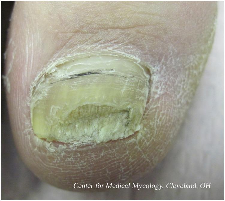
This clinical photograph shows a close-up view of a hallux (great toe) exhibiting classic features of distal subungual onychomycosis, a common fungal infection often caused by dermatophytes like Trichophyton rubrum. The nail plate demonstrates significant subungual hyperkeratosis, resulting in marked thickening and a crumbly, dystrophic texture at the distal edge. The nail is discolored with a yellowish-green hue and shows evidence of being trimmed to manage the bulk. A thin, horizontal dark line is visible near the proximal nail plate, possibly representing a splinter hemorrhage or subungual debris. The surrounding periungual skin displays secondary changes, including xerosis, scaling, and fine white fissuring, which may indicate concomitant tinea pedis or a compromised epidermal barrier. This image serves as a clinical example of chronic nail fungal infection, illustrating the diagnostic challenges posed by nail plate penetration and the necessity for long-term antifungal therapy.
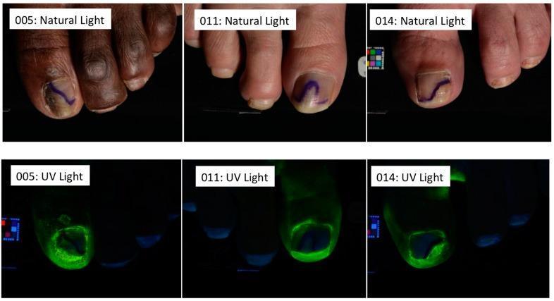
Clinical photographs depicting three subjects (005, 011, and 014) with onychomycosis, shown in a comparative side-by-side format under natural and UV light. The top row (Natural Light) displays hallmark signs of fungal infection: subungual hyperkeratosis, nail plate thickening, and yellow-brown chromonychia. Blue ink markings on the nail plates delineate the proximal boundary of the treatment area. The bottom row (UV Light) demonstrates the distribution of a fluorescently-labeled efinaconazole vehicle solution. The intense green fluorescence is concentrated at the hyponychium and subungual space, illustrating the delivery of the topical antifungal to the nail bed and matrix. Subject 005 exhibits the highest degree of disease severity with 55% nail involvement, while subjects 011 and 014 show 35% and 40% involvement, respectively. This comparison highlights the penetration capabilities of specialized vehicle formulations in penetrating the keratinized nail unit to reach the site of infection.
Onychomycosis (Tinea Unguium) - "Sorosis" of the Nails
Note: "Sorosis" is not a standard medical/dermatological term for nail disease. Based on the image provided, this appears to be onychomycosis (fungal nail infection). If you meant a different condition, please clarify.
What the Image Shows
- Yellowish-brown discoloration throughout all nail plates
- Subungual hyperkeratosis - buildup of crumbly keratin debris under the nail
- Onycholysis - nail plate separation from the nail bed (visible at distal and lateral edges)
- Nail plate thickening, friability, and surface roughness
- Longitudinal splitting and fissuring, especially the middle finger
- Exposed pinkish nail bed where the plate has partially detached
What is Onychomycosis?
Clinical Types
| Type | Features | Main Pathogen |
|---|---|---|
| Distal subungual (most common) | Starts at distal corner, yellow streak, spreads proximally, subungual hyperkeratosis, onycholysis | T. rubrum |
| White superficial | White friable patches on nail surface (toenails only) | T. mentagrophytes, Aspergillus |
| Proximal subungual | Leukonychia starting from proximal nail fold; consider HIV | T. rubrum |
| Candida | Massive nail destruction + nail bed hyperkeratosis | C. albicans |
Differential Diagnoses to Consider
- Nail psoriasis - can look identical (oil drop sign, pitting, salmon patch help distinguish)
- Traumatic onychodystrophy - history of injury
- Contact dermatitis or chronic paronychia
- Yellow nail syndrome
Treatment
- Terbinafine 250 mg/day - 2 months for fingernails, 3 months for toenails (most effective for dermatophytes) - Goldman-Cecil Medicine, p. 1784
- Itraconazole - pulse therapy (400 mg/day for 1 week/month × 2 cycles for fingers, × 3 cycles for toes)
- Fluconazole - 150-450 mg weekly for 3-6 months - Fitzpatrick's Dermatology, p. 4409
- Efinaconazole, tavaborole, ciclopirox lacquer - 48-week courses required
- Lower cure rates than systemic therapy
Reference Images
Practical Recommendations
- Confirm the diagnosis with KOH prep or fungal culture before starting systemic treatment
- Prescribe oral terbinafine (first-line for dermatophyte infection)
- Check baseline LFTs before systemic antifungals (hepatotoxicity risk)
- Advise the patient on relapse prevention: keep nails dry, use antifungal powder in footwear, treat concurrent tinea pedis
- Cure rates for fingernail onychomycosis are higher (~80%) than toenails (~50-70%) with systemic therapy
and the patient has developed Patches all over the bodyy Black patches looking like wound healing
Addison disease hyperpigmentation dark patches skin nails
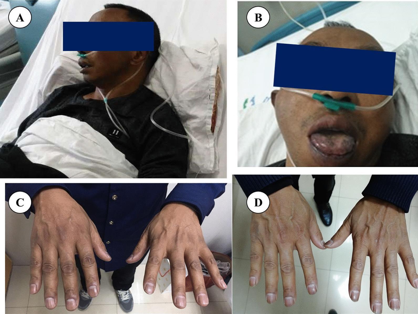
A series of clinical photographs documenting mucocutaneous hyperpigmentation in a patient with Addison’s disease and its partial resolution after treatment. Panel A displays diffuse, dark-brown hyperpigmentation of the facial skin. Panel B focuses on the oral cavity, revealing characteristic dark-brown macules and patches of varying sizes on the tongue and lips. Panel C shows the dorsal aspect of both hands, demonstrating diffuse, dark-brown pigmentation particularly prominent over the knuckles and small joints. Panel D presents the dorsal aspect of the hands after two months of hormone replacement therapy, showing a notable reduction in pigment intensity and a more even skin tone, though mild residual hyperpigmentation persists. These findings illustrate the clinical hallmark of primary adrenal insufficiency (Addison’s disease), where elevated ACTH levels lead to increased melanocyte stimulation, and provide a visual baseline for assessing treatment response.
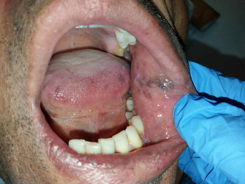
A clinical photograph of the oral cavity of an adult male, demonstrating hallmark signs of Addisonian melanoglossia and mucosal hyperpigmentation. The view is facilitated by a gloved hand retracting the left cheek. Distinctive dark brown to slate-gray macules and confluent patches are visible on the left buccal mucosa, exhibiting irregular borders and varying intensity. Similar hyperpigmented lesions are observed on the lateral and posterior surfaces of the tongue, presenting as mottled, reddish-purple to dark brown areas. The surrounding gingiva and hard palate appear relatively unaffected. The perioral skin also shows signs of generalized darkening. This image illustrates the oral manifestations of primary adrenal insufficiency (Addison's disease), where increased adrenocorticotropic hormone (ACTH) leads to excessive melanin production by melanocytes. This material is useful for medical students and clinicians in identifying dermatological markers of systemic endocrine disorders and adrenal crisis risk assessment.
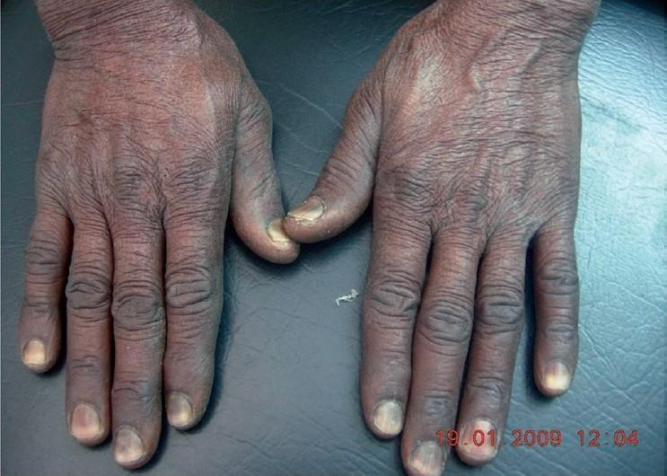
Clinical photograph showing the dorsal aspect of both hands, demonstrating diffuse, uniform cutaneous hyperpigmentation and specific melanonychia. The skin on the hands and fingers exhibits a generalized darkening, particularly accentuated over the knuckles and interphalangeal creases, with a slightly dry, xerotic texture. All ten fingernails show significant hyperpigmentation involving the proximal one-half to two-thirds of the nail plates, presenting as dark brown-to-black transverse bands that start at the cuticle and progress distally. The distal margins of the nails remain relatively spared. This visual presentation is characteristic of drug-induced pigmentation, commonly associated with chemotherapeutic agents such as hydroxyurea. The image serves as an educational reference for identifying adverse dermatological reactions to systemic medications and differentiating them from other causes of generalized hyperpigmentation like Addison's disease or Vitamin B12 deficiency.
Clinical Analysis: Onychomycosis + Widespread Dark Patches
Top Differential Diagnoses
1. HIV/AIDS (HIGHEST PRIORITY to exclude)
"Fungal nail infections are particularly common in HIV disease... proximal subungual onychomycosis is highly specific for advanced immunocompromised patients and should prompt an HIV test in patients whose HIV status is unknown." - Fitzpatrick's Dermatology, p. 4919
- Onychomycosis - severe, multi-nail, caused by dermatophytes, Candida, and non-dermatophyte molds
- Widespread pigmented patches - can result from:
- Post-inflammatory hyperpigmentation from recurrent skin infections
- Drug-induced pigmentation from antiretrovirals (e.g., zidovudine causes nail and skin darkening)
- Extensive tinea corporis that healed with hyperpigmentation
- Eosinophilic folliculitis (leaves dark macules on healing)
- Kaposi's sarcoma (violaceous/brown-black patches)
2. Addison's Disease (Primary Adrenal Insufficiency)
"Addison disease is characterized by hyperpigmentation of the skin, particularly of the elbows, knees, nail beds, nipples, areolae, and on recent scars. Hyperpigmentation is a result of increased levels of ACTH (which contains the α-MSH fragment)." - Costanzo Physiology, p. 262
- Diffuse skin darkening - can look like "tanning" or dark patches
- Accentuation at sites of prior trauma/scars - explains the "wound-healing" appearance
- Nail darkening (melanonychia)
- The immunosuppressive state increases susceptibility to skin/nail fungal infections
3. Pityriasis Versicolor (Hyperpigmented Type)
- Co-existing with onychomycosis is entirely plausible (same host susceptibility)
- More common in tropical climates, hot/humid conditions
- Patches typically have a fine scale when scratched (Besnier's sign)
- Confirmed with Wood's lamp (golden-yellow fluorescence) or KOH showing "spaghetti and meatballs" hyphae
4. Drug-Induced Hyperpigmentation
- Minocycline - blue-grey patches on scars and skin
- Hydroxyurea - diffuse darkening + nail pigmentation
- Antimalarials (chloroquine) - grey-blue patches
- Amiodarone - slate-grey photodistributed pigmentation
5. Post-Inflammatory Hyperpigmentation (PIH) from Recurrent Fungal/Other Skin Infections
Urgent Red Flags - What Makes This Clinically Concerning
| Red Flag Feature | Points Toward |
|---|---|
| Severe onychomycosis across ALL fingers | Immunosuppression |
| Dark patches resembling healed wounds | PIH from recurrent infection, Addison's, HIV |
| Multiple body areas involved | Systemic process, not just local |
| Patient appears to be young-to-middle-aged adult | HIV age group, Addison's |
Recommended Workup (Priority Order)
- HIV test (ELISA + Western blot) - cannot be deferred given this presentation
- CD4 count + viral load if HIV positive
- Cortisol (8 AM fasting) + ACTH level - rule out Addison's disease
- KOH mount + fungal culture of nail and skin lesions
- Full blood count, LFTs, RFTs, blood glucose
Key Clinical Message
Do NOT start antifungal therapy until HIV status and adrenal function are assessed. An immunosuppressed patient on terbinafine without treating the underlying cause will relapse and may have drug interactions (especially with antiretrovirals or azole antifungals). The underlying cause must be identified first.
patches r developed in legs arms etc
How do the dark patches look? · Any associated symptoms? · Any known medical history?
psoriasis dark skin hyperpigmented thick scaly plaques legs arms extensor
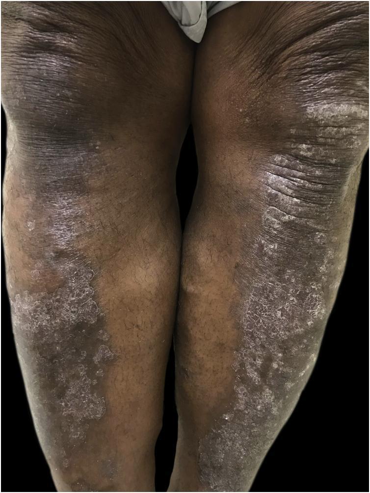
Clinical photograph of the anterior surface of the bilateral lower extremities in a dark-skinned patient, demonstrating chronic plaque psoriasis. The image shows extensive, well-demarcated hyperchromic plaques concentrated over the pretibial regions and knees. These plaques are characterized by thick, micaceous, silvery-white scales that contrast sharply with the dark, hyperpigmented base of the skin. There is evidence of lichenification, with prominent skin markings and a thickened, rough texture within the affected areas, suggesting chronic inflammation and mechanical friction. The presentation highlights the clinical appearance of psoriasis in skin of color, where erythema is often masked by hyperpigmentation and scaling may appear more prominent. The symmetrical distribution over the extensor surfaces is characteristic of the disease. This image serves as an educational resource for identifying dermatological manifestations in highly pigmented skin and understanding the morphological features of moderate-to-severe plaque psoriasis.
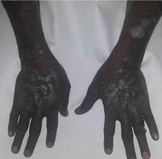
This clinical photograph displays widespread dermatological lesions on the dorsal and extensor surfaces of the hands and forearms of a patient with a dark skin phototype. The lesions present as a combination of well-demarcated, hyperpigmented plaques and raised papules covered with characteristic silvery-white, micaceous scales. On the dorsal aspect of the hands, the lesions are densely clustered, forming thick, rough, and confluent plaques that follow the anatomical contours of the metacarpals. On the forearms, the lesions appear larger, more discrete, and irregularly shaped, prominently featuring the silvery scaling against a background of post-inflammatory hyperpigmentation. The distribution on the extensor surfaces and the morphological features—specifically the silvery scale and plaque formation—are highly suggestive of chronic plaque psoriasis. This image serves as an educational tool for identifying typical psoriatic manifestations and observing how the condition appears in diverse skin tones, emphasizing the importance of recognizing silvery scales even when the underlying erythema may be masked by darker pigmentation.
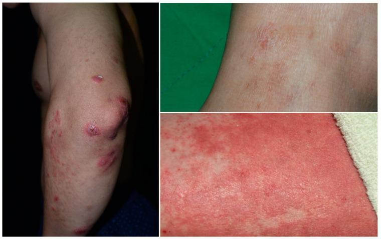
Clinical photographs depicting a patient with overlapping features of psoriasis and atopic dermatitis. The primary image shows the extensor surface of the arm and elbow, displaying multiple well-demarcated, erythematous, thick scaly plaques characteristic of psoriasis. These plaques are distributed predominantly around the olecranon and forearm. In contrast, the high-magnification close-up images reveal areas of more diffuse, poorly defined erythema and fine, granular papules without the heavy micaceous scaling typical of isolated psoriasis. Some regions exhibit signs of excoriation, crusting, and lichenification, suggesting a pruritic, eczematous component (atopic dermatitis). This visual evidence demonstrates 'PsEma' (psoriasis-eczema overlap), where classic psoriatic morphology coexists with the spongiotic patterns of dermatitis. The images serve as an educational tool for differentiating between pure psoriatic lesions and complex, concurrent inflammatory skin diseases that may require integrated therapeutic approaches.
Updated Diagnosis: Psoriasis with Nail Psoriasis
Why This Fits So Well
The Skin Lesions
"Psoriasis most frequently affects the elbows, knees, lumbosacral areas... The typical lesion is a well-demarcated, pink to salmon-colored plaque covered by loosely adherent silver-white scale... In darker skin, erythema is often masked by hyperpigmentation and scaling appears more prominent." - Robbins Pathologic Basis of Disease, p. 3855
The Nails
"Nail changes occur in 30% of cases of psoriasis and consist of yellow-brown discoloration (often likened to an oil slick), with pitting, dimpling, separation of the nail plate from the underlying bed (onycholysis), thickening, and crumbling." - Robbins Pathologic Basis of Disease, p. 3855
- Yellow-brown discoloration ✓
- Onycholysis (nail separation) ✓
- Subungual hyperkeratosis (debris under nail) ✓
- Thickening and crumbling ✓
- Multiple nails involved ✓
The Pain/Burning
- Lesions are cracked or fissured
- Located over pressure areas
- The patient has psoriatic arthritis (joint involvement with pain)
Diagnostic Approach
| Test | Purpose |
|---|---|
| Clinical examination - look for Auspitz sign (pinpoint bleeding when scale is removed) | Confirms psoriasis |
| Skin biopsy | Definitive - shows acanthosis, parakeratosis, neutrophilic infiltrate in stratum corneum (Munro's microabscesses) |
| KOH scraping of nail | Rule out secondary fungal co-infection |
| Fungal culture | If KOH doubtful |
| Joint exam + X-rays | Screen for psoriatic arthritis |
| HIV test | Still recommended - HIV can unmask/worsen psoriasis dramatically |
| HLA-B27 if arthritis suspected |
Treatment Plan
Skin Psoriasis
- Potent corticosteroids (betamethasone dipropionate 0.05%) - first line for plaques
- Vitamin D analogues (calcipotriol) - reduce hyperkeratosis
- Combination calcipotriol + betamethasone cream - very effective
- Coal tar preparations - good for thick scaly plaques
- Emollients - mandatory to reduce cracking and pain
- Methotrexate 7.5-25 mg/week - first-line systemic, also treats nail psoriasis
- Acitretin (retinoid) - especially good for thick/hyperkeratotic plaques
- Cyclosporine - rapid onset for severe disease
- Biologics (TNF-inhibitors: adalimumab, etanercept; IL-17 inhibitors: secukinumab) - for severe/refractory disease
Nail Psoriasis specifically
- Intralesional triamcinolone injected into nail matrix/bed - most effective for isolated nail psoriasis
- Topical calcipotriol + steroid under occlusion
- Tazarotene gel applied to nail
- Systemic therapy (methotrexate, biologics) - for nail psoriasis combined with skin disease