
Diatoms FMT pedogogy
I'll look into diatoms, FMT (presumably "Flipped/Mastery Teaching" or another pedagogy acronym), and their intersection. Let me clarify what you're looking for and search simultaneously.
Asking for Preferences
What does FMT stand for in your question? · What is the context of your question?
Asking for Preferences
What does FMT stand for in the forensic medicine context?
Got it - you want the pedagogy/teaching content on diatoms as it's taught in Forensic Medicine & Toxicology for MD students. Let me search the medical library and web simultaneously.
Searching sources...
Reading File
Searching the Web
diatom test drowning forensic medicine toxicology MD teaching
Now let me search the medical library for diatoms in forensic medicine textbooks in parallel with a PubMed search.
Finding Sources
Finding Sources
Searching PubMed
diatom test drowning forensic diagnosis
Excellent - found rich content in three major FM&T textbooks. Let me now read the core sections in parallel.
Reading File
Reading File
Reading File
Let me read the remaining diatom content from the Essentials textbook (2026 edition) and fetch the images.
Reading File
Loading Image
Loading Image
Loading Image
I now have comprehensive content from three major FM&T textbooks plus recent PubMed evidence. Let me also grab the Parikh's content and the bone marrow image, then compile the full response.
Reading File
Loading Image
I now have everything needed to compile a thorough FM&T teaching note. Here is the complete pedagogy:
Diatoms in Forensic Medicine & Toxicology - Teaching Note
1. What Are Diatoms?
Diatoms are microscopic, unicellular (or colonial) algae belonging to the class Bacillariophyceae. They are found ubiquitously in freshwater, saltwater, moist soils, cultivated land, and even in the atmosphere. Approximately 25,000 species are known.
Key biological features:
- Cell walls are strongly impregnated with silica, forming a hard, box-like outer skeleton called a frustule
- The frustule resists heat, acid, and putrefaction - which gives the diatom test its forensic utility
- They contain chlorophyll and diatoxanthin (a brown pigment)
- Size varies: 2 microns to 1 mm; most species are 10-80 microns in length
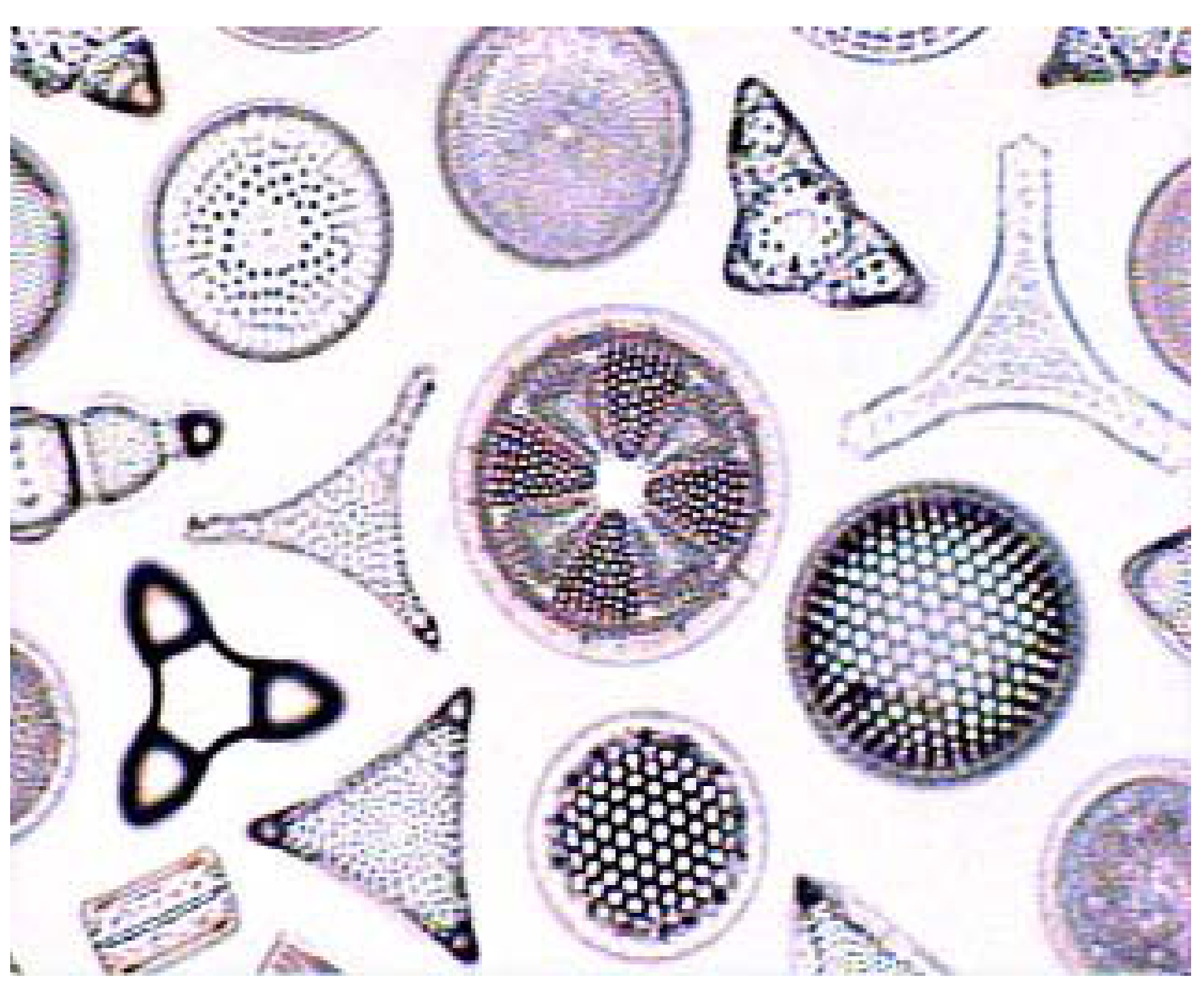
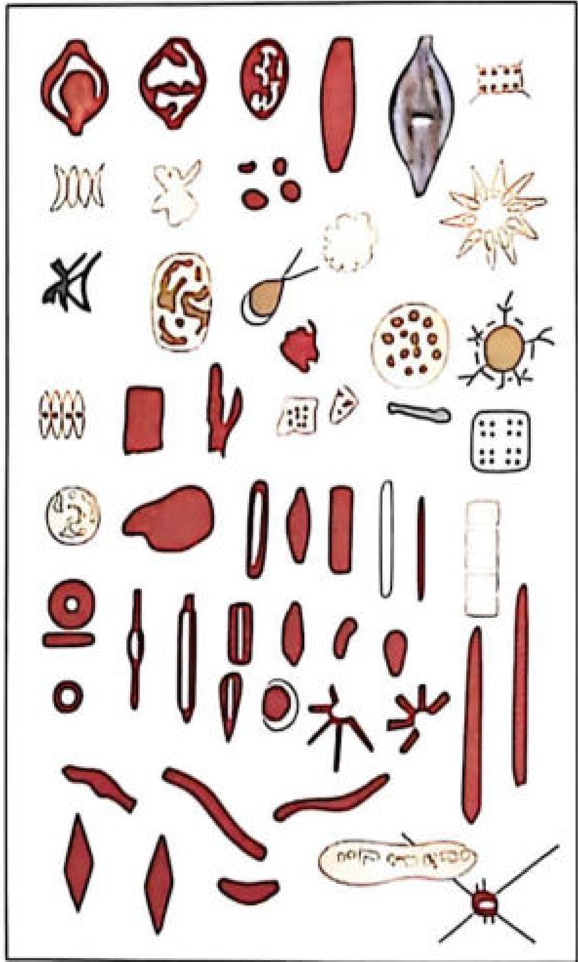
- Shapes: circular, triangular, oval, rectangular, linear, crescentic, boat-shaped, star-shaped (see below)
Microscopic appearance of diatom species:
2. Historical Background
| Year | Milestone |
|---|---|
| 1904 | Revenstrof - first to use diatoms as a test for drowning |
| 1941 | Incze - demonstrated that diatoms enter the systemic circulation via the lungs during drowning |
3. Principle of the Diatom Test
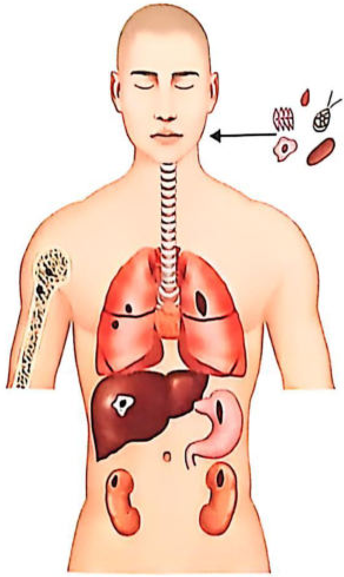
Core concept: When a living person drowns, active respiration aspirates diatom-laden water into the alveoli. Diatoms up to 60 microns in diameter (and fragments of larger ones) penetrate the alveolar walls, enter lymphatics and pulmonary veins, reach the left heart, and are then distributed via the systemic circulation to:
- Brain
- Bone marrow (femur, tibia, humerus, sternum)
- Liver
- Kidneys
- Skeletal muscle
- Bile and urine
This circulation only occurs in a living person with an intact heartbeat. When a dead body is thrown into water, passive percolation may bring diatoms into the lungs - but the absence of a beating heart prevents systemic dissemination.
4. Methods of Demonstration
A. Acid Digestion (Gold Standard for Deep Organs)
This is the most commonly taught and applied method in Indian FM&T exams:
- Specimens: 5 g of bone marrow, liver, kidney, or brain placed in separate test tubes
- Digest with concentrated nitric acid (or nitric + sulphuric acid), left at room temperature for 1-2 days, or heated in a water bath overnight
- The acid destroys all organic matter, leaving behind the siliceous diatom frustules intact
- Centrifuge, pour off supernatant, replace with distilled water; repeat 2-3 times
- Examine the deposit under phase-contrast or dark-ground illumination
- Diatom silica skeletons are birefringent under polarized light
Note: Some Japanese workers advocate enzyme/detergent digestion instead of acid - this preserves soft-bodied algae and protozoa as well.
B. Direct Microscopic Examination (Lungs only)
- Water squeezed from lungs, centrifuged, sediment examined
- Limited to fresh lung specimens
C. Microscopic Examination of Tissue Sections
- Optically empty sections are prepared and examined
D. Incineration Method
- Material incinerated in an electrical oven, then ashes dissolved in nitric acid for examination
5. Best Specimen for the Diatom Test
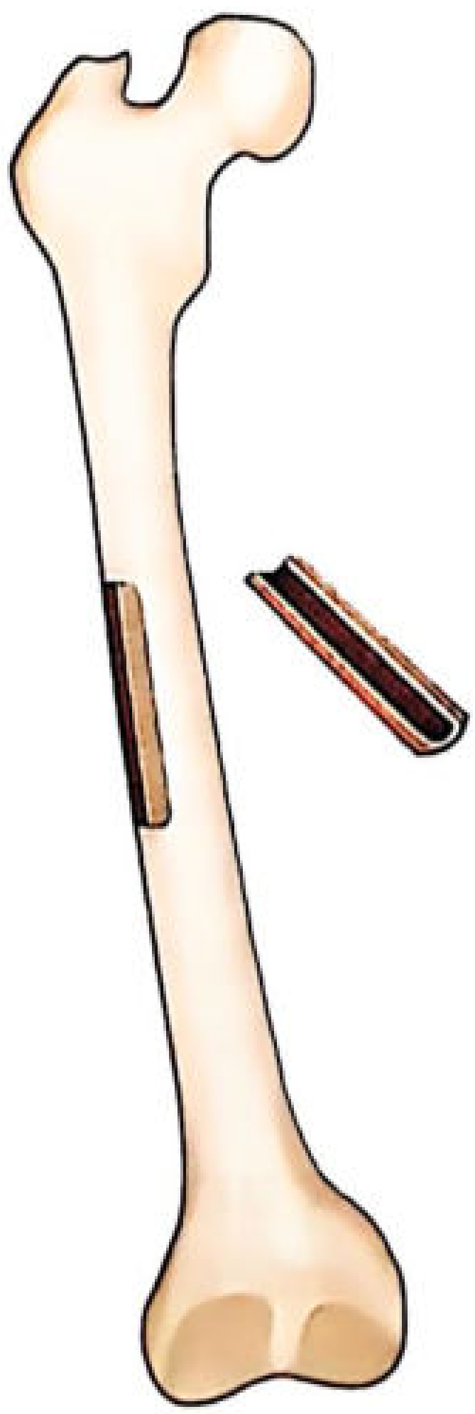
Bone marrow of long bones (femur, tibia, humerus) or sternum is the most suitable and reliable specimen because:
- It is a closed, protected system - less susceptible to post-mortem contamination
- The sternum is washed in distilled water, periosteum removed, a rectangular piece excised, marrow curetted out
Kidney (encapsulated, from non-decomposed body) is also preferred for similar reasons (DiMaio).
6. Control Sample
~2 litres of water from the suspected drowning site must be collected for comparison. Procedure:
- Add ~15 mL of iodine solution to kill microorganisms
- Allow to settle overnight
- Pour off bulk water, centrifuge remainder to recover diatoms
- Compare species, proportions, and size range with those found in body tissues
Positive result = Same species of diatoms found in drowning water AND in deep tissues at comparable proportions.
7. Interpretation
When the Test Is Valid (Parikh's Criteria)
The diatom test is valid only if:
- The deceased did not drink the water immediately before submersion
- Species recovered from tissue are all present in the drowning site water sample
- The various species are present in the same order of dominance and approximately the same proportions (for admissible size range)
Test Results Summary
| Result | Interpretation |
|---|---|
| Positive (matching species in deep organs) | Supports antemortem drowning |
| Negative | Does NOT exclude drowning |
| Diatoms in lungs only | Inconclusive (passive percolation possible) |
| Diatoms in deep organs (bone marrow, brain, kidney) | Strong evidence of vital immersion |
When Diatom Test Is Negative
- Drowning in heavily polluted water (algae do not grow in polluted water)
- Dry drowning (laryngospasm prevents water entry)
- Dead body thrown into water (postmortem submersion)
- Decomposed bodies (though diatoms themselves resist putrefaction, the test may still have value here)
- Low diatom count in the drowning water
- False negatives due to strong acid digestion reducing yield
8. Limitations and Controversies
| Issue | Detail |
|---|---|
| Ubiquity | Diatoms are found in air, food (shellfish), soils - risk of false positives |
| Contamination | Glassware, distilled water, reagents can contaminate; triple-distilled water and specially cleaned instruments are required |
| Three routes of entry | Inhalation (airborne), ingestion, and aspiration of water - antemortem aspiration is the only forensic-relevant one |
| Sensitivity/specificity | Both are debated; cannot be used as a standalone confirmatory test |
| Post-mortem contamination | Open organs may passively acquire diatoms from the surrounding water |
| Reliability | "Diatom test is often negative in undoubted cases of drowning in water full of diatoms" (Essentials FM&T, 36th ed.) |
DiMaio (Forensic Pathology, 3rd ed.) explicitly states: "a negative result does not rule out drowning."
9. Recent Evidence (PubMed, 2025-2026)
A 2025 systematic review in Int J Legal Med (PMID 39808189 - Tyr, Lunetta, Zilg) provides the most current consensus on medico-legal interpretation of diatom findings for fatal drowning diagnosis. Two additional 2025 reviews (PMID 39556128 and 40223726) address extraction methods and the long-standing controversies around the test's validity.
Bottom line from recent literature: Diatom analysis remains an adjunct tool - findings must be integrated with complete autopsy, histopathology, toxicology, and scene investigation. It is not a standalone confirmatory test.
10. The NMC/Exam High-Yield Points (FM3.4 Competency)
Per the NMC competency FM3.4 (listed in Essentials of FM&T, 36th ed.): "Describe and discuss types, pathophysiology, clinical features, postmortem findings and medicolegal aspects of drowning, diatom test, and Gettler test."
| Exam Topic | Key Fact |
|---|---|
| Class | Bacillariophyceae |
| Outer skeleton | Frustule (siliceous) |
| Size entering pulmonary circulation | Up to 60 microns |
| First to use diatom test | Revenstrof (1904) |
| Best specimen | Bone marrow (femur/sternum) |
| Test principle | Vital circulation carries diatoms to deep organs |
| Negative test | Does NOT exclude drowning |
| Digestion method | Concentrated nitric acid |
| Examination method | Phase-contrast / polarized light |
| Not applicable in | Dead body in water / dry drowning |
Sources:
- The Essentials of Forensic Medicine and Toxicology, 36th ed. (2026), pp. 364-366
- P C Dikshit Textbook of Forensic Medicine and Toxicology, pp. 316-317
- Parikh's Textbook of Medical Jurisprudence, Forensic Medicine and Toxicology, pp. 227-228
- DiMaio's Forensic Pathology, 3rd ed., p. 395
- Tyr A, Lunetta P, Zilg B. Int J Legal Med 2025 Mar [PMID: 39808189] - systematic review
Age estimation from bones , like skull pelvis and right humerus, for forensic medicine viva ,anatomy, landmarks ossifications , gender and forensic relevance for MD FMT viva
Searching sources...
Finding Sources
Finding Sources
Finding Sources
Let me broaden the search terms:
Finding Sources
Finding Sources
Finding Sources
Found key sources. Now reading all core sections in parallel - Essentials FM&T (METHODS OF AGE ESTIMATION), Forensic Anthropology (subadult + adult age estimation), and the skull suture/pelvis content:
Reading File
Reading File
Reading File
Reading File
Reading File
Reading File
Excellent. Now let me get the skull suture closure content, pubic symphysis / auricular surface content, and the ossification of bones table from Essentials FM&T:
Reading File
Reading File
Reading File
Reading File
Now let me get the skull sutures section and humerus ossification from the Essentials FM&T, plus fetch key images:
Reading File
Finding Sources
Loading Image
Excellent! That epiphyseal union diagram is perfect. Now let me grab the humerus ossification image, skull suture image, and read the Dikshit sutural closure + Parikh's pelvis/gender sections in parallel:
Loading Image
Reading File
Finding Sources
Loading Image
Reading File
Now I have everything needed. Here is the comprehensive FM&T viva teaching note:
Age Estimation from Bones - FM&T MD Viva Teaching Note
Overview: Methods of Age Estimation
Age can be estimated from:
- General physical features (height, weight) - reliable only until puberty
- Teeth - most reliable in children (0-14 years)
- Ossification of bones - most reliable up to ~25 years
- Secondary sex characters
- Skull suture closure - adults (rough estimate only, decade range)
- Pubic symphysis / auricular surface - adults
Key principle: Multiple criteria must always be used together. Skeletal age must be expressed as a range (e.g., 18 ± 2 years).
PART 1 - THE SKULL
A. Fontanelles (Infancy - useful for estimating age <2 years)
| Fontanelle | Closure Age |
|---|---|
| Lateral & occipital fontanelles | 2 months after birth |
| Posterior fontanelle | 6-8 months |
| Anterior fontanelle | 1.5-2 years |
B. Early Sutural Fusions (important viva points)
| Event | Age |
|---|---|
| Metopic suture closure | ~3 years (persists in 5-10% as normal variant) |
| Condylar portions of occipital fuse with squama | 3rd year |
| Condylar portions fuse with basioccipital | 5th year |
| Basioccipital fuses with basisphenoid (spheno-occipital synchondrosis) | 18-21 years |
The spheno-occipital synchondrosis is a key landmark - its fusion (18-21 years) indicates that an individual has reached adulthood. It fuses from the endocranial surface outward.
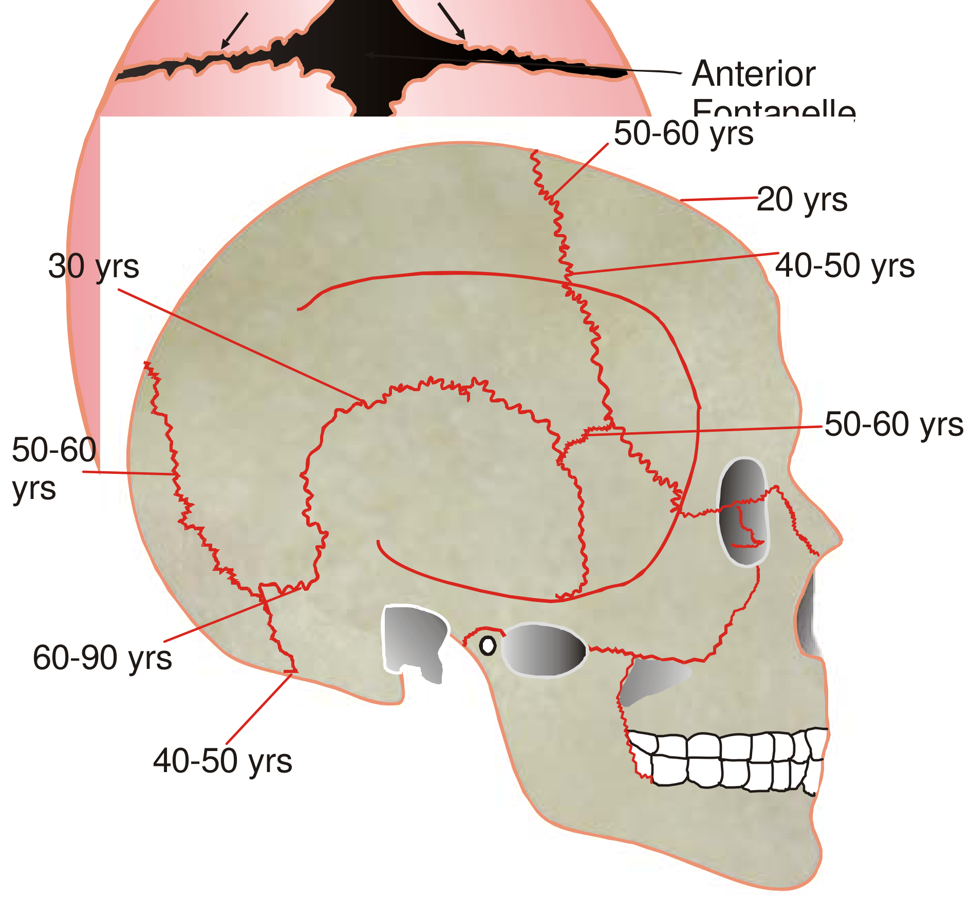
C. Cranial Vault Suture Closure (Adults - age estimation in decade range only)
Critical rule: Endocranial (inner) surface closes 5-10 years EARLIER than ectocranial (outer) surface.
Ectocranial suture closure sequence:
| Suture | Endocranial Start | Ectocranial/Complete Closure |
|---|---|---|
| Sagittal (posterior 1/3) | ~25 years | 30-40 years |
| Sagittal (anterior 1/3) + lower coronal | ~25 years | 40-50 years |
| Middle sagittal + upper coronal | - | 50-60 years |
| Lambdoid (starts near lambda) | 25-35 years | 45-50 years |
| Pterion | 40 years | 65 years |
| Mastoid-occipital | 45 years | 80 years |
| Asterion | - | 80 years |
| Squamous temporal | - | 60 years |
Order of most reliable estimation: Sagittal > Lambdoid > Coronal
Forensic limitations:
- Absence of ANY suture closure = age likely < 30 years
- Age can only be estimated in decades (±10 years range)
- Suture closure occurs earlier in males than females
- Lapsed union = ectocranial closure never occurs (normal variant) - do not misinterpret as young age
- Skull suture closure has been largely abandoned as a primary age marker due to high variability (Brogdon's Forensic Radiology)
D. Gender Determination from the Skull
| Feature | Male | Female |
|---|---|---|
| Overall size | Larger, heavier | Smaller, lighter |
| Supraorbital ridges | Prominent, well-marked | Poorly marked/absent |
| Mastoid process | Large, prominent | Small |
| Occipital condyles | Large | Small |
| External occipital protuberance | Prominent | Absent/less prominent |
| Forehead (frontal bone) | Sloping, receding | Vertical, rounded |
| Orbital margins | Rounded, blunt | Sharp, well-defined |
| Palate | Large, U-shaped | Smaller, parabolic |
| Mandible | Large, wide, square chin | Small, pointed chin |
| Zygomatic arches | Extend beyond mastoid | Do not extend beyond mastoid |
| Skull capacity | ~1450 cc | ~1300 cc |
PART 2 - THE HUMERUS (Right Humerus)
A. Ossification Centers of the Humerus
Proximal end (shoulder):
| Center | Appearance | Fusion |
|---|---|---|
| Head of humerus | 1st year (birth-1 yr) | 14-16 years (F); 18 years (M) |
| Greater tuberosity | 2-3 years | Fuses with head at 3-5 years |
| Lesser tuberosity | 5 years | Fuses with head; all three unite into one epiphysis |
| Combined proximal epiphysis fuses to shaft | - | ≥16 years (F), ≥18 years (M) |
Distal end (elbow) - mnemonic CRITOE:
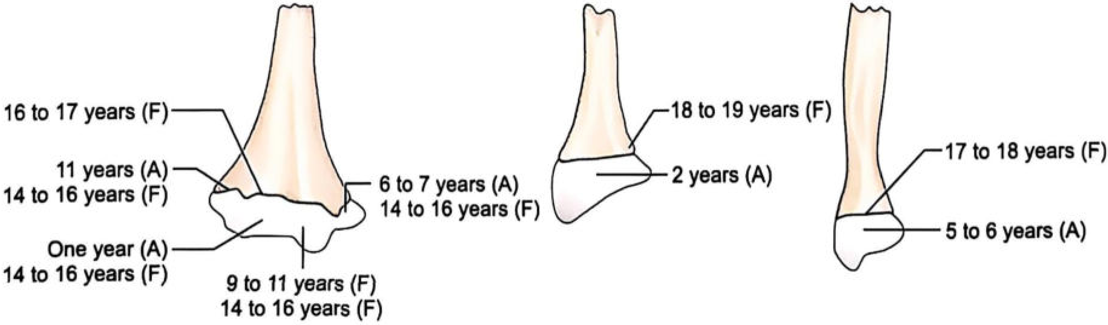
| Letter | Center | Appearance | Physeal Fusion |
|---|---|---|---|
| C | Capitellum | 1 year | 14 years |
| R | Radial head | 4-5 years | 16 years |
| I | Medial epicondyle (Internal) | 5-7 years | 15 years |
| T | Trochlea | 8-9 years | 14 years |
| O | Olecranon | 8-10 years | 14 years |
| E | Lateral epicondyle (External) | 11-12 years | 16 years |
Important viva trap: The medial epicondyle fuses last among the distal ossification centers. It appears at 5-7 years but does not fuse until ~15 years. If seen as a separate fragment, it may be confused with an avulsion fracture.
B. Key Rules for the Humerus
- "Elbow earlier, wrist later" - in upper limb long bones, union occurs earlier at elbow and later at wrist
- Head of humerus is the last long bone epiphysis to unite (~18-19 years in females, up to 20-22 years in males)
- Distal epiphysis of humerus fuses at ≥15 years (F) and ≥12 years (F) / ≥15 years (M) - complete fusion by ~14-16 years
- Proximal humerus fusion: ≥18 years (M), ≥16 years (F)
C. Shoulder Ossification Centers (for completeness)
| Structure | Appearance | Fusion |
|---|---|---|
| Head of humerus | 2-4 months | 3 years - puberty (merges with shaft) |
| Greater tuberosity | 7-10 months | Puberty |
| Lesser tuberosity | 5 years | Puberty |
| Coracoid process | 3 months | 15-17 years |
| Subcoracoid (glenoid) | 8-10 years | 14-17 years |
| Acromion secondary centers | 14-16 years | 18-25 years |
Acromion os acromiale - failure of acromial ossification centers to fuse (normal variant, present in ~8% of population). Important to distinguish from fracture on imaging.
PART 3 - THE PELVIS
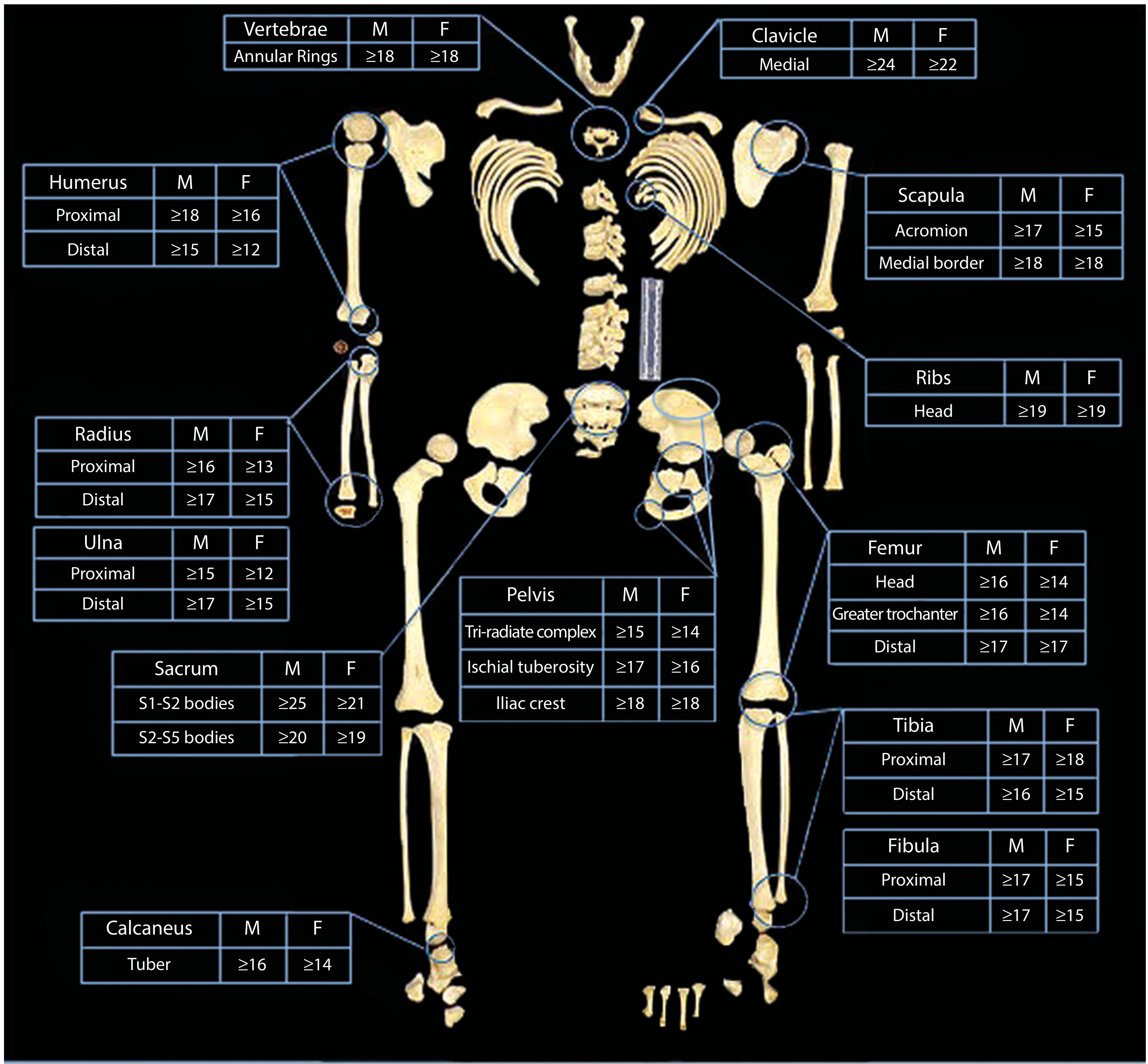
A. Epiphyseal Fusion in the Pelvis (Age Estimation)
| Structure | Fusion Age (Male) | Fusion Age (Female) |
|---|---|---|
| Tri-radiate complex (ilium + ischium + pubis at acetabulum) | ≥15 years | ≥14 years |
| Ischial tuberosity | ≥17 years | ≥16 years |
| Iliac crest | ≥18 years | ≥18 years |
The iliac crest epiphysis is one of the last to fuse (~18 years in both sexes). An unfused iliac crest means age is likely under 18 years - medico-legally significant.
B. Pubic Symphysis (Adult Age Estimation - Todd's Method / Suchey-Brooks)
The pubic symphysis is the best single criterion for determining age from the 3rd to 5th decade. Requires bones free of cartilage.
| Age | Pubic Symphyseal Appearance |
|---|---|
| < 20 years | Layer of compact bone near surface; marked ridges transversely across articular surface |
| ~20 years | Markedly irregular/uneven; transverse ridges prominent |
| 24-36 years | Ridges gradually disappear; granular appearance; ventral and dorsal margins become defined |
| Early 5th decade (~40-49 years) | Oval, smooth surface with raised upper and lower ends |
| Late 5th decade (~45-50 years) | Narrow-beaded rim develops on margins |
| 6th decade (50-59 years) | Erosion of surface; breakdown of ventral margin begins |
| 7th decade (60+ years) | Surface becomes irregularly eroded |
Critical viva point: If male criteria are applied to a female pubis, the age will be under-estimated by ~10 years, because the female pubis reaches full maturity about 10 years later than the male.
Suchey-Brooks method (most widely used today): 6 morphological phases for males and females separately, based on 1225 individuals from the Los Angeles ME's office.
C. Auricular Surface (Ilium - Lovejoy et al.)
The sacroiliac auricular surface of the ilium shows reliable age-related degenerative changes:
| Age Phase | Auricular Surface Features |
|---|---|
| Young (<30 years) | Billowing in transverse/horizontal orientation (low relief striae) |
| 30-40 years | Billows replaced by fine striae; fine-grained texture |
| 40-50 years | Loss of transverse organization; coarse-grained texture; microporosity |
| 50-60 years | Macroporosity; surface densification; apical lipping |
| >60 years | Subchondral erosion; retroauricular activity; irregular surface |
Three key regions assessed:
- Morphology of the auricular surface itself
- Apex of the auricular surface
- Retroauricular area (ligamentous attachment zone)
D. Gender Determination from the Pelvis (Most Reliable Method)
The pelvis is the most reliable bone for sex determination (accuracy ~95%).
| Feature | Male | Female |
|---|---|---|
| Overall shape | Narrow, funnel-shaped | Wide, basin-shaped |
| Pelvic inlet | Heart-shaped | Round/oval |
| Sub-pubic angle | <90° (acute, <70°) | >90° (obtuse, >100°) |
| Greater sciatic notch | Narrow (<68°) | Wide (>68°) |
| Sacrum | Narrow, long, curved | Wide, short, flat |
| Obturator foramen | Large, oval | Small, triangular |
| Acetabulum | Large (faces laterally) | Small (faces laterally + slightly anteriorly) |
| Iliac fossa | Deep | Shallow |
| Pubic arch | Narrow | Wide |
| Ischial tuberosities | Inverted (turned inward) | Everted (turned outward) |
| Pre-auricular sulcus | Absent | Often present (parturition groove) |
PART 4 - OTHER KEY BONES (Viva Bonus)
Sternum
| Event | Age |
|---|---|
| Four pieces of sternal body fuse upward | 14-25 years |
| Xiphoid unites with body | ~40 years |
| Manubrium fuses with body | Old age, ~60 years |
Clavicle (Medial Epiphysis)
- Last skeletal growth plate to fuse in the entire body
- Well-defined medial flake: 16-21 years
- Covers majority of medial surface: 24-29 years
- Complete fusion: 22-30 years (M ≥24, F ≥22)
- If medial clavicle epiphysis is fully fused → individual is likely >25-30 years
Hyoid Bone
- Greater cornu unites with body: 40-60 years
- Medico-legally important: fracture of hyoid in strangulation vs age-related fusion
Sacrum
| Fusion | Age |
|---|---|
| S4-S5, S3-S4 | ~12 years |
| S2-S3 | ~15-17 years |
| S1-S2 bodies | ≥25 years (M), ≥21 years (F) |
PART 5 - KEY Rules & Forensic Relevance
General Rules of Ossification
- Appearance of ossification centers is more accurate for age estimation than union of epiphyses
- Secondary centers that appear first are the last to fuse (and vice versa)
- Upper limb: union earlier at elbow, later at wrist
- Lower limb: union later at knee, earlier at hip and ankle
- Skeletal development in females is 1-2 years ahead of males
- Always express skeletal age as a range: e.g., 18 ± 2 years
Forensic Relevance: Why Age Estimation Matters in FMT
| Medico-legal Question | Bone Used |
|---|---|
| Is the person > 18 years? (POCSO, juvenile justice) | Iliac crest, medial clavicle, proximal humerus, wisdom teeth |
| Is the person > 21 years? (Age of majority, marriage, contracts) | Medial clavicle (fuses 22-30 years), S1-S2 |
| Identify unknown skeletal remains (decomposed/mutilated bodies) | All bones combined |
| Osseous age in living accused (bone radiograph) | Wrist X-ray (carpal bones), elbow, hip |
| Mass disasters and unidentified human remains | Dental + bone + DNA |
| Age of victim in sexual assault cases | X-ray of wrist, hip, pelvis, teeth |
Factors That Affect Ossification Timing
- Nutrition - malnutrition delays fusion
- Endocrine disorders - hypothyroidism delays; hyperthyroidism accelerates
- Hereditary/genetic factors
- Race - warmer climates: slightly earlier
- Sex - females 1-2 years ahead
- Infections, metabolic disorders, physical activity
PART 6 - Viva Quick-Fire Answers
| Q | A |
|---|---|
| Most reliable bone for sex determination? | Pelvis (95% accuracy) |
| Second most reliable bone for sex determination? | Skull |
| Most reliable indicator for age 0-14 years? | Teeth (dentition) |
| Best bone for adult age estimation (3rd-5th decade)? | Pubic symphysis |
| Last growth plate to fuse in the body? | Medial clavicle (22-30 years) |
| Last long bone epiphysis to fuse? | Head of humerus |
| In which direction does skull suture closure proceed? | Endocranial → ectocranial (inner first) |
| Which sex shows earlier skull suture closure? | Males |
| Which sex shows earlier epiphyseal union? | Females (1-2 years earlier) |
| Absence of any suture closure indicates? | Age < 30 years |
| Significance of unfused iliac crest? | Age < 18 years (important for POCSO/juvenile justice) |
| What is lapsed union? | Suture closure occurs endocranially but NOT ectocranially - normal variant, do not misinterpret as "young age" |
| Which suture is most reliable for age estimation? | Sagittal suture |
| What is CRITOE? | Mnemonic for elbow ossification centers: Capitellum-Radial head-Internal epicondyle-Trochlea-Olecranon-External epicondyle |
| Age to close anterior fontanelle? | 1.5-2 years |
| Age of spheno-occipital synchondrosis fusion? | 18-21 years (marks adulthood) |
| Gustafson's method is used for? | Age estimation from teeth (6 criteria: attrition, periodontosis, secondary dentine, cementum apposition, root resorption, root transparency) |
Sources:
- The Essentials of Forensic Medicine and Toxicology, 36th ed. (2026), pp. 78-88
- P C Dikshit Textbook of Forensic Medicine and Toxicology, pp. 73-76
- Parikh's Textbook of Medical Jurisprudence, Forensic Medicine and Toxicology
- Forensic Anthropology: A Comprehensive Introduction, 2nd ed. (Chapters 10-11)
- Imaging Anatomy: Bones, Joints, Vessels and Nerves (Tables 2.1-2.2 - CRITOE/shoulder ossification)This is a complete FM&T viva reference covering all three bones you asked about plus the broader context. Here's a quick orientation to the note:
What's covered:
- Skull - fontanelles (infancy), early sutural fusions (metopic, spheno-occipital), full cranial vault suture closure table with exact ages and sequence, gender determination features, forensic limitations of skull age estimation
- Humerus - proximal ossification centers (head + tubercles, appearance and fusion ages), distal elbow CRITOE table with appearance and fusion ages for all 6 centers, shoulder girdle ossification, key rules ("head of humerus is last long bone epiphysis to fuse")
- Pelvis - tri-radiate complex/iliac crest/ischial tuberosity fusion ages, pubic symphysis age changes by decade (Todd/Suchey-Brooks), auricular surface changes, gender determination table (most reliable bone ~95% accuracy)
- Bonus sections - sternum, clavicle (last growth plate to fuse in the body), hyoid, sacrum
- Viva quick-fire Q&A at the end with 20 high-yield answers
- Forensic relevance table - which bone answers which medico-legal question (POCSO, juvenile justice, unidentified remains, sexual assault cases)
This is a shared conversation. Sign in to Orris to start your own chat.