
Write a five mark short note on Pleomorphic adenoma Make it compatible, compact but high yielding. So that... I can easily remember and, uh, write it in a... Systematic way being a 2nd year resident. make it in a way its interesting and easy to learn visually and Create a high density, high yielding image based(generate image) Flashcard I can use Before exam to revise and remember all the important points regarding this short-note. but it should be in a way so its easy to understand and remember also use flowcharts or mindmaps in the response wherever possible dont use code
pleomorphic adenoma parotid gland histology microscopy
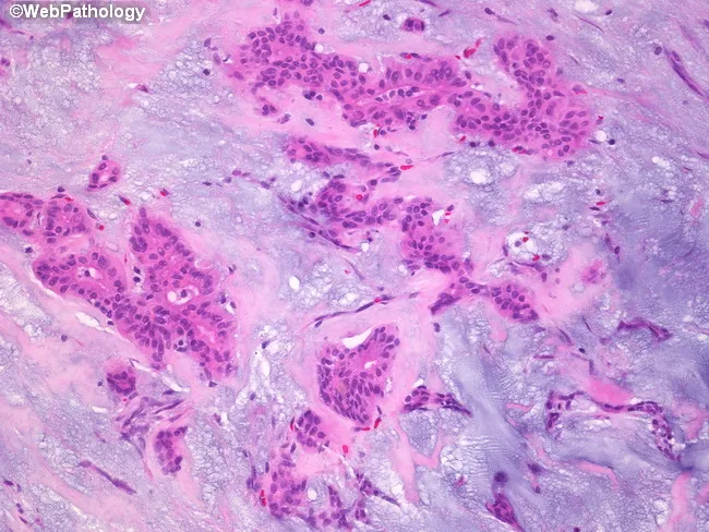
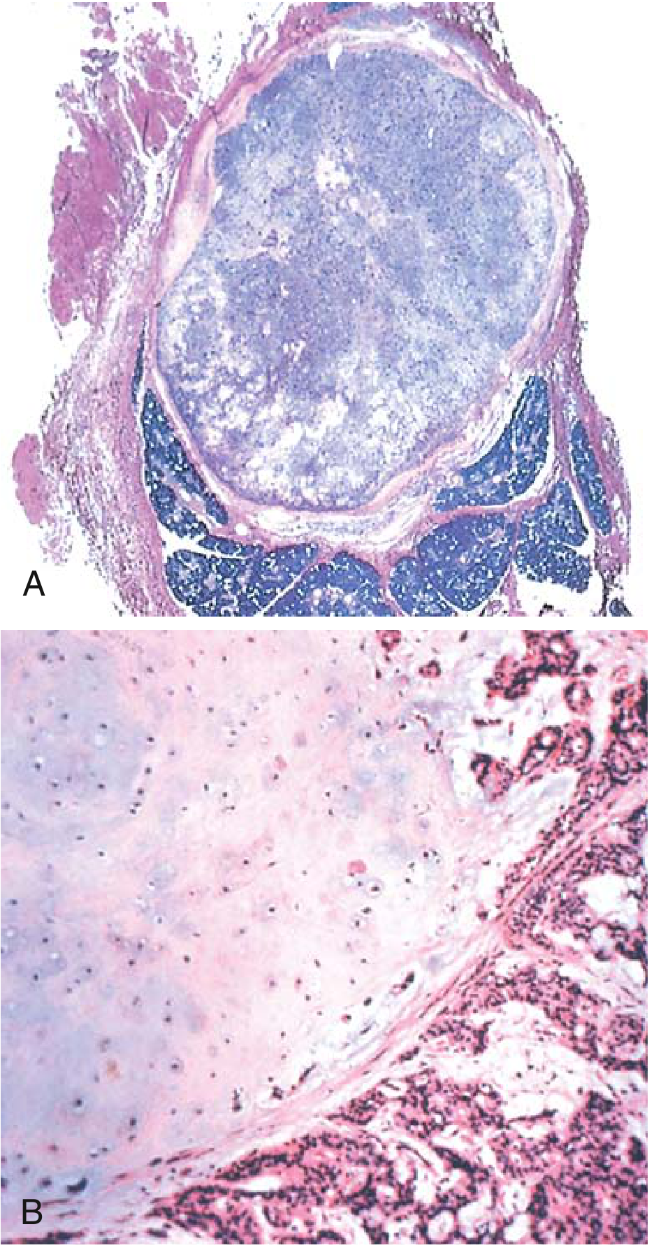
Pleomorphic adenoma histology, parotid salivary gland, examined by bright-field microscopy on Hematoxylin and Eosin stained sections. Imaging modality: Light microscopy; technique: Hematoxylin and Eosin staining. The sample reveals a classic biphasic neoplasm comprising epithelial ductal structures and myoepithelial cells embedded in an abundant stromal backdrop. Epithelial components form cords, ducts, and small island formations lined by cuboidal to columnar cells with eosinophilic cytoplasm and hyperchromatic nuclei. Intermixed myoepithelial cells display plasmacytoid or spindle morphologies, often surrounding epithelial elements. The stroma is a prominent feature, ranging from basophilic, myxoid to more eosinophilic, hyalinized areas. The myxoid matrix is rich in glycosaminoglycans and imparts a loose, gelatinous appearance, while hyalinized zones contribute dense eosinophilic bands. Overall architecture is often well circumscribed with minimal pleomorphism and low mitotic activity, consistent with benign behavior. Clinical significance includes differentiation from malignant salivary tumors; prognosis is favorable with complete surgical excision. Differential considerations include mucoepidermoid carcinoma, adenoid cystic carcinoma, basal cell adenoma, and Warthin tumor. This image is useful for educational purposes in pathology, otolaryngology, and head-and-neck surgery, illustrating quintessential pleomorphic adenoma features: epithelial and myoepithelial proliferation in a chondromyxoid stroma, with duct-like structures and a variable stromal composition. This histologic pattern aids diagnosis and education.
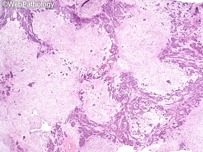
Histology of a pleomorphic adenoma involving the parotid gland. This light-microscopy image derives from a hematoxylin and eosin–stained paraffin section. It demonstrates a biphasic neoplasm with epithelial ductal–acinar elements embedded in a myxoid to chondromyxoid stroma. Epithelial components arrange as trabeculae, tubules, and duct-like acini, often lined by a two-layered cast: inner cuboidal to columnar luminal cells and outer basal/myoepithelial cells with plasmacytoid or spindle morphology. The surrounding stroma is variably myxoid, sometimes chondroid, producing a lobulated architecture with intimately interwoven cellular and stromal zones. Clefts and interspersed myxoid pools may be evident; ossification or calcifications are occasional. The lesion is typically well circumscribed within the parotid, showing no frank invasion of adjacent parenchyma on histology. Mitotic activity is rare. The overall appearance—myxoid/chondromyxoid matrix with epithelial tubules and ducts—supports pleomorphic adenoma; however, histologic overlap with other salivary gland neoplasms necessitates clinical correlation and, when indicated, ancillary immunohistochemistry (e.g., cytokeratin, S-100, GFAP) or molecular testing. Clinically, pleomorphic adenoma presents as a slow-growing, painless parotid mass in adults, with a favorable prognosis after complete surgical excision, though recurrence and potential for malignant transformation exist if spillage or capsular rupture occurs.
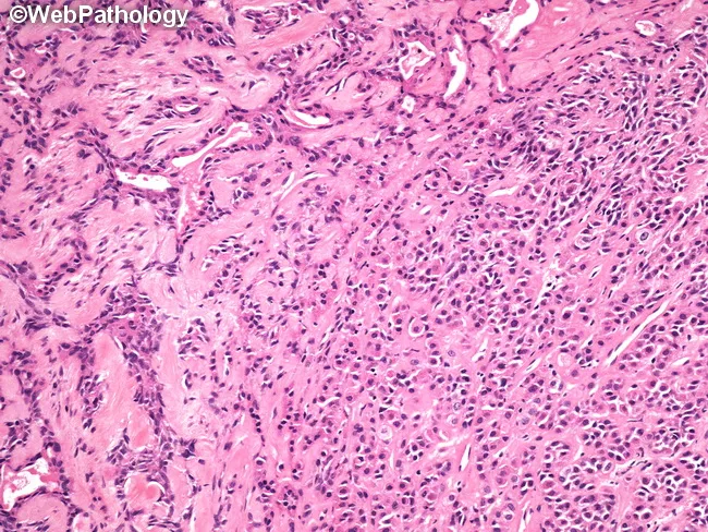
Imaging modality: light microscopy of Hematoxylin and Eosin stained histology section from a salivary gland lesion. Anatomical site: parotid region; histologic plane: peripheral-to-central glandular architecture. The slide reveals classic pleomorphic adenoma features with biphasic tumor components in a variably hyalinized, myxoid stroma. Epithelial nests and anastomosing cords are embedded in a chondromyxoid matrix, while duct-like tubules are evident in places. In the lower-right field, small nests and cords form an Indian file pattern, illustrating cellular diversity within a single tumor. The left third shows compressed and distorted tubular structures amid dense stromal hyalinization, contributing to a mosaic of microarchitectures. The myoepithelial element contributes spindle- to plasmacytoid cells with variable cytoplasmic appearance; the overall cytology lacks significant atypia or invasion. Clinically, these features correlate with a slow-growing, painless parotid mass and favorable prognosis after complete excision. Diagnostic significance rests on recognizing both epithelial and myoepithelial components within a mucoid to cartilaginous stroma, and on identifying variant patterns to avoid misclassification as malignant salivary neoplasms. Differential diagnoses include adenoid cystic carcinoma, mucoepidermoid carcinoma, and basal cell adenoma; accurate histologic interpretation guides management and recurrence risk assessment. Awareness of this histologic diversity improves diagnostic accuracy and informs surgical planning.
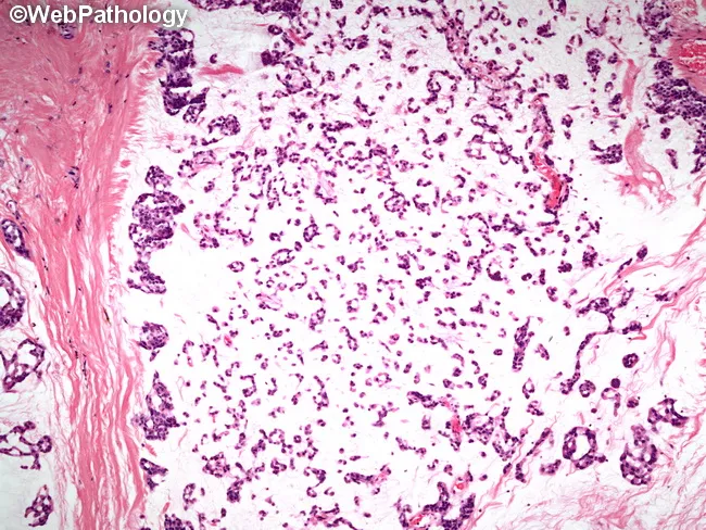
Histology image of a salivary gland tumor demonstrating classic pleomorphic adenoma (mixed tumor). Prepared as a hematoxylin and eosin stained slide and evaluated under light microscopy. The specimen represents salivary gland tissue in the parotid region. Epithelial elements are organized into small nests and rudimentary tubular structures, reflecting ductal differentiation. These epithelial components are embedded in a substantial myxoid background with scattered chondromyxoid areas. Myoepithelial cells contribute to the cellular admixture, accounting for variable cell shapes and occasional plasmacytoid appearances. The stroma ranges from loose, myxoid to more fibrous or hyaline areas, producing a heterogeneous, biphasic architecture typical of pleomorphic adenoma. The overall pattern is benign-appearing with well-demarcated margins in this field; however, the lesion is known for potential recurrence if incompletely excised. Pleomorphic adenoma is a benign salivary gland neoplasm featuring epithelial and myoepithelial components within mucoid and cartilaginous stroma. Diagnostic significance rests on recognizing the biphasic morphology and chondromyxoid matrix, which helps distinguish from malignant mimics such as mucoepidermoid carcinoma or adenoid cystic carcinoma. Clinically, these findings correlate with a slow-growing, painless parotid mass in middle-aged adults. This image is valuable for education, differential diagnosis practice, and radiology-pathology correlation in salivary gland tumors. for clinical education and research.
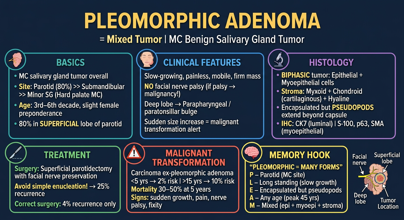
A high-yield medical exam flashcard for "Pleomorphic Adenoma" (also called Mixed Tumor / Benign Mixed Tumor). Design as a clean, visually rich study card with a dark navy blue background and bright white/yellow text. Organize content in clearly separated sections with icons: TITLE at top center: "PLEOMORPHIC ADENOMA" in large bold gold letters, subtitle "= Mixed Tumor | MC Benign Salivary Gland Tumor" LEFT COLUMN - BASICS box (teal border): • MC salivary gland tumor overall • Site: Parotid (80%) >> Submandibular >> Minor SG (Hard palate MC) • Age: 3rd–6th decade, slight female preponderance • 80% in SUPERFICIAL lobe of parotid CENTER COLUMN - CLINICAL FEATURES box (blue border): Slow-growing, painless, mobile, firm mass NO facial nerve palsy (if palsy → malignancy!) Deep lobe → Parapharyngeal / paratonsillar bulge Sudden size increase = malignant transformation alert RIGHT COLUMN - HISTOLOGY box (purple border) with small microscope icon: • BIPHASIC tumor: Epithelial + Myoepithelial cells • Stroma: Myxoid + Chondroid (cartilaginous) + Hyaline • Encapsulated but PSEUDOPODS extend beyond capsule • IHC: CK7 (luminal) | S-100, p63, SMA (myoepithelial) BOTTOM LEFT - TREATMENT box (green border): Surgery: Superficial parotidectomy with facial nerve preservation Avoid simple enucleation! → 25% recurrence Correct surgery: 4% recurrence only BOTTOM CENTER - MALIGNANT TRANSFORMATION box (red/orange border) with warning icon: Carcinoma ex-pleomorphic adenoma <5 yrs → 2% risk | >15 yrs → 10% risk Mortality 30–50% at 5 years Signs: sudden growth, pain, nerve palsy, fixity BOTTOM RIGHT - MEMORY HOOK box (yellow border): "PLEOMORPHIC = MANY FORMS" P – Parotid (MC site) L – Long standing (slow growth) E – Encapsulated but pseudopods A – Any age (peak 45 yrs) M – Mixed (epi + myoepi + stroma) Small schematic diagram of parotid gland showing superficial vs deep lobe with tumor location. Overall design: colorful, organized, medical student-friendly, easy to scan in 60 seconds before exam.
🧠 PLEOMORPHIC ADENOMA — 5 Mark Short Note
Also called: Mixed Tumor | Benign Mixed Tumor
🔶 QUICK IDENTITY MAP
PLEOMORPHIC ADENOMA
        │
        ├── MC BENIGN salivary gland tumor (60% of all parotid tumors)
        ├── MC salivary gland tumor OVERALL
        ├── "Pleomorphic" = MANY FORMS → hence the name
        └── Alt name: "Mixed Tumor" → because it has MIXED components
📍 SITES — "80-80-80 Rule"
Parotid gland ────────────── 80% of cases
  └── Superficial lobe ──── 80% of parotid PAs
        └── In parotid: 80% is benign (Parotid = mostly benign)

Then: Submandibular > Minor salivary glands
        └── Minor SG → Hard palate is MC site
Memory trick: Parotid Gland = Party Gland (MC site, 80% benign!)
👤 EPIDEMIOLOGY
| Feature | Detail |
|---|---|
| Age | 3rd–6th decade (avg 45 yrs) |
| Sex | Slight female preponderance |
| Etiology | Radiation exposure (known risk factor) |
🩺 CLINICAL FEATURES
Typical Presentation:
──────────────────────
Painless ──── Slow-growing ──── Mobile ──── Firm/Rubbery mass

Location in Parotid:
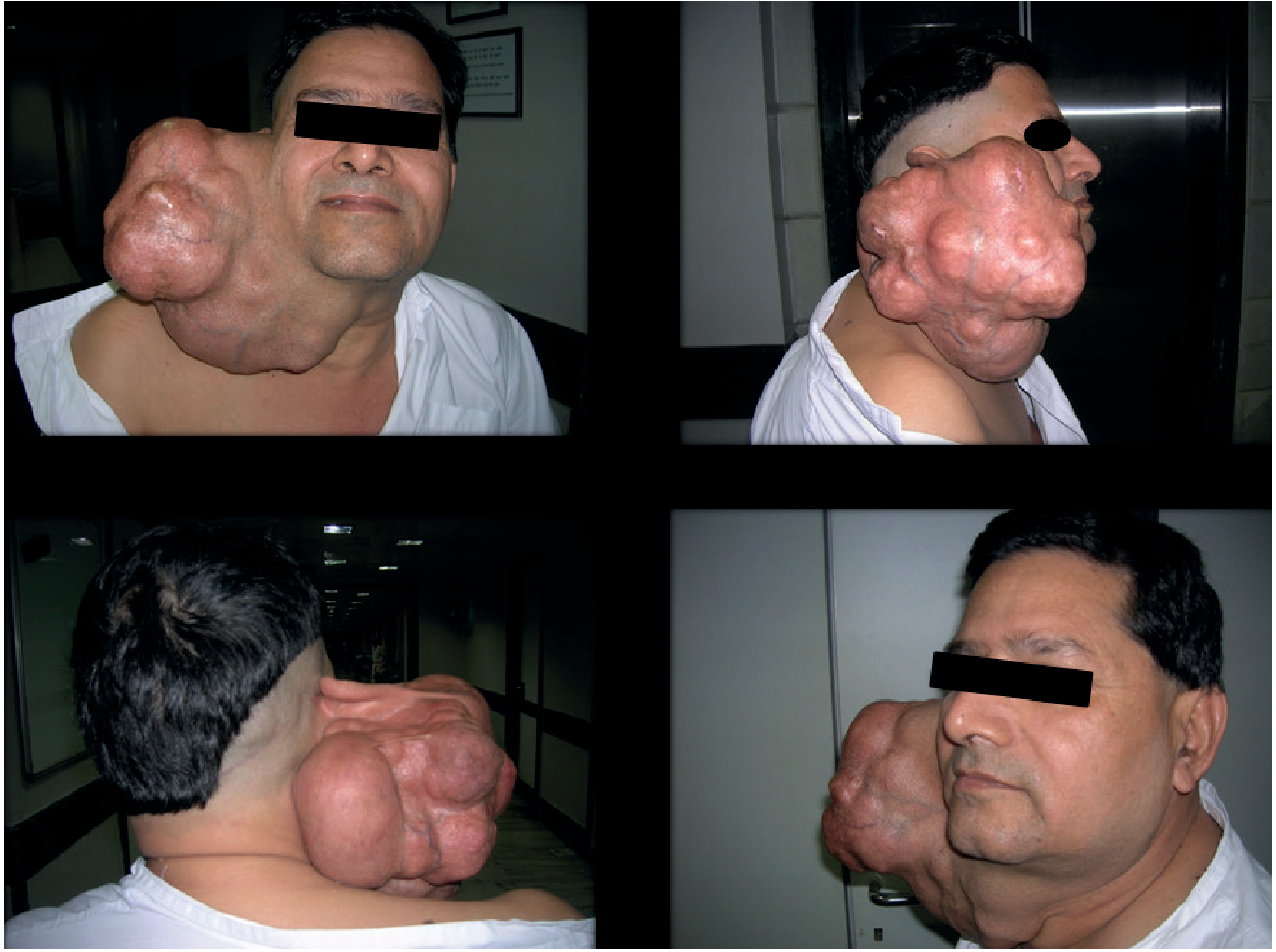
  Superficial lobe → Mass below/in front of ear (most common)
  Deep lobe → Parapharyngeal space → Paratonsillar BULGE
              ("dumbbell tumor" if extends through stylomandibular tunnel)

KEY NEGATIVES (in benign PA):
  ✗ NO pain
  ✗ NO facial nerve palsy
  ✗ NO fixity to skin/bone
⚠️ ALARM SIGNS for malignant transformation: Sudden increase in size + Pain + Facial nerve palsy + Skin fixity
🔬 PATHOLOGY
- Well-circumscribed, encapsulated, firm mass
- Cut surface: Gray-white with blue translucent chondroid areas + myxoid areas
- Capsule may be incomplete (especially in palate) → pseudopods extend beyond!
EPITHELIAL component
  ├── Ductal cells (cuboidal to columnar, form ducts/acini/tubules)
  └── Myoepithelial cells (spindle, plasmacytoid, clear cell morphology)

MESENCHYMAL (stromal) component
  ├── Myxoid stroma (MC)
  ├── Chondroid (cartilaginous) tissue ← PATHOGNOMONIC
  ├── Hyaline stroma
  └── Rarely: osseous (bone) tissue
| Cell Type | Markers |
|---|---|
| Luminal (ductal) cells | CK7 (strong, diffuse) |
| Myoepithelial cells | p63, S-100, SOX10, SMA (smooth muscle actin) |
🔪 TREATMENT
Treatment of Choice:
  PAROTID PA → Superficial Parotidectomy with facial nerve PRESERVATION
                        ↓
          Adequate cuff of normal tissue around tumor
                        ↓
          Recurrence = 4% (acceptable)

  AVOID:
  ❌ Simple Enucleation → capsule breach → tumor spillage → 25% recurrence!

  ORAL CAVITY PA (minor SG) → Complete surgical excision transorally
  SUBMANDIBULAR PA → Submandibular gland excision
⚡ RECURRENCE & MALIGNANT TRANSFORMATION
Recurrence after adequate surgery: ~4%
Recurrence after enucleation: ~25%

MALIGNANT TRANSFORMATION → "Carcinoma ex-Pleomorphic Adenoma"

  Duration < 5 years  →  2% risk
  Duration > 15 years → 10% risk

  Histology of malignant form:
  → Adenocarcinoma or Undifferentiated carcinoma (MC)

  Prognosis:
  → Mortality 30–50% at 5 years (very aggressive once malignant!)
🔑 HIGH-YIELD EXAM FACTS (one-liners)
- MC salivary gland tumor = Pleomorphic adenoma
- MC site = Parotid (superficial lobe)
- MC benign tumor of minor salivary glands = Pleomorphic adenoma (hard palate)
- The capsule has pseudopods - reason why enucleation causes recurrence
- Two-cell pattern: epithelial (CK7+) + myoepithelial (S-100, p63, SMA+)
- Chondroid stroma = hallmark / most distinctive feature
- Malignant transformation: <5yrs = 2%, >15yrs = 10%
- Surgery of choice = superficial parotidectomy (NOT enucleation!)
- Deep lobe PA = presents as paratonsillar bulge / dumbbell tumor
- Sudden facial palsy in known PA = malignancy until proven otherwise
📇 HIGH-YIELD EXAM FLASHCARD
🧩 VISUAL MINDMAP SUMMARY
                    PLEOMORPHIC ADENOMA
                           │
         ┌─────────────────┼─────────────────┐
         ▼                 ▼                 ▼
     BASICS           PATHOLOGY          CLINICAL
     ──────           ─────────          ────────
  MC benign SG     Biphasic tumor     Painless, mobile
  tumor overall    ├ Epithelial       Slow growing
                   │ (ductal+myoepi)  3rd–6th decade
  Parotid 80%      └ Stroma           Female > Male
  └ Superficial      ├ Myxoid (MC)
    lobe 80%         ├ Chondroid ←★   Deep lobe →
                     └ Hyaline        Paratonsillar
  Minor SG →                          bulge
  Hard palate        Capsule with
                     PSEUDOPODS!
         │                 │                 │
         └─────────────────┼─────────────────┘
                           ▼
                       TREATMENT
                       ─────────
                  Superficial Parotidectomy
                  (preserve facial nerve)
                  ❌ NOT enucleation (25% recurrence!)
                  ✅ Adequate cuff (4% recurrence)
                           │
                           ▼
                 MALIGNANT TRANSFORMATION
                 ──────────────────────────
                 <5 yrs = 2% | >15 yrs = 10%
                 Ca ex PA → 30–50% mortality at 5yr
                 Signs: Pain, Palsy, Fixity, Sudden growth
💡 MEMORY MNEMONICS
- Pseudopods (capsule is incomplete!)
- Long-standing tumor (slow growth, years)
- Epithelial + myoEpithelial cells (2 cell types)
- Always benign... unless it's been there >15 years
- Myxoid + chondroid stroma (the mesenchymal magic)
- Pain (new onset)
- Sudden size increase
- Cutaneous fixity
- Motor nerve (facial) palsy