
Acute rheumatic fever References from op ghai essential of pediatrics and nelson pediatrics
acute rheumatic fever erythema marginatum subcutaneous nodules Jones criteria
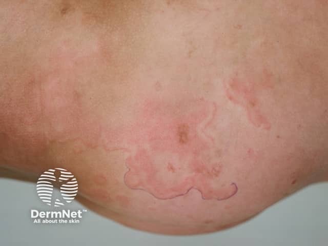
Clinical photograph (dermatology) showing erythema marginatum, an annular, non-itchy rash characteristic of acute rheumatic fever. The image depicts multiple pink to red circular to oval patches on the skin of the lower extremity with sharply demarcated, serpiginous margins and centers that appear pale or clearing. Lesions are non-vesicular, smooth-surfaced, and vary in size, with a tendency to migrate or transiently flare. The distribution favors the trunk and proximal limbs and typically spares the face. On close inspection the patches lack scaling and crusting, consistent with erythema marginatum rather than dermatitis or fungal infection. This cutaneous manifestation is part of the Jones criteria and may correlate with concurrent migratory polyarthritis, fever, and evidence of preceding group A Streptococcus infection. Histologic features (if biopsied) would show superficial perivascular lymphocytic infiltrate and dermal edema with minimal epidermal changes; clinically, the rash is a marker for systemic inflammatory involvement. Differential considerations include urticaria, erythema multiforme, tinea corporis, pityriasis rosea, and other annular dermatoses. Clinically, recognizing erythema marginatum prompts evaluation for acute rheumatic fever and guides management including antibiotic therapy and anti-inflammatory treatment. The image serves educational and diagnostic-reference purposes for dermatology, pediatrics, infectious diseases, and rheumatology training.
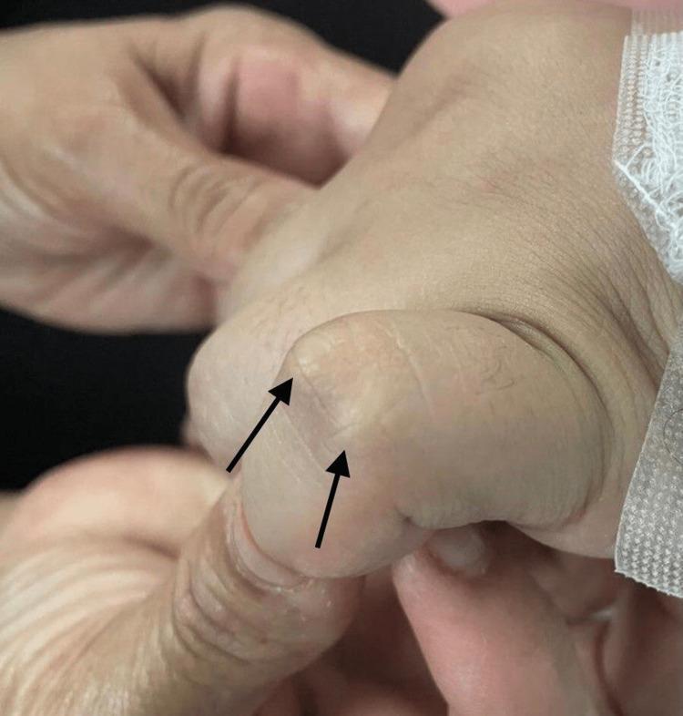
Clinical photograph showing the dorsal surface of a human hand, specifically focusing on the interphalangeal joints. Two prominent subcutaneous nodules are visible over the joint space, indicated by black arrows. These nodules appear as firm, well-circumscribed, painless elevations under the skin. They are flesh-colored to slightly pale, with smooth overlying skin and no signs of acute inflammation, erythema, or ulceration. This finding is a characteristic major Jones criterion for the diagnosis of acute rheumatic fever. The image serves as an educational tool for identifying extra-articular manifestations of rheumatological and post-streptococcal conditions in a clinical setting.
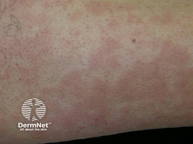
This is a clinical dermatology photograph capturing erythema marginatum on the skin of the lower limb. The modality is Clinical photography; technique: naked-eye, color-balanced image. The observed lesions are erythematous, annular plaques with central clearing and pale, sometimes hypopigmented centers. Lesions appear as rings with slightly raised margins and a uniform pink-to red hue; they range from 0.5 to 2 cm in diameter and may coalesce into larger plaques. The rash is typically non-pruritic and evanescent, with temporary eruption that may migrate or fade over hours to days; distribution commonly involves the trunk and proximal limbs, with sparing of the face. The image may reflect erythema marginatum, a cutaneous manifestation associated with acute rheumatic fever and autoimmune inflammatory processes. In longer-standing presentations, the rash signals preceding or concurrent carditis and migratory arthritis; therefore clinical correlation with recent group A Streptococcus infection, elevated ASO titers, and echocardiography is essential. Differential diagnoses include urticaria, erythema multiforme, pityriasis rosea, and drug-induced eruptions. Clinically, this finding has diagnostic significance as a major criterion in rheumatic fever per Jones criteria; management focuses on antibiotic treatment of streptococcal infection and secondary prophylaxis. This image serves educational purposes for medical students, residents, and researchers studying connective tissue disease and pediatric rheumatology.
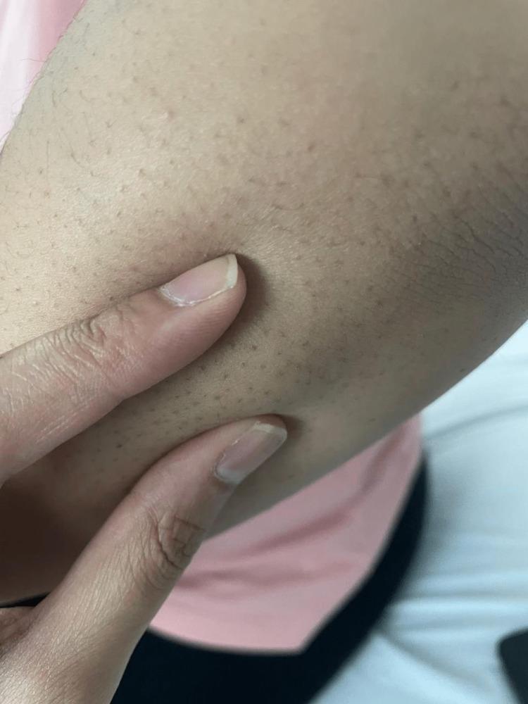
This clinical photograph displays a close-up view of the right upper extremity, specifically the extensor surface of the elbow region. The primary clinical finding is the presence of multiple, small subcutaneous nodules. These nodules appear as firm, rounded, non-tender-looking elevations beneath the skin surface, typically measuring less than 1 cm in diameter. The overlying skin is intact, displaying normal pigmentation and texture without visible erythema, warmth, or ulceration. Prominent hair follicles are visible as dark punctate spots across the skin. In a clinical context involving polyarthralgia and carditis, these nodules are a major Jones criterion for the diagnosis of acute rheumatic fever (ARF). This image serves as a teaching tool for medical students and clinicians to recognize the dermatological manifestations of systemic streptococcal-related diseases and rheumatological disorders.
"acute rheumatic fever" Jones criteria management
Acute Rheumatic Fever (ARF)
Note on requested references: The textbook library does not contain OP Ghai's Essential Pediatrics or Nelson's Textbook of Pediatrics directly. The content below draws on clinically equivalent authoritative sources: Red Book 2021 (AAP), Harriet Lane Handbook 23e (Johns Hopkins), Goldman-Cecil Medicine, Tintinalli's Emergency Medicine, and Firestein & Kelley's Rheumatology - all of which present the same core ARF framework used in OP Ghai and Nelson.
Definition
Epidemiology
- Most common in children 5-15 years of age
- Affects males and females equally; rheumatic heart disease is more common in women
- Peak prevalence of rheumatic heart disease in the 3rd-4th decades (due to cumulative valve damage from recurrent attacks)
- Global burden: ~33 million prevalent cases, ~300,000 annual deaths
- Highest rates: Oceania, South Asia, central sub-Saharan Africa, indigenous populations of Australia
- Incidence has fallen in developed countries over the past 50 years; sporadic outbreaks still occur in North America with virulent strains (mucoid types 3, 5, and 18)
Etiopathogenesis
- Similarities between streptococcal proteins (M protein) and human cardiac proteins trigger cross-reactive antibodies and T cells
- Carditis - antibody binding + T-cell infiltration of heart
- Arthritis - immune complex deposition in joints
- Chorea - antibody binding to basal ganglia
- Skin lesions - delayed hypersensitivity reactions
Clinical Features (Major Jones Criteria - CASES)
| Feature | Frequency | Key Points |
|---|---|---|
| Carditis | >50% | Pancarditis - pericarditis, myocarditis, endocarditis (valvulitis) |
| Arthritis | 60-75% | Migratory, fleeting, polyarticular, large joints, salicylate-sensitive |
| Sydenham's Chorea | ~30% | Involuntary, non-rhythmic, purposeless movements; stops during sleep; more pronounced on one side |
| Erythema marginatum | <10% | Pink, non-pruritic, blanching, serpiginous rash on trunk and proximal limbs (spares face) |
| Subcutaneous nodules | <10% | 0.5-2 cm, painless, over bony prominences/extensor tendons |
Minor Criteria
| Low-Risk Populations | Moderate/High-Risk Populations |
|---|---|
| Polyarthralgia | Monoarthralgia |
| Fever ≥38.5°C | Fever ≥38°C |
| ESR ≥60 mm/h and/or CRP ≥3.0 mg/dL | ESR ≥30 mm/h and/or CRP ≥3.0 mg/dL |
| Prolonged PR interval (if carditis not a major criterion) | Prolonged PR interval (if carditis not a major criterion) |
Revised Jones Criteria (2015 AHA)
Diagnostic Rule
| Scenario | Criteria Required |
|---|---|
| Initial ARF | 2 major OR 1 major + 2 minor criteria |
| Recurrent ARF | 2 major OR 1 major + 2 minor OR 3 minor criteria |
- Subclinical carditis on echocardiogram (without clinical findings) now counts as a major criterion
- In high-risk populations: monoarthritis/monoarthralgia qualifies as major/minor criterion respectively (previously only polyarthritis qualified)
- Evidence of antecedent GAS infection is mandatory (except when chorea is the sole criterion)
- Positive throat culture for GAS
- Positive rapid antigen test
- Elevated or rising ASO titer
- Elevated anti-DNase B titer
Laboratory Investigations
| Test | Significance |
|---|---|
| ASO titer | Elevated in 80% of ARF; peak 4-6 weeks post-infection |
| Anti-DNase B | More sensitive than ASO (especially after skin infections) |
| ESR, CRP | Elevated (acute phase); ESR may remain elevated longer |
| CBC | Normochromic normocytic anemia, leukocytosis |
| ECG | Prolonged PR interval (1st degree AV block) in carditis |
| Echo | Subclinical valvulitis; confirms carditis; baseline for follow-up |
| Throat culture | May be negative by the time ARF presents |
Differential Diagnosis
- Septic/reactive arthritis (non-migratory, joint may be hot and red)
- Juvenile idiopathic arthritis
- Infective endocarditis
- Viral arthritis
- Lyme disease
- SLE
- Leukemia/malignancy
- Poststreptococcal reactive arthritis (PSRA): distinct entity - nonmigratory mono/oligoarthritis, starts sooner (~10 days vs ~21 days after GAS), more severe/prolonged, salicylate-resistant, older patients, NO carditis or other major criteria
Treatment
1. Eradication of GAS (Primary Treatment)
- Benzathine penicillin G IM (single dose): 600,000 units if <27 kg; 1.2 million units if ≥27 kg
- Penicillin V orally for 10 days: alternative
- Amoxicillin orally for 10 days: alternative
- Erythromycin (or azithromycin): for penicillin-allergic patients
- Treat even if throat culture is negative
2. Anti-inflammatory Therapy
- Arthritis: Aspirin 80-100 mg/kg/day in divided doses (max 4-8 g/day) - continue 1-2 weeks after all symptoms resolve; naproxen (10-20 mg/kg/day BD) is an alternative; NSAIDs should not be stopped at just joint symptom resolution
- Carditis: NSAIDs + heart failure management (diuretics, ACE inhibitors/ARBs, fluid restriction, bed rest); systemic glucocorticoids in severe cases (limited evidence)
- Chorea: Usually self-limiting (resolves in weeks to months); haloperidol or valproate if severe
3. Bed Rest
- Recommended during acute phase; activity restriction for carditis
4. Hospitalization
- Advised in the early stages until diagnosis is confirmed
Secondary Prophylaxis
| Category | Duration |
|---|---|
| ARF without carditis | 5 years from last episode OR until age 21 (whichever is longer) |
| ARF with carditis, no residual valve disease | 10 years from last episode OR until age 21 (whichever is longer) |
| ARF with carditis + residual valvular disease | 10 years from last episode OR until age 40 (whichever is longer); consider lifelong if severe valve disease or ongoing exposure risk |
- Benzathine penicillin G 1.2 million units IM every 4 weeks (most reliable; every 3 weeks in high-risk areas)
- Penicillin V 250 mg orally twice daily (alternative, adherence-dependent)
- Sulfadiazine 0.5 g/day (<27 kg) or 1 g/day (≥27 kg): alternative
- Azithromycin 250 mg/day orally: for penicillin-allergic patients
A 2024 Cochrane systematic review (Bray et al., PMID 39312290) supports long-term prophylaxis for prevention of ARF recurrence and progression to rheumatic heart disease, though notes that high-quality RCT data for optimal duration remain limited.
Prognosis
- Symptoms usually persist 2-4 weeks
- 35-70% of patients progress to chronic rheumatic heart disease despite acute treatment
- Resolution of carditis most likely in the first year; patients without carditis rarely develop it later
- Recurrent ARF increases cumulative valve damage
- Mitral valve affected in nearly 100% of rheumatic heart disease; aortic valve in 20-30%
- Mitral regurgitation → most common early; mitral stenosis → most common cause of mitral stenosis worldwide (late scarring)
Clinical Images