
Draw a diagram showing different layers of the eyelids.
Generating Image
A detailed anatomical cross-section diagram of the eyelid layers, clearly labeled, showing all distinct tissue layers from anterior to posterior: skin (epidermis and dermis), subcutaneous areolar tissue, orbicularis oculi muscle, orbital septum, tarsal plate (tarsus), meibomian glands within the tarsus, conjunctiva (palpebral), and the eyelid margin with eyelashes and their follicles. Include arrows pointing to each layer with clean bold labels. Medical illustration style, white background, high detail, educational diagram.
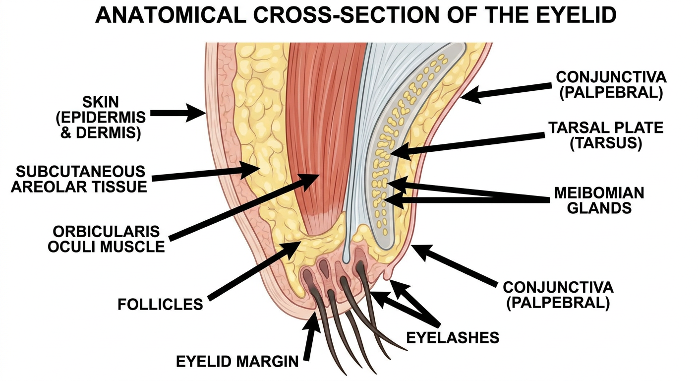
Here is a cross-sectional diagram of the eyelid layers, from anterior (front) to posterior (back):
- Skin - The thinnest skin in the body, consisting of epidermis and a thin dermis. Contains eyelashes and their follicles at the margin.
- Subcutaneous areolar tissue - Loose connective tissue with no fat, which allows fluid to accumulate easily (explaining why eyelid edema is so prominent).
- Orbicularis oculi muscle - The sphincter muscle responsible for closing the eye (innervated by CN VII - facial nerve).
- Orbital septum - A fibrous sheet that acts as a barrier between the eyelid and the orbital contents.
- Tarsal plate (Tarsus) - A dense fibrous plate that gives the eyelid its structural rigidity. Contains the Meibomian glands (modified sebaceous glands that secrete the lipid layer of the tear film).
- Palpebral conjunctiva - The innermost layer, a mucous membrane that lines the inner surface of the eyelid and reflects onto the globe.
A useful mnemonic for the layers is SLMT-C: Skin, Loose tissue, Muscle, Tarsus/septum, Conjunctiva.
This is a shared conversation. Sign in to Orris to start your own chat.