
Primitive streak. 2. Gastrulation
gastrulation AND embryogenesis
NCBI Bookshelf embryology gastrulation primitive streak
diagram primitive streak gastrulation bilaminar embryonic disc germ layers
https://www.ncbi.nlm.nih.gov/books/NBK554394
1. Primitive streak
Formation and parts
- Epiblast cells proliferate and migrate toward the midline, producing the primitive streak.
- Its cranial end forms the primitive node.
- The center of the node contains the primitive pit.
- A longitudinal depression in the streak is the primitive groove.
Functions
- Initiates gastrulation.
- Establishes the embryonic body axes:
- Cranial-caudal
- Dorsal-ventral
- Right-left
- Provides the site through which epiblast cells migrate inward to form definitive endoderm and intraembryonic mesoderm.
- The primitive node contributes to formation of the notochordal process and subsequently the notochord.
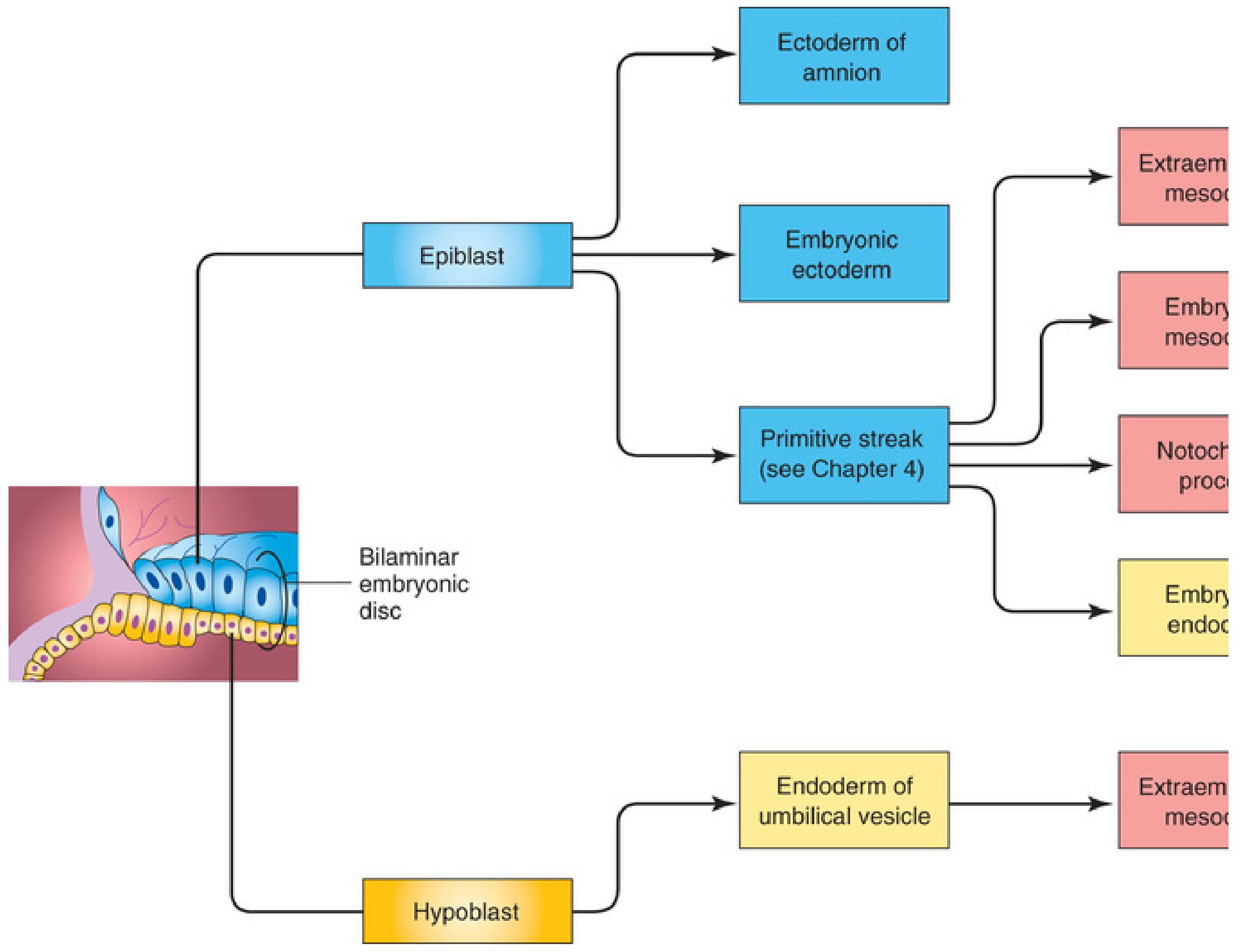
Fate and clinical importance
2. Gastrulation
Sequence of events
- Primitive streak forms in the epiblast.
- Epiblast cells move toward the streak, detach, and migrate inward, a process called ingression.
- The first wave of ingressing cells displaces the hypoblast and forms the definitive endoderm.
- The next wave spreads between ectoderm and endoderm to form the intraembryonic mesoderm.
- Epiblast cells remaining on the surface become ectoderm.
Result: three germ layers
| Germ layer | Main derivatives |
|---|---|
| Ectoderm | Epidermis, central and peripheral nervous systems, eyes, internal ear, neural crest derivatives |
| Mesoderm | Muscle, bone, cartilage, connective tissues, dermis, blood and vessels, cardiovascular system, kidneys, gonads, serous membranes |
| Endoderm | Epithelial lining of gastrointestinal and respiratory tracts, and parenchyma/epithelium of associated glands such as liver and pancreas |
Importance
- Begins morphogenesis, the development of body form.
- Establishes embryonic axes.
- Produces the germ layers from which all tissues and organs develop.
- Sets up the midline through formation of the notochordal process and notochord.
Primitive streak is the gateway for gastrulation; gastrulation transforms a bilaminar disc into a trilaminar disc consisting of ectoderm, mesoderm, and endoderm.
Notochord: Development, functions and clinical aspects
notochord development congenital anomalies
NCBI Bookshelf notochord embryology development clinical significance chordoma
human embryology diagram development of notochord notochordal process primitive node
https://www.ncbi.nlm.nih.gov/books/NBK554394
Notochord
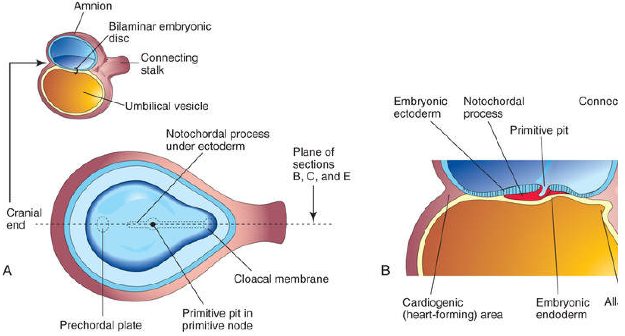
Development
1. Formation of notochordal process
- Cells migrate inward through the primitive node and primitive pit.
- These prenotochordal cells migrate cranially in the midline, between ectoderm and endoderm.
- They form a cellular cord called the notochordal process.
- A lumen develops within it, the notochordal canal.
- The process grows toward the prechordal plate, close to the future oropharyngeal membrane.
2. Formation of notochordal plate
- The floor of the notochordal canal fuses temporarily with the underlying endoderm.
- This creates a flattened layer of cells called the notochordal plate.
- For a short period, the amniotic cavity communicates with the yolk sac through the neurenteric canal.
3. Formation of definitive notochord
- The notochordal plate folds inward.
- Its margins fuse and separate from the endoderm.
- A solid midline rod, the definitive notochord, is formed.
Langman's Medical Embryology, pp. 78-79.
Functions of the notochord
1. Establishes the embryonic midline and body axis
2. Induces neurulation
- Sonic hedgehog (SHH)
- Noggin
- Chordin
3. Organizes axial skeleton formation
- Sclerotomal cells migrate around the notochord and neural tube.
- These cells form the vertebral bodies and other parts of the vertebral column.
- Thus, the notochord acts as a central template around which the vertebral column develops.
4. Contributes to intervertebral discs
Clinical aspects
1. Chordoma
- Skull base, particularly the clivus
- Vertebral column
- Sacrum
2. Neurenteric cyst and neurenteric fistula
- Neurenteric cysts
- Neurenteric fistulae
- Associated vertebral and spinal-cord abnormalities
3. Vertebral anomalies
- Hemivertebrae
- Fused vertebrae
- Vertebral segmentation defects
- Scoliosis associated with congenital vertebral malformations
4. Persistent notochordal remnants
Exam summary
| Heading | Key point |
|---|---|
| Origin | Prenotochordal cells migrating through primitive node and pit |
| Time | Third week of development |
| Sequence | Notochordal process → notochordal plate → definitive notochord |
| Position | Midline, between ectoderm/neural tube and endoderm |
| Major roles | Midline organizer, neural induction, axial skeleton patterning |
| Main adult remnant | Nucleus pulposus of intervertebral disc |
| Clinical relevance | Chordoma, neurenteric anomalies, vertebral malformations |
Node cells form the notochord, which induces the neural plate, guides vertebral formation, and persists only as the nucleus pulposus.
2. Pharyngeal arches
pharyngeal arches congenital anomalies
NCBI Bookshelf pharyngeal arches derivatives cranial nerves embryology
human embryology pharyngeal arches diagram derivatives cranial nerves
Pharyngeal arches
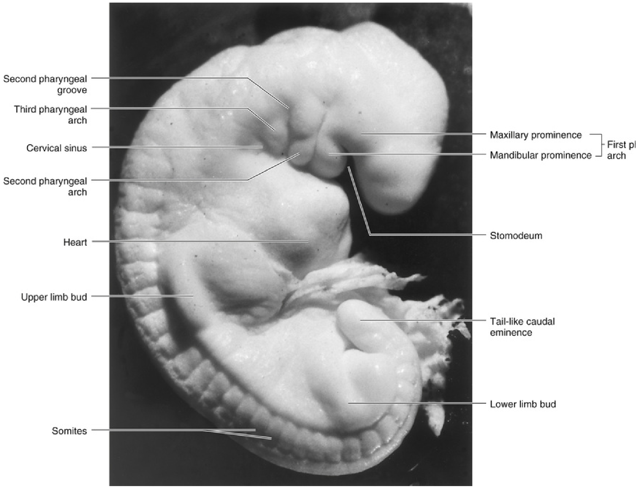
Basic structure
- External ectoderm
- Internal endoderm
- A mesenchymal core derived from mesoderm and neural crest cells
- A cartilage component
- A muscular component
- A cranial nerve
- An arterial component, linked to an aortic arch
Derivatives of pharyngeal arches
| Arch | Nerve | Skeletal/cartilage derivatives | Muscular derivatives | Arterial derivative |
|---|---|---|---|---|
| 1st - mandibular arch | Trigeminal nerve, mainly V3 | Malleus, incus, mandible, maxilla, zygomatic bone, part of temporal bone, sphenomandibular ligament | Muscles of mastication, mylohyoid, anterior belly of digastric, tensor tympani, tensor veli palatini | Maxillary artery |
| 2nd - hyoid arch | Facial nerve, VII | Stapes, styloid process, stylohyoid ligament, lesser horn and upper body of hyoid | Muscles of facial expression, stapedius, stylohyoid, posterior belly of digastric | Stapedial artery, mostly disappears |
| 3rd arch | Glossopharyngeal nerve, IX | Greater horn and lower body of hyoid | Stylopharyngeus | Common carotid artery and proximal internal carotid artery |
| 4th arch | Vagus nerve, X via superior laryngeal branch | Contributes to laryngeal cartilages, particularly thyroid cartilage | Cricothyroid, levator veli palatini, pharyngeal constrictors | Left: part of arch of aorta. Right: proximal right subclavian artery |
| 6th arch | Vagus nerve, X via recurrent laryngeal branch | Laryngeal cartilages, especially cricoid, arytenoid, corniculate, and cuneiform cartilages | Intrinsic muscles of larynx except cricothyroid | Proximal pulmonary arteries; left distal portion becomes ductus arteriosus |
Important individual arches
1st pharyngeal arch
- Maxillary prominence: maxilla, zygomatic bone, part of temporal bone
- Mandibular prominence: mandible and related structures
1st arch = muscles of mastication = trigeminal nerve (V3).
2nd pharyngeal arch
- Stapes
- Styloid process
- Stylohyoid ligament
- Lesser horn and upper body of hyoid bone
2nd arch = facial expression = facial nerve (VII).
3rd pharyngeal arch
- Greater horn of hyoid
- Lower part of body of hyoid
- Stylopharyngeus muscle
3rd arch = stylopharyngeus = glossopharyngeal nerve (IX).
4th and 6th pharyngeal arches
- 4th arch: cricothyroid muscle and pharyngeal constrictors, supplied by the superior laryngeal nerve.
- 6th arch: intrinsic laryngeal muscles except cricothyroid, supplied by the recurrent laryngeal nerve.
- 4th = superior laryngeal
- 6th = recurrent laryngeal
Relation to pharyngeal pouches and clefts
- Arches: mesenchymal swellings that form muscle, cartilage, arteries, and nerves.
- Pouches: internal endoderm-lined outpocketings.
- Clefts/grooves: external ectoderm-lined depressions.
Clinical aspects
1. First-arch syndromes
- Treacher Collins syndrome: malar and mandibular hypoplasia, down-slanting palpebral fissures, external-ear anomalies, and possible conductive hearing loss.
- Pierre Robin sequence: micrognathia, posterior displacement of the tongue, and airway obstruction, often with cleft palate.
- Hemifacial microsomia / oculo-auriculo-vertebral spectrum: asymmetric underdevelopment of facial structures and ear anomalies.
2. Branchial cyst, sinus, or fistula
3. DiGeorge syndrome
- Thymic hypoplasia or aplasia, causing T-cell immunodeficiency
- Hypocalcemia from parathyroid hypoplasia
- Conotruncal cardiac defects
- Characteristic facial features
4. Aortic-arch abnormalities
- Right-sided aortic arch
- Double aortic arch
- Aberrant right subclavian artery
- Patent ductus arteriosus or ductus-related anomalies
Quick recall
| Arch | Main nerve | One classic muscle |
|---|---|---|
| 1st | V3 | Masseter |
| 2nd | VII | Facial-expression muscles |
| 3rd | IX | Stylopharyngeus |
| 4th | X, superior laryngeal | Cricothyroid |
| 6th | X, recurrent laryngeal | Intrinsic laryngeal muscles |
“Chew, smile, stylopharyngeus, swallow, speak”
- 1st: Chew
- 2nd: Smile
- 3rd: Stylopharyngeus
- 4th: Swallow
- 6th: Speak
Langman’s Medical Embryology, pp. 305-306.
thorax at t4 level
Which T4 thorax topic do you need?
1. Primitive streak
Formation and parts
- Epiblast cells proliferate and migrate toward the midline, producing the primitive streak.
- Its cranial end forms the primitive node.
- The center of the node contains the primitive pit.
- A longitudinal depression in the streak is the primitive groove.
Functions
- Initiates gastrulation.
- Establishes the embryonic body axes:
- Cranial-caudal
- Dorsal-ventral
- Right-left
- Provides the site through which epiblast cells migrate inward to form definitive endoderm and intraembryonic mesoderm.
- The primitive node contributes to formation of the notochordal process and subsequently the notochord.
Fate and clinical importance
2. Gastrulation
Sequence of events
- Primitive streak forms in the epiblast.
- Epiblast cells move toward the streak, detach, and migrate inward, a process called ingression.
- The first wave of ingressing cells displaces the hypoblast and forms the definitive endoderm.
- The next wave spreads between ectoderm and endoderm to form the intraembryonic mesoderm.
- Epiblast cells remaining on the surface become ectoderm.
Result: three germ layers
| Germ layer | Main derivatives |
|---|---|
| Ectoderm | Epidermis, central and peripheral nervous systems, eyes, internal ear, neural crest derivatives |
| Mesoderm | Muscle, bone, cartilage, connective tissues, dermis, blood and vessels, cardiovascular system, kidneys, gonads, serous membranes |
| Endoderm | Epithelial lining of gastrointestinal and respiratory tracts, and parenchyma/epithelium of associated glands such as liver and pancreas |
Importance
- Begins morphogenesis, the development of body form.
- Establishes embryonic axes.
- Produces the germ layers from which all tissues and organs develop.
- Sets up the midline through formation of the notochordal process and notochord.
Primitive streak is the gateway for gastrulation; gastrulation transforms a bilaminar disc into a trilaminar disc consisting of ectoderm, mesoderm, and endoderm.
Development
1. Formation of notochordal process
- Cells migrate inward through the primitive node and primitive pit.
- These prenotochordal cells migrate cranially in the midline, between ectoderm and endoderm.
- They form a cellular cord called the notochordal process.
- A lumen develops within it, the notochordal canal.
- The process grows toward the prechordal plate, close to the future oropharyngeal membrane.
2. Formation of notochordal plate
- The floor of the notochordal canal fuses temporarily with the underlying endoderm.
- This creates a flattened layer of cells called the notochordal plate.
- For a short period, the amniotic cavity communicates with the yolk sac through the neurenteric canal.
3. Formation of definitive notochord
- The notochordal plate folds inward.
- Its margins fuse and separate from the endoderm.
- A solid midline rod, the definitive notochord, is formed.
Langman's Medical Embryology, pp. 78-79.
Functions of the notochord
1. Establishes the embryonic midline and body axis
2. Induces neurulation
- Sonic hedgehog (SHH)
- Noggin
- Chordin
3. Organizes axial skeleton formation
- Sclerotomal cells migrate around the notochord and neural tube.
- These cells form the vertebral bodies and other parts of the vertebral column.
- Thus, the notochord acts as a central template around which the vertebral column develops.
4. Contributes to intervertebral discs
Clinical aspects
1. Chordoma
- Skull base, particularly the clivus
- Vertebral column
- Sacrum
2. Neurenteric cyst and neurenteric fistula
- Neurenteric cysts
- Neurenteric fistulae
- Associated vertebral and spinal-cord abnormalities
3. Vertebral anomalies
- Hemivertebrae
- Fused vertebrae
- Vertebral segmentation defects
- Scoliosis associated with congenital vertebral malformations
4. Persistent notochordal remnants
Exam summary
| Heading | Key point |
|---|---|
| Origin | Prenotochordal cells migrating through primitive node and pit |
| Time | Third week of development |
| Sequence | Notochordal process → notochordal plate → definitive notochord |
| Position | Midline, between ectoderm/neural tube and endoderm |
| Major roles | Midline organizer, neural induction, axial skeleton patterning |
| Main adult remnant | Nucleus pulposus of intervertebral disc |
| Clinical relevance | Chordoma, neurenteric anomalies, vertebral malformations |
Node cells form the notochord, which induces the neural plate, guides vertebral formation, and persists only as the nucleus pulposus.## Pharyngeal arches
Basic structure
- External ectoderm
- Internal endoderm
- A mesenchymal core derived from mesoderm and neural crest cells
- A cartilage component
- A muscular component
- A cranial nerve
- An arterial component, linked to an aortic arch
Derivatives of pharyngeal arches
| Arch | Nerve | Skeletal/cartilage derivatives | Muscular derivatives | Arterial derivative |
|---|---|---|---|---|
| 1st - mandibular arch | Trigeminal nerve, mainly V3 | Malleus, incus, mandible, maxilla, zygomatic bone, part of temporal bone, sphenomandibular ligament | Muscles of mastication, mylohyoid, anterior belly of digastric, tensor tympani, tensor veli palatini | Maxillary artery |
| 2nd - hyoid arch | Facial nerve, VII | Stapes, styloid process, stylohyoid ligament, lesser horn and upper body of hyoid | Muscles of facial expression, stapedius, stylohyoid, posterior belly of digastric | Stapedial artery, mostly disappears |
| 3rd arch | Glossopharyngeal nerve, IX | Greater horn and lower body of hyoid | Stylopharyngeus | Common carotid artery and proximal internal carotid artery |
| 4th arch | Vagus nerve, X via superior laryngeal branch | Contributes to laryngeal cartilages, particularly thyroid cartilage | Cricothyroid, levator veli palatini, pharyngeal constrictors | Left: part of arch of aorta. Right: proximal right subclavian artery |
| 6th arch | Vagus nerve, X via recurrent laryngeal branch | Laryngeal cartilages, especially cricoid, arytenoid, corniculate, and cuneiform cartilages | Intrinsic muscles of larynx except cricothyroid | Proximal pulmonary arteries; left distal portion becomes ductus arteriosus |
Important individual arches
1st pharyngeal arch
- Maxillary prominence: maxilla, zygomatic bone, part of temporal bone
- Mandibular prominence: mandible and related structures
1st arch = muscles of mastication = trigeminal nerve (V3).
2nd pharyngeal arch
- Stapes
- Styloid process
- Stylohyoid ligament
- Lesser horn and upper body of hyoid bone
2nd arch = facial expression = facial nerve (VII).
3rd pharyngeal arch
- Greater horn of hyoid
- Lower part of body of hyoid
- Stylopharyngeus muscle
3rd arch = stylopharyngeus = glossopharyngeal nerve (IX).
4th and 6th pharyngeal arches
- 4th arch: cricothyroid muscle and pharyngeal constrictors, supplied by the superior laryngeal nerve.
- 6th arch: intrinsic laryngeal muscles except cricothyroid, supplied by the recurrent laryngeal nerve.
- 4th = superior laryngeal
- 6th = recurrent laryngeal
Relation to pharyngeal pouches and clefts
- Arches: mesenchymal swellings that form muscle, cartilage, arteries, and nerves.
- Pouches: internal endoderm-lined outpocketings.
- Clefts/grooves: external ectoderm-lined depressions.
Clinical aspects
1. First-arch syndromes
- Treacher Collins syndrome: malar and mandibular hypoplasia, down-slanting palpebral fissures, external-ear anomalies, and possible conductive hearing loss.
- Pierre Robin sequence: micrognathia, posterior displacement of the tongue, and airway obstruction, often with cleft palate.
- Hemifacial microsomia / oculo-auriculo-vertebral spectrum: asymmetric underdevelopment of facial structures and ear anomalies.
2. Branchial cyst, sinus, or fistula
3. DiGeorge syndrome
- Thymic hypoplasia or aplasia, causing T-cell immunodeficiency
- Hypocalcemia from parathyroid hypoplasia
- Conotruncal cardiac defects
- Characteristic facial features
4. Aortic-arch abnormalities
- Right-sided aortic arch
- Double aortic arch
- Aberrant right subclavian artery
- Patent ductus arteriosus or ductus-related anomalies
Quick recall
| Arch | Main nerve | One classic muscle |
|---|---|---|
| 1st | V3 | Masseter |
| 2nd | VII | Facial-expression muscles |
| 3rd | IX | Stylopharyngeus |
| 4th | X, superior laryngeal | Cricothyroid |
| 6th | X, recurrent laryngeal | Intrinsic laryngeal muscles |
“Chew, smile, stylopharyngeus, swallow, speak”
- 1st: Chew
- 2nd: Smile
- 3rd: Stylopharyngeus
- 4th: Swallow
- 6th: Speak
*Langman’s Medical Embryology, pp. 305-306.*Do you mean the structures seen in a transverse section of the thorax at the T4 vertebral level, or the surface landmark at the T4 level?
yes
thoracic anatomy sternal angle T4
T4 T5 sternal angle anatomical landmarks tracheal bifurcation azygos arch
Thorax at T4 level
Surface landmark
- Sternal angle is palpable at the manubriosternal joint.
- It marks attachment of the 2nd costal cartilage, so it is used to count ribs and intercostal spaces.
Important structures/events at the T4-T5 plane
| Structure/event | Relationship at this level |
|---|---|
| Mediastinal division | Boundary between superior and inferior mediastinum |
| Trachea | Bifurcates at the carina into right and left main bronchi |
| Aortic arch | Begins and ends near this plane; descending thoracic aorta begins |
| Azygos vein | Arches over root of right lung to enter superior vena cava |
| Thoracic duct | Crosses from right to left behind the esophagus |
| Esophagus | Passes posterior to the aortic arch |
| Ligamentum arteriosum | Located near inferior aspect of aortic arch |
| Left recurrent laryngeal nerve | Loops under the arch of aorta near ligamentum arteriosum |
| Pulmonary trunk | Bifurcation is approximately at this level, though it varies with posture and respiration |
Structures in a transverse section around T4
- Sternum and anterior chest wall
- Great vessels in superior mediastinum, including superior vena cava and arch of aorta
- Tracheal bifurcation or main bronchi
- Esophagus
- Thoracic duct
- Azygos vein on the right side
- Descending thoracic aorta on the left side of the vertebral column
- T4 vertebral body, spinal canal, and spinal cord
High-yield memory line
Clinical relevance
- The carina is an important landmark during bronchoscopy and endotracheal-tube placement.
- Enlargement of the left atrium may widen the carinal angle on chest imaging.
- The aortic arch, ligamentum arteriosum, left recurrent laryngeal nerve, and left main bronchus are closely related. Aortic-arch pathology can therefore cause left vocal-cord paralysis through recurrent laryngeal nerve involvement.
- The sternal angle provides a reliable landmark for identifying the second rib clinically.
Show a labeled T4 transverse thorax diagram
labeled transverse section thorax at T4 T5 carina aortic arch azygos vein esophagus
labeled transverse section thorax T4 T5 diagram carina aortic arch azygos vein
Labeled transverse thoracic section at T4-T5 level
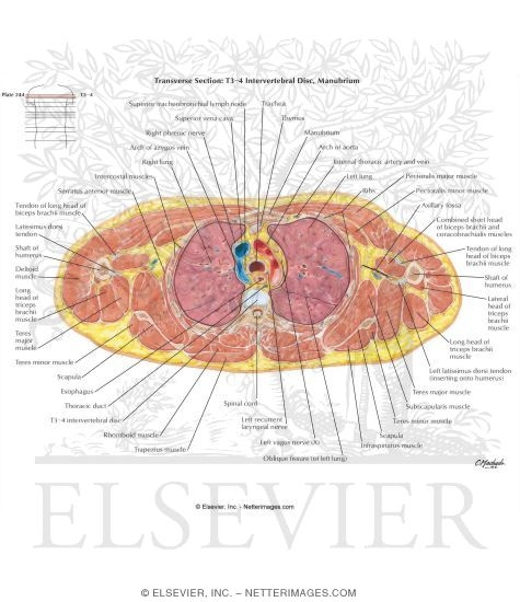
- Tracheal bifurcation (carina)
- Arch of aorta and start of descending thoracic aorta
- Azygos arch entering the superior vena cava
- Esophagus, posterior to the trachea
- Thoracic duct, crossing toward the left
- T4-T5 vertebral level
- Lungs and pleural cavities on both sides