
Pt with pulmonary embolism
Reading File
The query is about a patient with pulmonary embolism - this is a broad clinical scenario. Before I proceed, let me clarify what the user needs.
Asking for Preferences
What aspect of pulmonary embolism are you focusing on?
Full overview it is. Let me search the medical library and recent literature in parallel.
Finding Sources
Searching PubMed
"pulmonary embolism"[MeSH Terms] AND management
Good. Now let me read the most comprehensive PE sections in parallel - Rosen's Emergency Medicine (great for clinical management), Murray & Nadel (respiratory specialty), and Miller's Anesthesia (risk stratification).
Reading File
Reading File
Reading File
Good. Now let me read the core Rosen's PE section for management/diagnosis details, and the full Murray & Nadel PE chapter for pathophysiology.
Reading File
Reading File
Now let me get the pathophysiology (Murray & Nadel block 3), the treatment section from Rosen's (continuing from where I left off), and fetch the autopsy image that was referenced.
Reading File
Reading File
Loading Image
I now have comprehensive material to write a full overview. Let me now read the pathophysiology section from Murray & Nadel specifically on PE mechanisms.
Finding Sources
Reading File
I now have comprehensive information across all domains. Let me compile the full overview.
Pulmonary Embolism (PE) - Full Clinical Overview
Pathophysiology
PE is a life-threatening manifestation of venous thromboembolism (VTE). In most cases, a thrombus originating from deep veins of the lower extremities detaches, travels through the right heart, and lodges in the pulmonary vasculature.
Virchow's Triad - the three predisposing factors:
- Hypercoagulability (thrombophilia, cancer, oral contraceptives, immobility)
- Venous stasis (prolonged immobilization, heart failure, post-surgical)
- Venous injury/inflammation (trauma, surgery, IV catheters)
Hemodynamic consequences:
- Obstruction of the pulmonary vascular bed raises pulmonary vascular resistance (PVR)
- Acute pulmonary hypertension causes right ventricular (RV) pressure overload and dilation
- The dilated RV compresses the interventricular septum, reducing LV filling ("D-sign")
- Decreased LV cardiac output leads to systemic hypotension and ischemia
- In extreme cases, the thrombus can occlude the entire RV outflow system (as seen in the autopsy photo below), causing sudden cardiovascular collapse
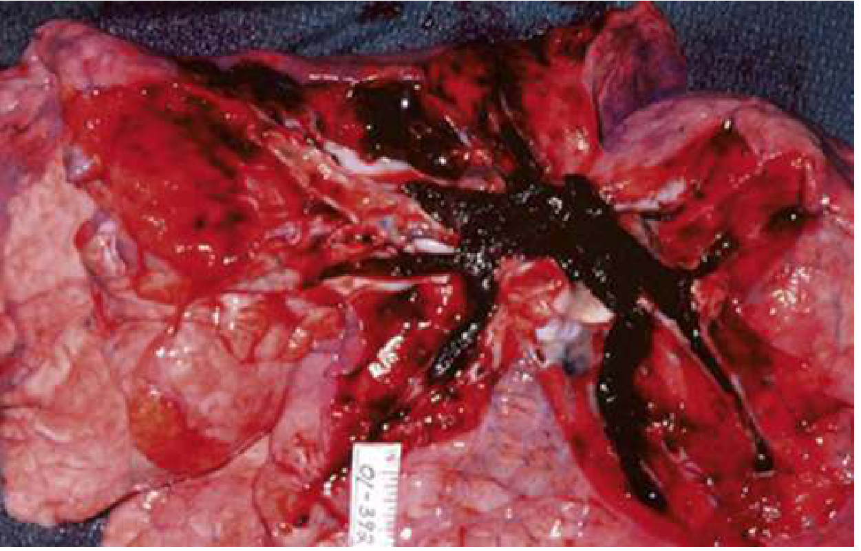
Gas exchange effects:
- V/Q mismatch is the primary cause of hypoxemia
- Dead space ventilation increases (perfused segments lost)
- Reflex bronchoconstriction and atelectasis contribute
- Pulmonary infarction can occur in peripheral PE, producing pleuritic pain, hemoptysis, and fever
Risk Factors
| Category | Examples |
|---|---|
| Surgical/Trauma | Lower extremity surgery, prolonged procedures, pelvic trauma |
| Immobility | Prolonged bed rest, long-haul flights ("economy class syndrome") |
| Malignancy | Lung, brain, pancreas, leukemia - especially active/treated cancers |
| Thrombophilia (inherited) | Factor V Leiden, prothrombin G20210A mutation, antithrombin III deficiency, protein C/S deficiency |
| Thrombophilia (acquired) | Antiphospholipid syndrome |
| Hormonal | OCP, HRT, fertility drugs, anti-androgen prostate cancer therapy |
| Prior VTE | Strongest single predictor of recurrence |
| Pregnancy/Postpartum | 5x risk vs. age-matched controls; peaks postpartum especially after C-section |
| Obesity, smoking, age >35 | Modifiable/additive factors |
Clinical Presentation
PE is called the "great masquerader" because it can present across a wide spectrum:
Symptoms (most to least common):
- Dyspnea - 75-80% of patients; can be constant, exertional, or vague ("can't take a full breath")
- Chest pain - second most common; can be pleuritic (sharp, worse with inspiration - seen in ~20%) or non-pleuritic (dull, aching)
- Cough, hemoptysis - with pulmonary infarction (peripheral PE)
- Unilateral leg swelling/pain - present in <30%, but relatively specific when combined with dyspnea
- Syncope - <5% of syncope patients have PE, but unexplained syncope with risk factors should prompt evaluation
- Low-grade fever - if high fever (>38.6°C/101.5°F), consider pneumonia instead
Signs:
- Tachycardia (most common sign)
- Tachypnea, hypoxemia
- Hypotension/shock (massive PE)
- Loud P2, right-sided S3/S4, signs of RV failure (raised JVP, peripheral edema)
- PEA arrest is the most common ECG finding in PE-related cardiac arrest
Pearl: Approximately 25% of sudden cardiac deaths are attributed to PE.
Diagnosis & Workup
Step 1 - Estimate Pre-Test Probability (PTP)
Wells Score for PE:
| Criterion | Points |
|---|---|
| Clinical signs/symptoms of DVT | 3 |
| PE is #1 diagnosis OR equally likely | 3 |
| Heart rate >100 bpm | 1.5 |
| Immobilization ≥3 days or surgery in prior 4 weeks | 1.5 |
| Prior DVT or PE | 1.5 |
| Hemoptysis | 1 |
| Malignancy (treated within 6 months or palliative) | 1 |
- Low: <2 points | Moderate: 2-6 | High: >6
Revised Geneva Score (fully objective):
- Low: 0-3 | Intermediate: 4-10 | High: >10
Step 2 - Apply PERC Rule (if low gestalt PTP)
If all 8 criteria are met AND gestalt PTP is low, PE can be ruled out without further testing:
Age <50 | Pulse <100 | SaO2 >94% | No unilateral leg swelling | No hemoptysis | No recent trauma/surgery | No prior PE/DVT | No hormone use
Step 3 - D-Dimer
- Sensitivity 95-98%, specificity 40-55%
- Use in non-high PTP patients (Wells <6 or Geneva <5)
- Negative D-dimer effectively excludes PE (NPV 99-100%)
- Age-adjusted D-dimer threshold = age × 10 mcg/L (for patients >50 years) - increases specificity without sacrificing sensitivity
Step 4 - Imaging
CT Pulmonary Angiography (CTPA) - first-line imaging modality
- Visualizes emboli directly in pulmonary arteries
- Also shows RV dilation (ratio >0.9 signals intermediate/high risk)
- Preferred over V/Q in most patients
V/Q Scan - preferred when:
- Contrast allergy or renal impairment
- Pregnancy (lower fetal radiation vs. CTPA)
- Young women (lower breast radiation)
Compression Ultrasonography (CUS) - for DVT; if positive in a patient with high PE suspicion, may be sufficient to start treatment without CTPA
Echocardiography - NOT recommended for initial PE diagnosis; valuable for risk stratification once PE is confirmed (RV strain, septal shift, McConnell's sign)
Ancillary Tests
| Test | Utility |
|---|---|
| ECG | Sinus tachycardia most common; S1Q3T3, new RBBB, T-wave inversions V1-V4 suggest RV strain |
| Troponin (I or T) | Elevated = myocardial injury from RV ischemia; predicts worse prognosis |
| BNP/NT-proBNP | Elevated = RV strain; guides risk stratification |
| ABG | Hypoxemia, hypocapnia, respiratory alkalosis; elevated A-a gradient |
| CXR | Usually non-specific; Hampton's hump (wedge-shaped opacity), Westermark's sign (oligemia), atelectasis |
Risk Stratification
| Category | Criteria | Mortality |
|---|---|---|
| Low-risk (submassive) | Hemodynamically stable, no RV dysfunction, normal troponin/BNP | <1% |
| Intermediate-low | Stable, but elevated troponin OR BNP OR RV dysfunction on imaging | ~3-5% |
| Intermediate-high (submassive) | Stable, but BOTH troponin elevated AND RV dysfunction | ~10-15% |
| High-risk (massive) | Hemodynamic instability (SBP <90 or drop >40 mmHg for >15 min), or cardiac arrest | >30% |
Shock index >1 (HR > SBP) indicates hemodynamic compromise.
Management
Anticoagulation (cornerstone of treatment)
DOACs (Direct Oral Anticoagulants) - preferred for most patients:
- Rivaroxaban: 15 mg BID x 21 days, then 20 mg daily
- Apixaban: 10 mg BID x 7 days, then 5 mg BID
- Dabigatran or Edoxaban: require 5-10 days of parenteral heparin first
Heparin (IV UFH or LMWH):
- Use if thrombolysis or surgical intervention is planned
- UFH is preferred pre-thrombolysis (short half-life, reversible)
- LMWH (enoxaparin) preferred for cancer-associated PE
Duration of anticoagulation:
- Provoked PE (reversible risk factor): 3 months
- Unprovoked PE: minimum 3 months; consider extended if low bleeding risk
- Cancer-associated PE: long-term (DOAC or LMWH preferred)
Treatment by Risk Category
Low-Risk PE:
- DOAC anticoagulation
- Outpatient management appropriate if Hestia criteria negative or PESI class I-II
- Follow-up at 1-2 weeks
Intermediate-Risk PE:
- Hospital admission (monitored bed); anticoagulation (DOAC or heparin)
- Thrombolysis reserved for clinical deterioration
- Activate PERT (PE Response Team) if available
High-Risk (Massive) PE:
- IV UFH immediately
- Systemic thrombolysis (alteplase 100 mg IV over 2 hours) - if no contraindications
- Surgical embolectomy - if thrombolysis contraindicated or failed
- Catheter-directed therapy (CDT) - alternative in intermediate-high risk or thrombolysis contraindications
- ECMO - as bridge to definitive therapy in refractory shock
Hemodynamic & Supportive Care
- Supplemental O2: target SpO2 >90%; avoid hypoxic pulmonary vasoconstriction
- Avoid aggressive fluid resuscitation - small boluses (250-500 mL) only; excessive fluids worsen RV dilation and LV compression
- Vasopressors: norepinephrine first-line for hypotension; dobutamine as adjunct (may worsen hypotension if used alone)
- Avoid intubation if possible - positive pressure ventilation increases intrathoracic pressure, reduces preload, and can precipitate hemodynamic collapse in severe PE. If intubation is unavoidable, optimize hemodynamics before induction.
IVC Filter
- Indicated when anticoagulation is absolutely contraindicated AND PE risk is high
- Retrievable filters preferred
- Does NOT replace anticoagulation - resume as soon as safe
Special Populations
Pregnancy
- Risk is ~5x that of non-pregnant women; peaks postpartum (especially post-C-section)
- Increased coagulation factors (V, VIII, X, vWF) and decreased protein S
- D-dimer is unreliable (normally elevated in pregnancy)
- Pregnancy-adapted YEARS algorithm may help guide testing
- Imaging: V/Q (lower breast radiation) vs. CTPA - both acceptable; halve the perfusion dose for V/Q
- Treatment: LMWH throughout pregnancy and postpartum (warfarin teratogenic; DOACs not approved)
Cancer
- LMWH (e.g., dalteparin) or DOACs (rivaroxaban, apixaban) preferred over warfarin
- Duration: indefinite while cancer is active
- Outpatient management increasingly supported - see recent meta-analysis (PMID 39603673)
Prognosis & Long-Term Complications
- 30-day mortality: <2% low-risk, up to 30-65% in massive PE with cardiac arrest
- Post-PE syndrome: dyspnea, exercise intolerance, decreased quality of life in ~50% at 1 year
- Chronic thromboembolic pulmonary hypertension (CTEPH): occurs in 0.5-5% of survivors; workup with V/Q scan at 3-6 months if persistent symptoms; definitive treatment is pulmonary endarterectomy
- Recurrence: ~10% at 1 year, ~30% at 10 years for unprovoked PE
PE Response Teams (PERT)
Multidisciplinary teams (emergency medicine, pulmonology, cardiology, cardiac surgery, hematology, interventional radiology) that streamline decision-making for intermediate and high-risk PE. A 2025 meta-analysis (PMID 40258457) assessed outcomes associated with PERT activation in acute PE.
Sources: Rosen's Emergency Medicine (10e), Murray & Nadel's Textbook of Respiratory Medicine, Miller's Anesthesia (10e), PubMed 2024-2026 literature.
This is a shared conversation. Sign in to Orris to start your own chat.