Polarity of epithelial tissue
Polarity of Epithelial Tissue
What is Epithelial Polarity?
- Yamada's Textbook of Gastroenterology, 7th ed.
- Medical Physiology
The Two Poles of an Epithelial Cell
| Feature | Apical Surface | Basolateral Surface |
|---|---|---|
| Faces | Lumen / external environment | Basement membrane + interstitium |
| Surface modifications | Microvilli (increase area ~20-fold) | Lateral interdigitations; basal infoldings |
| Key proteins | Digestive enzymes (sucrase, peptidases); GPI-linked proteins; ion channels for luminal uptake | Na⁺/K⁺-ATPase (almost all epithelia); receptors for hormones/growth factors |
| Lipid character | Glycolipid- and cholesterol-rich "rafts" | Phosphatidylcholine-rich |
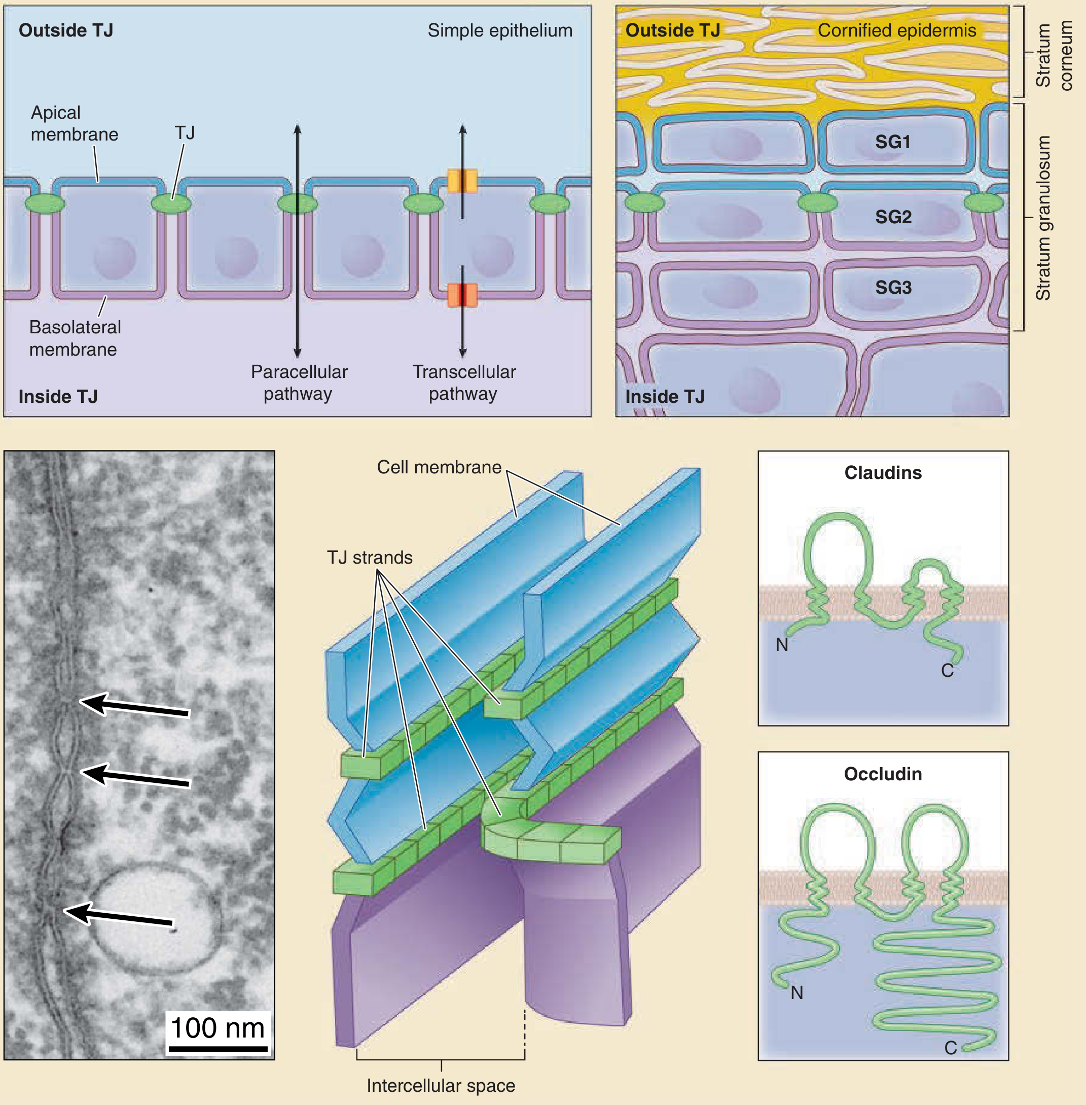
The "Fence" Function: How Polarity is Maintained
- Gate function - selectively restrict paracellular movement of ions and molecules between the lumen and interstitium (size- and charge-selective)
- Fence function - act as a physical barrier within the plane of the membrane, preventing diffusion and intermingling of apical and basolateral membrane proteins and lipids
- Claudins - 4-transmembrane proteins; primary "zip-lock" sealing the paracellular space; ion- and size-selectivity varies with claudin subtype
- Occludin - 4-transmembrane protein; regulates barrier tightness
- JAM-A (Junctional Adhesion Molecule A) - involved in junction assembly
- ZO-1, ZO-2, ZO-3 - intracellular scaffold proteins that link TJ strands to the actin cytoskeleton
Initiation of Polarity: How Cells Become Polarized
- E-cadherin (Ca²⁺-dependent adherens junction protein) is rapidly recruited to points of cell-to-cell contact - this is the critical initial trigger
- E-cadherin clustering recruits catenins (α-catenin, β-catenin) → reorganizes the perijunctional actin-myosin ring
- A membrane cytoskeletal scaffold assembles: fodrin, ankyrin, atypical PKC (aPKC)-PAR-6 complex, and SNAREs - these direct proteins to the appropriate membrane domain
- Tight junction proteins are then recruited, assembling the physical fence that locks in the nascent apical vs. basolateral domains
- Integrin-mediated binding to extracellular matrix reinforces and stabilizes polarity
- Par complex (Par-3/Par-6/aPKC) - defines the apical domain; originally discovered in C. elegans and Drosophila
- Crumbs (Crb) complex - transmembrane polarity regulator at the apical surface
- Scribble complex - defines the basolateral domain
Protein Sorting to Apical vs. Basolateral Domains
- Contained in the cytoplasmic tail of the protein
- Often include a tyrosine residue within conserved sequences (similar to endocytosis signals)
- Recognized by adaptins during clathrin-coated vesicle formation at the TGN
- Found in the transmembrane or extracellular domains
- GPI anchors cluster proteins into glycolipid/cholesterol rafts that bud from the Golgi toward the apical surface
- N- and O-linked glycans act as apical sorting signals; their specificity depends on glycosyltransferases expressed in each cell type
Functional Significance of Polarity
- Intestinal Na⁺ absorption: Apical Na⁺ entry (via Na⁺-glucose cotransporter SGLT1 or ENaC) + basolateral Na⁺/K⁺-ATPase extrusion → net Na⁺ movement from lumen to blood
- Gastric HCl secretion: H⁺/K⁺-ATPase restricted to apical membrane of parietal cells
- Hepatocyte bile transport: Basolateral uptake of bile salts (via NTCP/OATPs) + apical secretion into bile canaliculus (via BSEP/MRP2)
- Antigen sampling: Receptors detecting luminal antigens restricted to apical domain
- Signal transduction: Receptors for hormones/growth factors polarized to basolateral domain (facing the blood)
- Enzyme restriction: Brush-border enzymes kept at the apical surface to act only on luminal substrates
Polarity Loss and Disease
| Condition | Polarity Defect |
|---|---|
| Colorectal/gastric cancer | β-catenin and APC mutations → Wnt pathway activation; E-cadherin loss → epithelial-mesenchymal transition |
| Signet ring cell carcinoma | Germline or somatic E-cadherin mutations; loss of cell-cell adhesion |
| Peutz-Jeghers syndrome | LKB1 (Par-4 homolog) mutation → abnormal polarization + Wnt5a upregulation → arborizing polyps |
| Inflammatory bowel disease | Tight junction disruption → barrier loss; cytokine-mediated claudin dysregulation |
| Atopic dermatitis | Defective epidermal TJ barrier → antigen penetration → allergic sensitization |
Make a presentation on epithelial polarity, tight junctions, and disease
Polarity of epithelial tissue from laiq Hussain
Laiq Hussain histology textbook epithelial polarity
Polarity of Epithelial Cells
What is Polarity?
| Surface | Faces | Also Called |
|---|---|---|
| Apical | Lumen / free surface / external environment | Free surface |
| Lateral | Adjacent epithelial cells | Intercellular surface |
| Basal | Basement membrane | Base |
1. Apical Surface and Its Specializations
A. Microvilli
- Small, finger-like cytoplasmic projections from the apical surface
- Core of actin filaments (supported by villin, fimbrin, and myosin I)
- Function: increase apical surface area for absorption (up to 20-fold)
- Seen in LM as the striated border (intestinal epithelium) or brush border (renal tubules)
- Most developed in intestinal absorptive cells (enterocytes) and proximal convoluted tubule cells of the kidney
B. Stereocilia (Stereovilli)
- Unusually long microvilli (not true cilia - no microtubules, no movement)
- Core of actin filaments, like regular microvilli
- Found in: epididymis and vas deferens (absorption of testicular fluid) and sensory hair cells of the inner ear (mechanoreception)
C. Cilia
| Feature | Motile Cilia | Primary Cilia (Monocilia) |
|---|---|---|
| Microtubule arrangement | 9+2 (axoneme) | 9+0 |
| Movement | Yes - coordinated beat via dynein | No |
| Number per cell | Many | One per cell |
| Function | Move mucus/particles (respiratory tract), move ova (fallopian tube) | Chemosensors, osmosensors, mechanosensors |
| Location | Respiratory epithelium, uterine tube | Almost all eukaryotic cells |
2. Lateral Surface and Cell Junctions (Junctional Complex)
A. Zonula Occludens (Tight Junction)
- Located at the most apical end of the lateral membrane
- Composed of: claudins, occludin, JAM-A, tricellulin (transmembrane); ZO-1, ZO-2, ZO-3 (scaffolding)
- Two key functions:
- Gate function - restricts paracellular passage of molecules between cells (selective barrier, varies by claudin composition)
- Fence function - prevents lateral diffusion of membrane proteins/lipids between apical and basolateral domains, maintaining polarity
- When TJs are disrupted → apical and basolateral proteins intermingle → polarity is lost
B. Zonula Adherens (Adherens Junction)
- Just below the tight junction; forms a belt around the cell
- Composed of E-cadherin linked via α- and β-catenin to actin filaments
- Function: cell-to-cell adhesion; also the trigger for polarity initiation (E-cadherin clustering is the first step in polarization)
C. Macula Adherens (Desmosome)
- Spot-like, scattered along the lateral membrane below the zonula adherens
- Composed of desmogleins and desmocollins (cadherins) → connect to desmoplakin/plakophilin → anchored to intermediate filaments (keratin)
- Function: strong mechanical adhesion - resists shearing forces (prominent in skin, cardiac muscle)
D. Gap Junctions (Nexus / Communicating Junctions)
- Accumulation of transmembrane channels composed of connexins (two hexameric connexons forming a pore)
- Allow passage of ions, small metabolites, and second messengers (up to ~1,000 Da)
- Function: electrical and metabolic coupling between cells; important in cardiac muscle conduction and smooth muscle coordination
E. Lateral Interdigitations (Plicae)
- Tortuous infoldings of the lateral membrane of adjacent cells
- Prominent in fluid/electrolyte-transporting epithelia (intestine, gallbladder)
- Increase lateral surface area for placement of Na⁺/K⁺-ATPase to drive transcellular fluid transport
3. Basal Surface and Its Specializations
A. Basement Membrane
- Sits between the basal surface of the epithelium and underlying connective tissue
- Two layers:
- Basal lamina (produced by epithelium): lamina lucida + lamina densa (type IV collagen, laminin, nidogen, perlecan)
- Reticular lamina (produced by connective tissue): type III collagen fibers
- Functions: mechanical support, filtration, cell signaling, scaffold for regeneration
B. Cell-to-Matrix Junctions
- Hemidesmosomes: connect keratin intermediate filaments inside the cell to laminin in the basal lamina via α6β4 integrin and BP180 (collagen XVII)
- Focal adhesions: connect actin cytoskeleton to fibronectin/vitronectin in ECM via integrins (α5β1, αvβ3); associated with vinculin, talin, paxillin
C. Basal Infoldings
- Deep infoldings of the basal plasma membrane
- Trap mitochondria between the folds
- Function: increase basolateral surface area for Na⁺/K⁺-ATPase activity; prominent in renal tubules and salivary gland ducts
Summary: Polarity at a Glance
LUMEN / FREE SURFACE
        ↑
  APICAL domain
  - Microvilli (actin core, absorption)
  - Stereocilia (long microvilli, absorption/sensory)
  - Cilia (9+2 motile or 9+0 primary)
        |
  ──────────── TIGHT JUNCTION (fence + gate)
  ──────────── ADHERENS JUNCTION (E-cadherin/actin)
  ──────────── DESMOSOME (cadherin/intermediate filaments)
  ──────────── GAP JUNCTION (connexins, communication)
  LATERAL domain: interdigitations, Na⁺/K⁺-ATPase
        |
  BASAL domain
  - Basal infoldings (renal tubules)
  - Hemidesmosomes → basement membrane
  - Focal adhesions → ECM
        ↓
  BASEMENT MEMBRANE
  CONNECTIVE TISSUE
Clinical Relevance
| Disease | Polarity/Junction Defect |
|---|---|
| Pemphigus vulgaris | Autoantibodies against desmoglein-3 → desmosome disruption → skin blisters (acantholysis) |
| Bullous pemphigoid | Autoantibodies against BP180/BP230 at hemidesmosomes → subepidermal blistering |
| Kartagener syndrome (PCD) | Dynein arm defects → immotile cilia → bronchiectasis, situs inversus, male infertility |
| Barrett esophagus | Chronic acid reflux → squamous-to-columnar metaplasia with loss of normal polarity |
| Colorectal cancer | E-cadherin/β-catenin mutations → loss of adherens junction and polarity → EMT |
| Inflammatory bowel disease | Claudin dysregulation → tight junction barrier failure → increased paracellular permeability |
Note on Laiq Hussain's textbook: Medical Histology by Laiq Hussain Siddiqui (5th ed., Medtech) is not currently in the digital library, so this answer draws from the standard histology textbooks available (Ross, Junqueira, Medical Physiology) which cover the same topics with equivalent or greater depth. The content structure above mirrors Laiq Hussain's Chapter 3 organization as referenced in course materials.
Add diagram
A detailed scientific diagram of epithelial cell polarity showing a single polarized columnar epithelial cell with three clearly labeled domains: Apical domain at the top with microvilli (brush border), stereocilia, and motile cilia labeled separately; Lateral domain in the middle showing the junctional complex with tight junction (zonula occludens), adherens junction (zonula adherens), desmosome (macula adherens), and gap junction labeled from top to bottom with small inset molecular diagrams of claudin/occludin in tight junction and connexon in gap junction; Basal domain at the bottom showing basal infoldings with mitochondria trapped between folds, hemidesmosomes connecting to the basement membrane (with lamina lucida and lamina densa labeled), and focal adhesions connecting to the extracellular matrix. Arrows showing transcellular transport: Na+ entering apically, Na+/K+-ATPase on lateral/basal membrane. The lumen is labeled above, connective tissue below. Clean white background, professional medical textbook illustration style, blue and purple color scheme with clear annotations.