
Intussusception treatment
intussusception barium enema reduction pediatric
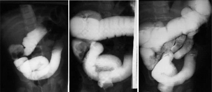
This sequence of three abdominal fluoroscopic images demonstrates a barium enema procedure being used for the therapeutic reduction of an ileocolic intussusception in a pediatric patient. In the first image, contrast highlights the classic 'coiled spring' appearance in the lower right quadrant, signifying the intussusceptum (ileum) being telescoped into the intussuscipiens (colon). As the therapeutic enema progresses in the second and third images, hydrostatic pressure from the barium contrast is shown gradually pushing the intussusceptum back towards the cecum. The series captures the proximal migration of the contrast material through the ascending and transverse colon. Educational focus is placed on recognizing the radiographic signs of intussusception reduction, including the resolution of the intussuscepted mass and the eventual free reflux of barium into the terminal ileum, which signifies a successful non-operative reduction of the bowel obstruction.
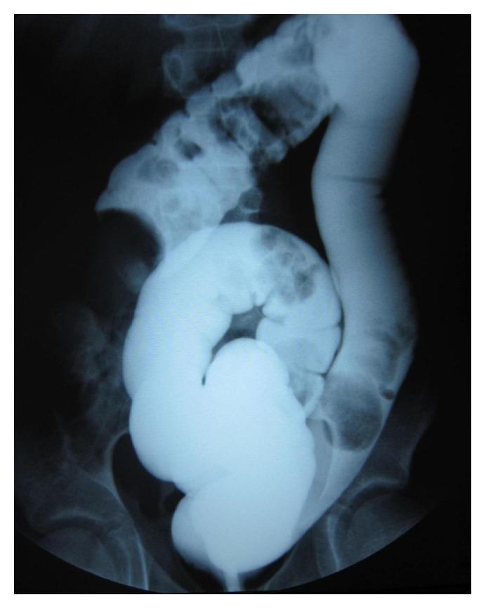
This abdominal X-ray radiography captures a diagnostic barium contrast enema procedure in a pediatric patient. The imaging modality highlights the colon using radiopaque contrast material, demonstrating an unsuccessful hydrostatic reduction of an ileocolic intussusception. A prominent, rounded, and lobulated filling defect is visible in the right upper quadrant, representing the intussusceptum (the prolapsed segment of the bowel) nested within the intussuscipiens. Proximal to this obstruction, the contrast distribution is irregular and narrowed. Distal to the lesion, the barium pools within the sigmoid colon and rectum, which are positioned over the pelvic bones. The image illustrates the classic 'coiled spring' or 'cup-shaped' sign typical of intussusception on contrast studies. This educational visual is highly relevant for pediatric radiology and surgery, focusing on the diagnosis of intestinal obstruction and malrotation-associated complications like Ladd's bands which may contribute to such clinical presentations.
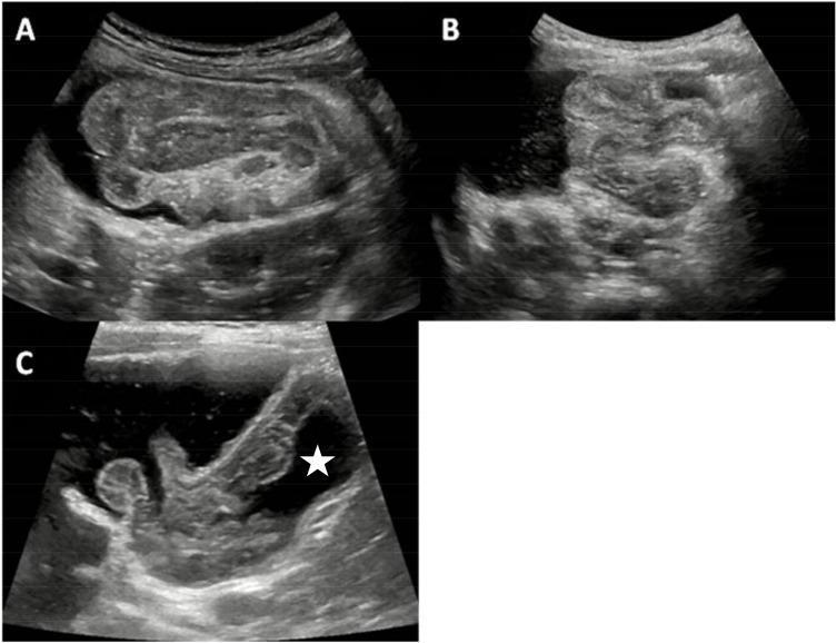
This medical visual consists of three transabdominal ultrasound panels (A, B, and C) demonstrating the progression of ultrasound-guided saline enema reduction for pediatric intussusception. Panel A shows a classic 'pseudokidney' or 'target' sign in longitudinal section, representing the intussusceptum (invaginated bowel) within the intussuscipiens, surrounded by anechoic saline. Panel B captures the dynamic reduction phase where the intussusception is visibly shortened as retrograde hydrostatic pressure from the saline enema pushes the bowel segment back toward its anatomical position. Panel C confirms successful reduction, characterized by the complete disappearance of the layered mass and the visualization of anechoic saline passing through the ileocecal valve into the terminal ileum (indicated by a white star). This sequence illustrates the therapeutic utility of real-time sonography in managing ileocolic intussusception, emphasizing key landmarks such as the concentric rings of the bowel wall and the return of fluid flow to obstructed segments.
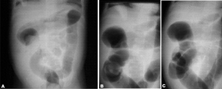
This series of diagnostic X-ray radiographs demonstrates the sequential non-operative reduction of an intussusception using an air enema in a pediatric patient. Panel A shows the intussusceptum as a soft tissue mass (filling defect) within the distal ascending colon, outlined by air. Panel B illustrates the progression of the procedure, with air pressure pushing the intussusceptum proximally toward the cecum, indicated by the shifting position of the mass and the expansion of the air column. Panel C shows the successful completion of the reduction, characterized by the disappearance of the soft tissue mass and the free passage of air into the terminal ileum, representing a resolved ileocolic intussusception. The images provide a clinical example of the 'crescent sign' and 'meniscus sign' often seen during pneumatic reduction, serving as an educational resource for pediatric radiology and emergency gastrointestinal interventions.
intussusception treatment reduction
Intussusception: Treatment
1. Initial Stabilization (All Patients)
- IV fluid resuscitation - repeated 20 mL/kg boluses of normal saline until adequate intravascular volume
- NPO status
- Nasogastric decompression (if obstructed/vomiting)
- Broad-spectrum antibiotics (standard pre-procedure/surgical prophylaxis)
- Prompt surgical consultation - must be available before attempting radiological reduction
- Bailey and Love's Short Practice of Surgery, 28th Ed.; Rosen's Emergency Medicine
2. Non-Operative Reduction (Pediatric - First Line)
Indications
- Hemodynamically stable, well-hydrated child
- No signs of peritonitis
- No free intraperitoneal air (on imaging)
Contraindications
- Free intraperitoneal air (perforation)
- Peritonitis / clinical shock
- Suspected bowel necrosis (relative - Doppler may not reliably exclude)
A. Pneumatic (Air) Reduction
- Air is insufflated per rectum under fluoroscopic guidance
- Pressure is applied retrograde until the intussusceptum is pushed back
- Success sign: free flow of air into the terminal ileum
- Serious complication: tension pneumoperitoneum from colonic perforation (rare but life-threatening - requires immediate needle decompression)
- Use of deep sedation has been shown to increase success rates without increasing complications in prospective study
B. Hydrostatic (Barium/Water-Soluble Contrast or Saline) Reduction
- Contrast or saline is run by gravity retrograde into the colon
- Can be guided by fluoroscopy (contrast enema) or real-time ultrasound (saline enema)
- Ultrasound-guided saline enema is increasingly used - avoids radiation and provides real-time visualization
- Success sign on barium: free reflux of contrast into terminal ileum + resolution of filling defect / coiled-spring appearance
C. Technique Choice
| Feature | Pneumatic (Air) | Hydrostatic (Contrast/Saline) |
|---|---|---|
| Guidance | Fluoroscopy | Fluoroscopy or Ultrasound |
| Radiation | Yes (fluoroscopy) | Yes (fluoroscopy) / No (US-guided) |
| Perforation risk | Tension pneumoperitoneum | Barium peritonitis (rare) |
| Success rate | ~80-90% | ~80-90% |
- Grainger & Allison's Diagnostic Radiology; Rosen's Emergency Medicine
3. Small Bowel Intussusception
- Often an incidental finding - may reduce spontaneously
- Not suitable for enema reduction
- If causing ileus or obstruction: requires surgical management
- Key on ultrasound: smaller intussusceptum diameter, rarely contains lymph nodes; edematous wall = less likely to reduce spontaneously
- Grainger & Allison's Diagnostic Radiology
4. Surgical Treatment
- Enema reduction fails or is contraindicated
- Peritonitis, perforation, or shock
- Evidence of bowel necrosis / gangrene
- Pathological lead point identified (Meckel's diverticulum, lymphoma, polyp, duplication cyst) - these are unlikely to reduce non-operatively
- Adult intussusception (almost always has a lead point, often malignant)
Approaches
- Open or laparoscopic manual reduction - performed by squeezing the distal bowel to deliver the intussusceptum back (not pulling)
- Resection with primary anastomosis - required for irreducible intussusception, infarcted/gangrenous bowel, or pathological lead point
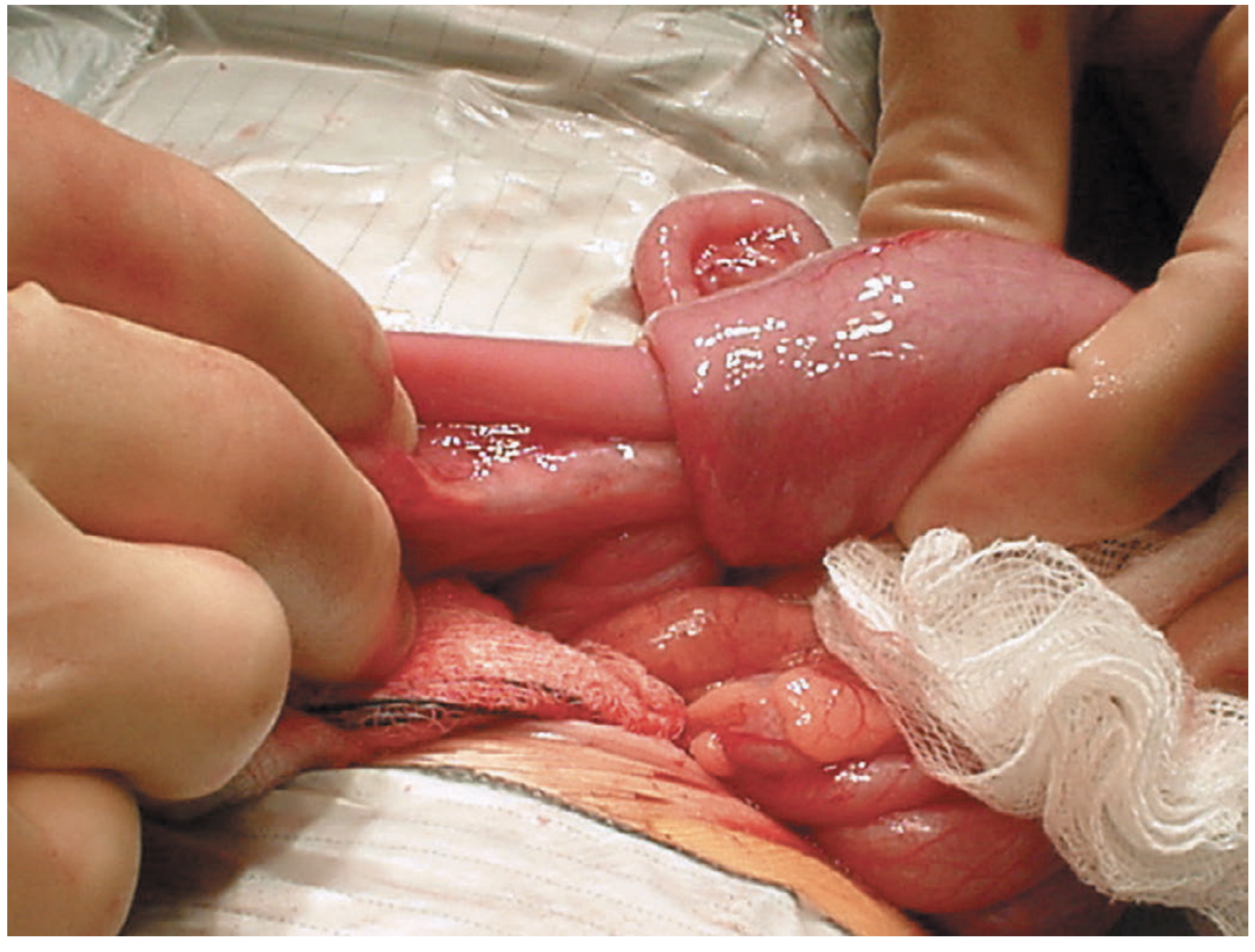
- Bailey and Love's Short Practice of Surgery
5. Adult Intussusception
- Usually has a pathological lead point - neoplasm or malignancy is common
- Surgical resection is nearly always required - non-operative reduction is not recommended as primary treatment
- Two systematic reviews (PMID 39085533, 2024; PMID 40831154, 2025) confirm surgical management is standard, with resection preferred to avoid missing underlying malignancy
- Rosen's Emergency Medicine; Sleisenger & Fordtran's GI and Liver Disease
6. Post-Reduction Management & Recurrence
- After successful non-operative reduction: 6-hour observation period is safe
- If tolerating oral fluids: may be discharged
- Recurrence rate: ~5% after non-operative reduction (usually within 48 hours)
- Recurrence or failure of repeat reduction -> surgical evaluation
- Pathological lead points are more likely in children >2 years and in recurrences
Summary Algorithm
Pediatric intussusception confirmed on US
        |
   Resuscitate: IV fluids, NPO, NG tube
        |
   Stable? No peritonitis? No free air?
       /       \
     YES         NO
      |           |
  Enema        Surgery
  reduction    (if perf/
  (air or     peritonitis)
  hydrostatic)
      |
  Successful?
   /       \
YES         NO
 |           |
6-hr obs   Repeat enema
Discharge  or Surgery