
• Describe different types of dressings and their indications based on wound characteristics. • Explain chronic wound management. • Describe the rationale and indications for contracture release and strategies to prevent recurrence.
wound dressing types comparison chart hydrocolloid alginate foam film
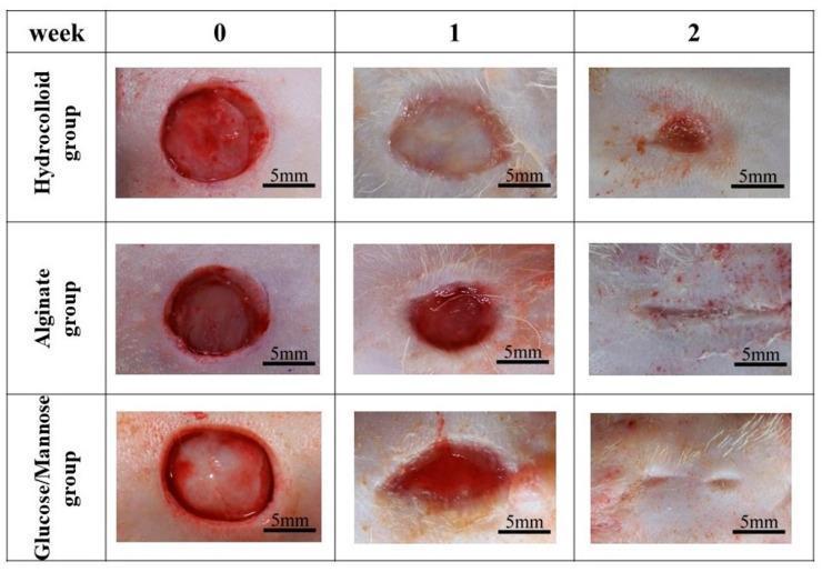
This clinical comparison chart presents macroscopic photographs of full-thickness circular skin wounds in a rat model across three treatment groups: Hydrocolloid, Alginate, and Glucose/Mannose (G/M) film. The image documents the progression of wound healing at week 0, week 1, and week 2 post-surgery. At week 0, all groups display uniform 10mm circular open wounds with exposed red subcutaneous tissue. By week 1, the hydrocolloid group shows a pale, slightly expanded wound bed with exudate, whereas the Alginate and G/M groups show wound contraction and deep red granulation tissue formation. By week 2, both the Alginate and G/M groups demonstrate near-complete wound closure with thin, linear scarring and re-epithelialization. In contrast, the Hydrocolloid group shows incomplete closure with a small, residual red scabbed area. Each photograph includes a 5mm scale bar for quantitative reference. This visual data illustrates the comparative efficacy of different dressing materials in promoting secondary intention healing and re-epithelialization speed.
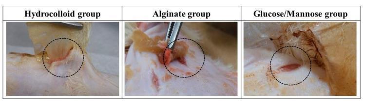
This clinical photograph displays a comparative analysis of wound dressing adhesion in a rat model at one week post-surgery. The image is divided into three panels representing different treatment groups: Hydrocolloid, Alginate, and Glucose/Mannose (G/M) film. In each panel, the surgical wound site is circled with a dashed line during the process of dressing removal using forceps. The Hydrocolloid group shows aggressive adhesion, with the dressing pulling on the fragile granulation tissue and causing visible skin elevation and irritation at the wound margins. The Alginate group exhibits moderate adhesion, with visible brownish-yellow dressing remnants and some tissue disruption. In contrast, the Glucose/Mannose group demonstrates non-adhesive properties, where the film is easily separated from the wound bed, leaving the underlying red granulation tissue and surrounding periwound skin relatively intact and calm. This comparison illustrates the clinical impact of dressing material on secondary trauma and the preservation of the wound bed during dressing changes.
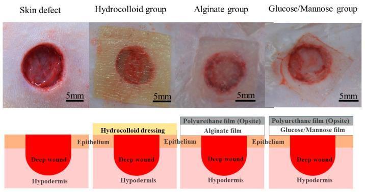
This composite educational graphic features clinical photographs and corresponding schematic diagrams comparing wound dressing applications on full-thickness skin defects in a Sprague-Dawley rat model. The top row displays four clinical photographs: a control 'Skin defect' (an open, circular deep wound with red tissue) and three treatment groups: Hydrocolloid, Alginate, and Glucose/Mannose (G/M). Each wound is approximately 10mm in diameter, indicated by a 5mm scale bar. The hydrocolloid dressing appears as a yellow, semi-opaque square; the alginate and G/M groups show translucent films that allow visualization of the underlying red granulation tissue. The bottom row provides cross-sectional schematics of the wound architecture. The 'Skin defect' shows a deep wound extending through the epithelium into the hypodermis. The 'Hydrocolloid group' illustrates a single-layer dressing directly contacting the epithelium and wound. In contrast, the 'Alginate' and 'Glucose/Mannose' groups utilize a dual-layer system featuring a primary film (Alginate or G/M) secured by a secondary Polyurethane film (Opsite) to maintain a moist healing environment.
contracture release Z-plasty scar burn surgery
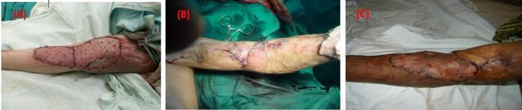
A series of three intraoperative clinical photographs (A, B, C) demonstrating different surgical techniques for reconstructing the anterior elbow following burn contracture release. Panel A shows a split-thickness skin graft (STSG) applied as a solitary covering; the graft is reddish-pink with a meshed appearance and is secured along its irregular borders with dark silk sutures. Panel B displays a Z-plasty local flap technique, visible by characteristic zigzag-shaped incisions and suture lines without any grafted skin, illustrating the rearrangement of local tissue to lengthen the scar. Panel C demonstrates a combined approach using both Z-plasty flaps and STSG to cover a larger raw area, showing both the geometric suture patterns of the flap and the textured, grafted skin. The images serve as an educational comparison of plastic surgery reconstructive methods, including graft adherence, suture placement, and tissue mobilization in the context of post-burn contracture management.
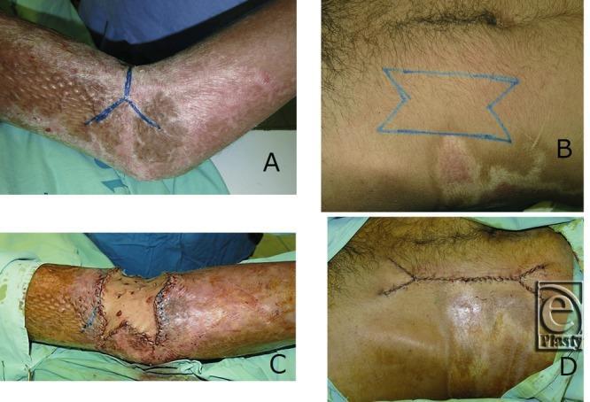
This four-panel clinical photographic series demonstrates a surgical technique for releasing a postburn scar contracture using a full-thickness skin graft with M-plasty donor site closure. Panel A shows the recipient site on the left elbow, featuring an extensive, hyperpigmented, and textured burn scar with a blue preoperative double-Y incision mark across the joint. Panel B illustrates the donor site on the abdomen, marked with a blue double M-plasty design tailored to match the recipient defect. Panel C displays the immediate postoperative result at the elbow following contracture release and successful placement of the full-thickness skin graft, which fits the defect precisely without additional trimming. Panel D shows the donor site after primary closure with nylon sutures in a two-layered technique. The M-plasty closure results in a shortened scar length and minimizes the formation of 'dog ears,' demonstrating an efficient tissue-sparing approach in reconstructive plastic surgery.
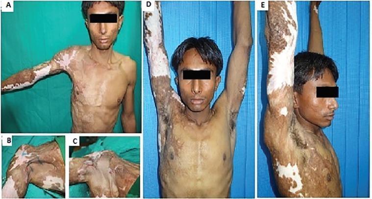
This composite of clinical photographs illustrates the surgical management of a Type 1a right axillary contracture resulting from thermal burns. Panel A shows the preoperative state, characterized by extensive hypertrophic scarring with mixed hyperpigmentation and hypopigmentation (vitiliginous areas) across the anterior chest and right arm, causing significant restriction in shoulder abduction. Panel B demonstrates the preoperative marking for a square flap technique, a modified Z-plasty designed to maximize tissue lengthening. Panel C displays the immediate postoperative appearance with the square flap advanced and triangular flaps transposed to release the contracture. Panels D (frontal) and E (lateral) show the long-term follow-up at 1.5 years, demonstrating a successful clinical outcome with full 180-degree shoulder abduction. The skin in the reconstructed axilla shows improved texture and elasticity, although residual post-burn pigmentary changes persist on the adjacent trunk and extremity. This sequence serves as an educational resource for plastic and reconstructive surgery, highlighting local flap techniques for post-burn scar contracture release.
1. Types of Wound Dressings and Their Indications
The Ideal Dressing
- Provide a moist environment to facilitate epidermal migration
- Enable gas exchange between wound and environment
- Protect against bacterial contamination
- Be non-adherent (to avoid trauma on removal), sterile, non-toxic, non-allergenic, and readily available at minimal cost
Selecting a Dressing: Key Wound Factors
| Wound Factor | Appropriate Dressing |
|---|---|
| Needs moisture (dry wound) | Hydrogel |
| Maintains moisture | Film, hydrocolloid |
| Absorbs moisture (exudative) | Foam, alginate, gelling fibers, superabsorbent |
| Reduces pain | Hydrogel, soft silicone, contact layers |
| Bleeding / hemostasis | Alginates |
| Superficial infection | Dressings impregnated with silver, chlorhexidine, PHMB, iodine, honey, copper oxide |
Choice of Dressing Based on Wound Depth and Drainage
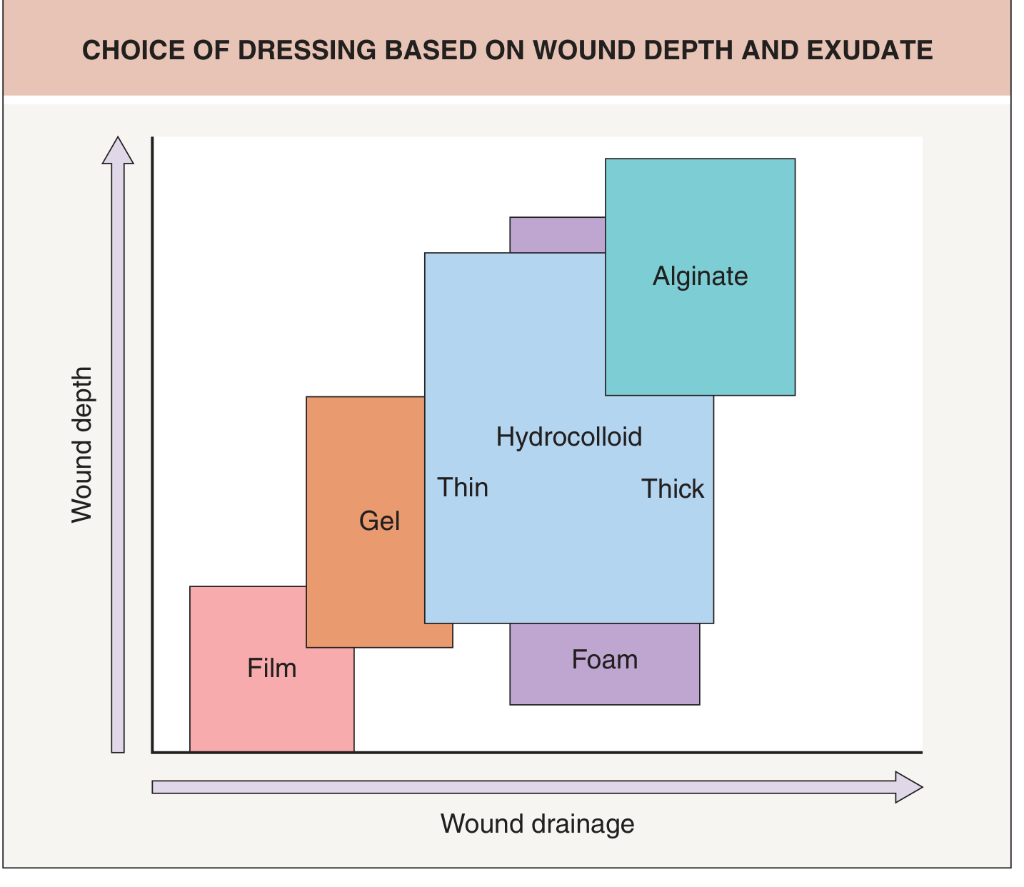
Types of Dressings
1. Traditional Gauze Dressings
- Composed of cotton plus cellulose acetate (for absorbency)
- Inexpensive, easy to use, but limited moisture retention; may adhere to wound
- Impregnated gauze (e.g. Jelonet, Vaseline gauze, Aquaphor): white petrolatum or paraffin wax added to reduce adherence; antimicrobial variants include povidone-iodine (Betadine), framycetin, chlorhexidine
- Indication: clean wounds with minimal exudate; shallow, healing wounds
- Limitation: require frequent replacement; risk of maceration if left in place too long
2. Film Dressings
- Thin, self-adhesive, transparent sheets of polyurethane or synthetic semipermeable material
- Gas-permeable (O2, CO2, water vapor) but impermeable to bacteria and large molecules
- Translucent - allows wound visualization without removal
- Enhance re-epithelialization of graft donor sites by 25-45%
- Indication: shallow, low-exudate wounds; post-operative wounds in epithelialization stage; graft donor sites
- Limitation: non-absorbent (fluid accumulates beneath); difficult to apply without wrinkling; avoid on fragile/thin skin
3. Hydrogel Dressings
- Water-based gels (cross-linked polymers); provide moisture to dry wounds
- Promote autolytic debridement; reduce pain due to cooling effect
- Indication: dry, necrotic wounds; wounds in debridement stage; painful wounds; granulating wounds requiring moisture
- Limitation: can macerate surrounding skin if excessive moisture; requires secondary dressing
4. Hydrocolloid Dressings (e.g. DuoDERM)
- Inner adhesive layer of hydrophilic colloid (pectin, carboxymethyl cellulose) + outer semipermeable polyurethane layer
- Form a gel in contact with wound exudate, resulting in autolytic debridement
- Stimulate angiogenesis; waterproof; do not require secondary dressing
- Indication: moderate exudate wounds; minor burns; pressure ulcers; venous leg ulcers; epithelialization stage
- Limitation: impermeable to oxygen (may be problematic in infected wounds); produce a malodorous gel (may be mistaken for infection); not for heavily exuding or infected wounds
5. Foam Dressings (e.g. Allevyn)
- Semipermeable polyurethane foam; absorb moderate-to-heavy exudate while maintaining moist interface
- Provide cushioning and thermal insulation; good conformity to body surfaces
- Indication: moderately to highly exuding wounds such as leg ulcers, pressure ulcers, cavity wounds, donor sites; graduation stage of healing
- Limitation: not suitable for dry or minimally exuding wounds
6. Alginate Dressings (e.g. Kaltostat)
- Derived from seaweed; contain calcium and sodium salts
- Interact with wound fluid ions to form a gel; hemostatic properties (calcium facilitates clot formation)
- Can be used post-debridement
- Indication: moderate-to-heavily exuding wounds; split-thickness skin graft donor sites; bleeding wounds; post-debridement
- Limitation: require secondary dressing; not for dry wounds or primary closure wounds
7. Gelling Fibers (Hydrofibers, e.g. Aquacel)
- Composed of carboxymethyl cellulose fibers; up to 3x more absorbent than alginates
- Vertical uptake of wound fluid reduces peri-wound maceration
- Aid in autolytic debridement
- Indication: heavily exuding wounds; wounds at risk of maceration
8. Superabsorbent Dressings
- Contain three-dimensional polyacrylic acid polymer networks; absorb hundreds of times their own weight
- Trap bacteria and proteolytic enzymes
- Indication: highly exuding wounds; wounds with malodorous exudate
- Note: swelling under compression can increase wound pressure - use cautiously with compression therapy
9. Silicone Dressings (e.g. Mepitel)
- Non-adherent perforated silicone sheet; designed for prolonged application (up to 2 weeks)
- Minimize trauma and pain on removal
- Indication: fragile skin; paediatric wounds; skin tears; partial-thickness burns; blistering conditions
10. Antimicrobial Dressings
- Ionic silver (e.g. Aquacel Ag): broad-spectrum activity against bacteria including MRSA and Pseudomonas
- Povidone-iodine (e.g. Inadine): broad-spectrum antiseptic
- Honey-impregnated: osmotic mechanism; antibiofilm
- DACC-impregnated: hydrophobic binding irreversibly captures bacteria
- Copper oxide: bactericidal plus pro-angiogenic
- Indication: contaminated or infected wounds; wounds with biofilm; colonized chronic wounds
11. Contact Layers
- Single-layer, non-adherent, woven polyethylene mesh impregnated with petrolatum, hydrocolloid, or silicone
- Protect wound bed during dressing changes; allow exudate to pass through
- Stay in place up to 2 weeks while secondary dressings are changed more frequently
- Indication: primary layer over advanced tissue products; fragile new granulation tissue
12. Composite Dressings
- Combine 2+ semi-occlusive dressings (e.g. a hydrogel/hydrocolloid contact layer + absorptive layer + film outer layer)
- Maximize absorbency, minimize maceration; no secondary dressing needed; waterproof
- Indication: complex wounds requiring multiple functions from one dressing
13. Negative Pressure Wound Therapy (NPWT / VAC)
- Intermittent or continuous topical negative pressure (up to -125 mmHg) through a sealed foam dressing
- Stimulates granulation tissue formation, reduces local edema and tissue exudate, reduces bacterial load
- Indication: open/complex acute wounds (e.g. open abdomen); securing skin grafts to recipient bed; chronic wounds (pressure ulcers, diabetic foot ulcers); wounds awaiting definitive cover
2. Chronic Wound Management
- Optimal blood flow - treat underlying vascular disease
- Control of infection - debridement, antimicrobials
- Removal of devitalized tissue - debridement
- Proper dressing - maintain moisture balance
- Coverage of the injured area
- Compression therapy where indicated (e.g. venous ulcers)
The TIME Framework
| Letter | Principle | Clinical Action |
|---|---|---|
| T | Tissue (non-viable) | Debridement (surgical, enzymatic, autolytic, mechanical, biological) |
| I | Infection / Inflammation | Antimicrobial dressings, systemic antibiotics; manage biofilm |
| M | Moisture imbalance | Match dressing to exudate level; avoid maceration and desiccation |
| E | Edge of wound (non-advancing) | Excision of wound margins; address epibole; advanced therapies |
Debridement Strategies
- Sharp/surgical debridement: fastest method; removes necrotic tissue and biofilm
- Enzymatic debridement: collagenase-based products digest devitalized tissue
- Autolytic debridement: via occlusive/semi-occlusive dressings (hydrogels, hydrocolloids) - harnesses endogenous enzymes
- Maggot (biological) therapy: sterile larvae (Lucilia sericata) selectively liquefy necrotic tissue and reduce biofilm
Management by Wound Aetiology
- Treat underlying venous hypertension with compression bandaging (four-layer or two-layer)
- Debride sloughy tissue; manage exudate with alginates, foams, or hydrofibers
- Oasis wound matrix: 55% healing at 12 weeks vs 34% compression alone
- Offloading pressure with total contact casting or custom orthotics
- Debride callus and necrotic tissue aggressively
- Manage infection (risk of osteomyelitis)
- INTEGRA dermal regeneration template: approved for diabetic foot ulcers
- Dermagraft (neonatal fibroblast construct): 30% wound closure at 12 weeks vs 18% conventional therapy
- Relieve pressure (repositioning, pressure-redistributing mattresses)
- Debride necrotic tissue; NPWT effective for stage III/IV ulcers
Advanced Wound Therapies
Topical Growth Factors
- Becaplermin (recombinant PDGF-BB / Regranex): promotes fibroblast migration and angiogenesis; approved for diabetic neuropathic ulcers
- Maintaining wound exudate (rich in EGF, PDGF, TGF-beta) via occlusive dressings augments healing
Cellular and Tissue-Based Products (CTPs)
- Dermoinductive (cell-active): provide cells and growth factors to activate healing
- Apligraf (bilayer living skin equivalent): 56% healing over 65 days vs 38% with conventional dressing
- DermaSkirt, Therasnap
- Thermoocclusive (scaffold-based): provide structural matrix for cell migration
- Integra: bovine type I collagen + shark chondroitin-6-sulfate + silicone layer; prepares wound bed for grafting; used for partial/deep wounds, exposed bone/tendons
- Oasis (porcine small intestine submucosa): acellular xenograft
Stem Cell-Based Therapies
- Derived from human amniotic membrane of placental tissue
- Products: Epiflex (MiMedx), Grafix (Osiris)
- Contain multiple growth factors (VEGF, PDGF, EGF, TGF) to stimulate healing and reduce scarring
- Significant drawback: extremely expensive; recommended weekly application
3. Contracture Release: Rationale, Indications, and Prevention of Recurrence
What is a Contracture?
- Functional impairment (restricted range of movement), especially across joints
- Deformity, disability, and psychological distress
- Aesthetic disfigurement
- Contractures may also arise from the differential growth pattern between scar tissue and the normal surrounding tissues (particularly important in children who grow)
Rationale for Contracture Release
- Joint motion is restricted and conservative measures have failed
- Contracture causes functional disability (e.g. inability to extend a finger, open the mouth, abduct the shoulder)
- Progressive deformity (especially in growing children)
- Aesthetic disfigurement causing psychological harm
- Mature scar (typically >12 months) that has not responded to non-surgical management
Surgical Techniques for Contracture Release
1. Z-Plasty (and Variants)
- The workhorse technique: two triangular flaps are transposed, lengthening the scar in the axis of contracture at the expense of width
- A standard Z-plasty with 60-degree angles gives ~75% gain in length
- Variants: multiple Z-plasties, W-plasty, Y-V, V-Y plasties
- Best for: linear band contractures that do not involve loss of tissue; web space contractures
- Advantage: uses local tissue with matching color and texture; no donor site required
2. Skin Grafts
- Split-thickness skin grafts (STSG): useful for large areas but contract more during healing; often require secondary procedures
- Full-thickness skin grafts (FTSG): better color/texture match and less secondary contraction than STSG - preferred when skin grafts are used
- In general, flaps are preferable to skin grafts because grafts themselves undergo secondary contracture
3. Local Flaps
- Rotation, advancement, transposition flaps use adjacent pliable tissue
- Preserve perfusion; superior outcomes to grafts at joint surfaces
4. Free Flaps
- Required for severe contractures with large tissue deficits
- Provide well-vascularized, pliable tissue; especially valuable in axillary, neck, and elbow contractures
5. Combined Approaches
- Z-plasty + STSG (for large releases with insufficient local tissue)
- Double Y-incision + FTSG with M-plasty donor site closure (tissue-sparing)

Strategies to Prevent Recurrence
1. Pressure Therapy
- Custom-fitted pressure garments generating 20-25 mmHg; worn for 23 hours/day for up to 2 years
- Mechanism: reduces oxygen tension by compressing small blood vessels → decreases myofibroblast proliferation, collagen synthesis, and fibroblast apoptosis via mechanoreceptors
- Initiated as soon as the wound is healed and the patient can tolerate pressure
- Critical in burns, skin grafts, and any wound healing >2-3 weeks
2. Silicone Therapy
- Silicone gel sheets applied continuously for 12+ hours/day
- Mechanism not fully elucidated; thought to rehydrate the stratum corneum and modulate cytokine activity
- Reduces scar thickness, redness, and pliability
- First-line preventive treatment; started as soon as wound is closed
- Should be continued as long as the scar is maturing
3. Splinting and Positioning
- Static or dynamic splints hold joints in the position of optimal function/extension opposite to contracture forces
- Especially important in burns involving the hand, neck, axilla, and elbow
- Maintained during sleep when the patient is not wearing pressure garments
- Passive and active range-of-motion exercises complement splinting
4. Physiotherapy and Exercise
- Regular stretching, range-of-motion exercises
- Hydrotherapy and scar massage promote elasticity
- Initiated early in the post-operative period
5. Intralesional Corticosteroids
- Triamcinolone acetonide 10-40 mg/mL injected into papillary dermis every 2-4 weeks
- First-line injectable treatment for hypertrophic scarring
- Reduces collagen synthesis and fibroblast action through anti-inflammatory and antiproliferative properties
- 50-100% response rate; up to 50% experience recurrence
- May be combined with 5-FU or bleomycin in resistant cases
6. Adjuvant Therapies at Surgical Site
- Postoperative silicone + pressure should begin immediately after wound closure
- Laser therapy: ablative fractional lasers improve scar texture and pliability
- 5-FU and bleomycin: adjuncts to corticosteroid; promote fibroblast apoptosis and anti-proliferative effects
- Radiation therapy: used post-excision for keloids (high recurrence risk), with caution due to carcinogenesis risk in younger patients
7. Tissue Choice at Reconstruction
- Using flaps rather than skin grafts reduces the risk of graft contracture
- When grafts are necessary, FTSG is preferred over STSG (less secondary contraction)
- Placing flaps with maximum pliability (free flaps with thin, supple donor tissue) at high-risk sites
Summary Box: Prevention of Contracture Recurrence
| Modality | Timing | Duration |
|---|---|---|
| Silicone gel sheeting | Immediately post-healing | 12+ months, until scar matures |
| Pressure garment | When wound is healed | Up to 2 years (23 hr/day) |
| Splinting | Intraoperative + post-op | Until full ROM restored |
| Physiotherapy | Early post-operative | Ongoing |
| Intralesional steroids | For hypertrophy (6 wk-3 mo) | Every 2-4 weeks until flat |
| Laser therapy | After 6 months if scar persists | Variable |
- Bailey & Love's Short Practice of Surgery, 28th Ed. - Wound Dressings, Contractures
- Dermatology 2-Volume Set, 5th Ed. - Traditional & Moisture-Retentive Dressings (Ch. 145)
- Sabiston Textbook of Surgery - Treatment for Hypertrophic Scars, Scar Contracture
- Schwartz's Principles of Surgery, 11th Ed. - Cellular and Tissue-Based Products in Chronic Wound Management