
Nervous system. Anatomy and physiology basic information
Reading File
Now let me search the medical library for nervous system anatomy and physiology content in parallel.
Finding Sources
Finding Sources
Finding Sources
Finding Sources
Reading File
Finding Sources
Reading File
Reading File
Reading File
Finding Sources
Reading File
Reading File
I now have excellent content from multiple authoritative textbooks. Let me fetch the neuron diagram image and read a bit more about physiology (action potentials, synapses) before composing the final answer.
Loading Image
Reading File
I now have comprehensive content from multiple authoritative textbooks. Let me compile the full answer.
The Nervous System: Anatomy and Physiology - Basic Overview
1. Overview and General Organization
The nervous system enables the body to respond to continuous changes in its external and internal environment. It controls and integrates the functional activities of all organs and organ systems.
Anatomically, the nervous system divides into two major parts:
- Central Nervous System (CNS) - the brain and spinal cord, located in the cranial cavity and spinal canal respectively. Covered by three protective membranes (meninges): the outer dura mater, the middle arachnoid, and the delicate inner pia mater.
- Peripheral Nervous System (PNS) - cranial nerves, spinal nerves, peripheral ganglia, and specialized nerve endings that lie outside the dura mater.
Functionally, the nervous system also divides into:
- Somatic Nervous System (SNS) - controls voluntary, conscious functions (skeletal muscle movement + conscious sensation from skin, joints, muscles). Under conscious voluntary control, except for reflex arcs.
- Autonomic Nervous System (ANS) - regulates involuntary visceral functions: heart rate, blood pressure, digestion, temperature regulation, reproductive function.
- Medical Physiology (Boron & Boulpaep), pp. 397-398
- Histology: A Text and Atlas, p. 905
2. Subdivisions at a Glance
| Subdivision | Components | Key Feature |
|---|---|---|
| Central (CNS) | Brain + spinal cord | Oligodendrocytes provide myelin; axons cannot regenerate |
| Peripheral (PNS) | Ganglia, sensory receptors, spinal/cranial nerves | Schwann cells provide myelin; axons can regenerate |
| Autonomic (ANS) | Portions of both CNS and PNS | Functionally distinct; controls viscera |
- Medical Physiology, Table 10-1
3. The Neuron - Basic Structural and Functional Unit
The neuron (nerve cell) is the fundamental signaling unit of the nervous system. The human brain contains approximately 100 billion (10¹¹) neurons, each interacting with thousands of others.
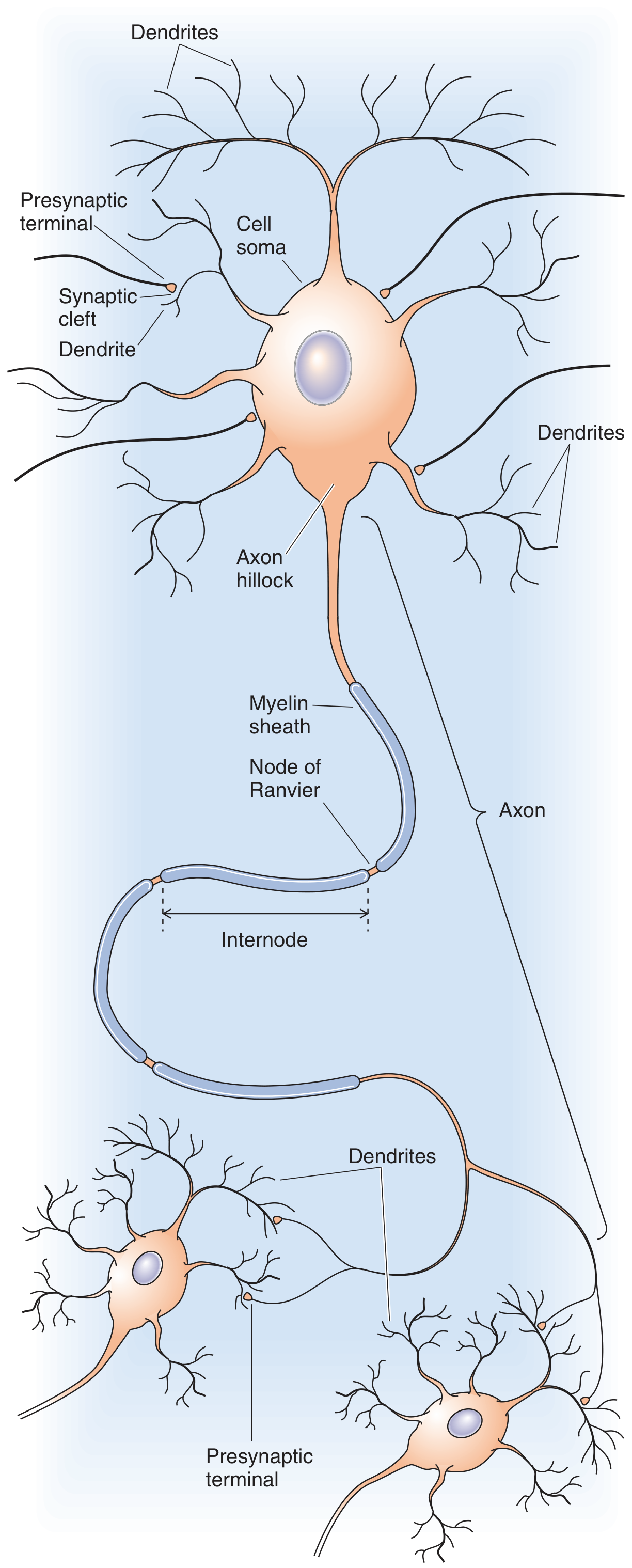
Figure: Morphology of a typical neuron - Medical Physiology, Fig. 10-1
Every neuron has four functional regions:
a. Cell Body (Soma / Perikaryon)
- Contains the nucleus and most cellular organelles (endoplasmic reticulum, Golgi complex).
- Responsible for housekeeping functions: protein synthesis and processing.
b. Dendrites
- Tapering branching processes arising from the cell body.
- The main area for receiving information from other neurons.
- Their membranes carry receptors for neurotransmitters released by neighboring cells.
c. Axon
- Arises from the cell body at the axon hillock, which transitions into the initial segment (also called the spike initiation zone - where action potentials normally originate).
- Thin, does not taper, and can extend more than a meter in length.
- Many axons are wrapped in myelin (insulating membrane layers from glial cells), with gaps called Nodes of Ranvier between segments.
- Unmyelinated axons: action potential travels by continuous propagation (slow).
- Myelinated axons: action potential "jumps" node to node - called saltatory conduction - which greatly speeds impulse conduction.
d. Presynaptic Terminals
- At the axon's target, multiple endings convert the electrical signal into a chemical signal.
- Release of neurotransmitters by exocytosis triggers communication with the next cell.
- Medical Physiology, pp. 399-401
4. Synaptic Transmission
A synapse is the junction between a presynaptic terminal and its target cell. It consists of:
- The presynaptic terminal (sends signal)
- The synaptic cleft (gap between cells)
- The postsynaptic membrane (receives signal)
When an action potential reaches the presynaptic terminal, neurotransmitter molecules are released into the synaptic cleft, diffuse across it, and bind to receptors on the postsynaptic membrane - converting the chemical signal back into an electrical (or biochemical) response.
Synapses can undergo long-term changes based on patterns of prior activity (synaptic plasticity), which is the basis of learning and memory.
- Medical Physiology, pp. 401-402
5. Glial Cells (Neuroglia)
Nervous tissue has two main cell types: neurons (primary signalers) and glial cells (supporting cells). Glial cells are not primary signaling cells but perform diverse, essential functions:
| Glial Cell Type | Location | Function |
|---|---|---|
| Oligodendrocytes | CNS | Produce myelin sheaths around CNS axons |
| Schwann cells | PNS | Produce myelin sheaths around PNS axons |
| Astrocytes | CNS | Blood-brain barrier maintenance, metabolic support |
| Microglia | CNS | Immune surveillance |
6. Gray Matter vs. White Matter
- Gray matter - contains neuron cell bodies. In the brain: predominantly on the outer cortical surface. In the spinal cord: located centrally (butterfly-shaped).
- White matter - rich in myelinated axons (myelin gives the white appearance). In the brain: deep inside; in the spinal cord: superficially surrounding the gray matter.
The cerebral cortex surface shows gyri (ridges) and sulci (grooves), which dramatically increase the surface area of the brain.
- Gray's Anatomy for Students, p. 1302
7. The Autonomic Nervous System (ANS)
The ANS regulates involuntary functions and has two main divisions:
Sympathetic Division ("Fight or Flight")
- Originates in spinal cord segments T1 to L2 (thoracolumbar outflow).
- Uses two-neuron pathways: preganglionic neuron (cell body in intermediolateral horn of spinal cord) → postganglionic neuron (in sympathetic chain ganglia or peripheral ganglia).
- Preganglionic fibers exit via white rami communicantes; postganglionic fibers return via gray rami communicantes.
- Effects: increased heart rate, dilated pupils, bronchodilation, decreased GI motility.
Parasympathetic Division ("Rest and Digest")
- Originates from cranial nerves (III, VII, IX, X) and sacral spinal cord (S2-S4) - craniosacral outflow.
- Also uses two-neuron pathways, but ganglia are located near or within the target organs.
- Effects: decreased heart rate, increased GI motility, pupil constriction, increased secretions.
- Guyton and Hall Textbook of Medical Physiology, pp. 754-757
- Katzung's Basic and Clinical Pharmacology, 16th Ed.
8. Afferent vs. Efferent Pathways
- Afferent (sensory) nerves - carry signals FROM the periphery TO the CNS (e.g., touch, pain, temperature, proprioception).
- Efferent (motor) nerves - carry signals FROM the CNS TO peripheral tissues (muscles, glands).
- Reflex arcs - neural pathways that mediate reflex actions; most involve the spinal cord without requiring direct brain input.
9. Major Brain Regions
| Region | Key Structures | Major Functions |
|---|---|---|
| Cerebral cortex | 4 lobes (frontal, parietal, temporal, occipital) | Voluntary movement, sensation, cognition, language |
| Frontal lobe | Motor cortex, prefrontal cortex | Motor control, planning, personality |
| Parietal lobe | Somatosensory cortex | Touch, proprioception, spatial awareness |
| Temporal lobe | Auditory cortex, hippocampus | Hearing, memory, emotion |
| Occipital lobe | Visual cortex | Vision processing |
| Brainstem | Midbrain, pons, medulla | Vital reflexes (breathing, heart rate), cranial nerves |
| Cerebellum | Cerebellar cortex, deep nuclei | Coordination, balance, fine motor control |
| Spinal cord | Gray + white matter | Relay center; reflex arcs |
The internal capsule (V-shaped white matter structure) carries axons between the cortex and deeper brain structures. The corona radiata fans out from it to the cortical surface.
- Gray's Anatomy for Students, p. 1302
Summary
The nervous system is the body's command and communication network. Its fundamental unit is the neuron, which transmits electrical signals (action potentials) and communicates chemically across synapses. Structurally it divides into CNS (brain + spinal cord) and PNS (all nerves outside). Functionally it divides into somatic (voluntary) and autonomic (involuntary) systems. The autonomic system further splits into sympathetic (activating) and parasympathetic (restoring) divisions. Regional specialization in the brain means each area has unique, non-substitutable functions - a fact that drives clinical neurology.
This is a shared conversation. Sign in to Orris to start your own chat.