
Explain SLE
Reading File
Now I'll search the medical library for SLE content across multiple angles simultaneously.
Finding Sources
Searching PubMed
"systemic lupus erythematosus"[MeSH Terms] AND management
Finding Sources
Searching PubMed
"lupus erythematosus, systemic"[MeSH Terms] AND treatment
I found the Rheumatology and Dermatology textbooks. Let me read the core SLE sections in parallel.
Reading File
Reading File
Reading File
Excellent. Now let me get deeper content from the Rheumatology textbook (the most authoritative source) and look for pathophysiology, diagnosis, and management.
Finding Sources
Finding Sources
Reading File
Now let me get the core SLE pathophysiology, systemic features, and management from the major rheumatology sources.
Finding Sources
Finding Sources
Reading File
I have rich content. Let me get a bit more on pathogenesis, treatment, and fetch key images.
Reading File
Reading File
Loading Image
I now have comprehensive material to write a thorough explanation. Let me also get the treatment section quickly.
Reading File
I now have comprehensive, authoritative content from multiple textbooks. Here is the full explanation of SLE:
Systemic Lupus Erythematosus (SLE)
Definition and Overview
Systemic lupus erythematosus is a chronic, multisystem autoimmune disease characterized by widespread tissue injury mediated by autoantibodies and immune complex deposition. It follows a relapsing-remitting course and can affect virtually every organ. The disease primarily affects young to middle-aged women, with a female-to-male ratio of approximately 9:1. It is more common and more severe in people of African, Hispanic, and Asian ancestry.
Pathogenesis
SLE arises from a breakdown in immune self-tolerance, driven by a combination of genetic susceptibility, environmental triggers, and dysregulated immune responses.
Genetic Factors
Multiple non-HLA genes are implicated, including FCGR2A (Fc receptor for IgG), BLK (B-lymphocyte kinase), IRF5 (interferon regulatory factor 5), PTPN22, and STAT4. HLA-DR2 and HLA-DR3 are the most consistently associated HLA alleles. Deficiencies in early complement components (C1q, C2, C4) strongly predispose to lupus by impairing clearance of apoptotic debris and immune complexes. - Goldman-Cecil Medicine, p. 2812
Autoantibodies
The hallmark of SLE is production of autoantibodies against nuclear antigens. In the preclinical phase, anti-Ro antibodies appear first, followed by anti-dsDNA and finally anti-Smith (Sm) and anti-RNP antibodies around the time of diagnosis. Key autoantibodies include:
| Antibody | Clinical Association |
|---|---|
| Anti-dsDNA | Highly specific for SLE; correlates with disease activity and nephritis |
| Anti-Sm | Highly specific for SLE |
| Anti-Ro/SSA | Subacute cutaneous LE, neonatal lupus, congenital heart block |
| Anti-La/SSB | Associated with anti-Ro |
| Anti-phospholipid (aPL) | Thrombosis, pregnancy loss (antiphospholipid syndrome) |
| ANA | Sensitive (>95%) but not specific |
Approximately one-third of SLE patients have antiphospholipid antibodies, including anticardiolipin antibodies, lupus anticoagulant, and anti-beta2-glycoprotein I. - Goldman-Cecil Medicine, p. 2812
Immune Complex-Mediated Injury
Tissue damage is driven by deposition or in situ formation of immune complexes, activation of the classical complement pathway, and recruitment of neutrophils and monocytes. Paradoxically, while complement is activated and consumed (explaining low C3/C4 levels during flares), complement deficiency also predisposes to SLE by impairing clearance of apoptotic cells. Type I interferons play a central role - an "interferon signature" is detectable in the blood of most SLE patients. - Goldman-Cecil Medicine, p. 2812
Clinical Manifestations
SLE is protean. The table below shows approximate frequencies from Goldman-Cecil Medicine:
| Manifestation | Frequency (%) |
|---|---|
| Cutaneous | 88 |
| Arthritis/arthralgias | 76 |
| Neuropsychiatric | 66 |
| Pleuritis/pericarditis | 63 |
| Anemia | 57 |
| Raynaud phenomenon | 44 |
| Vasculitis | 43 |
| Atherosclerosis | 37 |
| Nephritis | 31 |
| Thrombocytopenia | 30 |
| Cardiac valvular disease | 18 |
| Pulmonary alveolar hemorrhage | 12 |
Skin (88%)
The classic malar (butterfly) rash - fixed erythema over the cheeks and bridge of the nose, sparing the nasolabial folds - is one of the most recognizable findings:
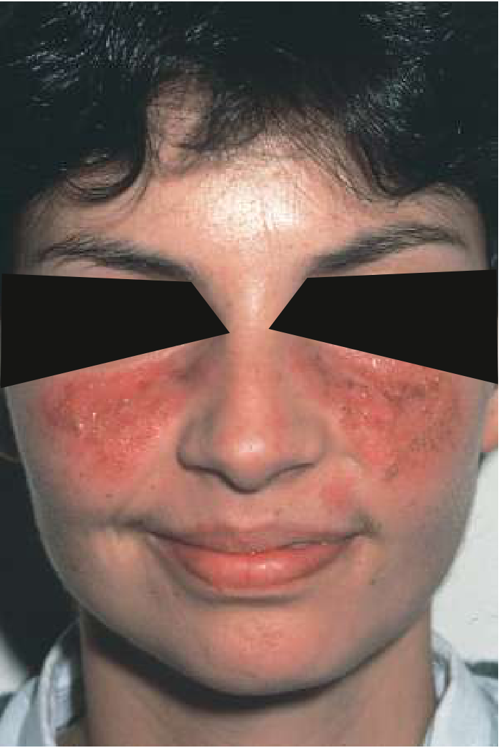
Malar rash in SLE. Note the rash does not cross the nasolabial fold. - Goldman-Cecil Medicine
Other cutaneous features include:
-
Discoid lupus - scarring, hyperpigmented plaques with follicular plugging on the face/scalp
-
Subacute cutaneous LE (SCLE) - annular or papulosquamous lesions, associated with anti-Ro; often drug-induced
-
Photosensitivity - exacerbation of skin disease with UV exposure
-
Oral ulcers (21%) - typically painless
-
Bullous lupus - responds dramatically to dapsone
-
Diffuse non-scarring alopecia, "lupus hairs" (short fragile frontal hairs), periungual telangiectasia, Raynaud phenomenon
-
Andrews' Diseases of the Skin, p. 189
Musculoskeletal
Non-erosive arthritis or arthralgias are the most common presenting complaint. The arthritis is typically symmetric, affecting small joints of the hands, wrists, and knees. Jaccoud arthropathy (reducible deformities from tendon laxity) can occur, unlike the fixed erosive disease of rheumatoid arthritis.
Renal (Lupus Nephritis)
Clinically apparent renal involvement occurs in ~31% but subclinical disease is more common. Renal biopsy is essential for classification and management. The ISN/RPS classification:
- Class I: Minimal mesangial
- Class II: Mesangial proliferative
- Class III: Focal glomerulonephritis (<50% glomeruli)
- Class IV: Diffuse glomerulonephritis (≥50% glomeruli) - the most severe
- Class V: Membranous - presents with nephrotic syndrome
- Class VI: Advanced sclerotic (>90% globally sclerosed glomeruli)
Class III and IV carry the worst prognosis and require aggressive immunosuppression. - Goldman-Cecil Medicine, p. 2817
Neuropsychiatric
Neuropsychiatric SLE (NPSLE) encompasses seizures, psychosis, cognitive dysfunction, mononeuritis multiplex, stroke (often from antiphospholipid antibodies), aseptic meningitis, and transverse myelitis. These may be related to autoantibody-mediated injury, vasculitis, or thrombosis. - Bradley and Daroff's Neurology in Clinical Practice
Cardiovascular
- Libman-Sacks endocarditis - sterile, verrucous valvular vegetations (mitral > aortic), associated with antiphospholipid antibodies
- Pericarditis and myocarditis
- Premature atherosclerosis - a major cause of long-term morbidity independent of traditional risk factors
- Raynaud phenomenon in up to 60%
Pulmonary
- Pleuritis (most common, ~30%) - pleuritic chest pain with exudative effusions
- Shrinking lung syndrome - diaphragm dysfunction without parenchymal disease
- Alveolar hemorrhage (12%) - life-threatening
- Pulmonary hypertension
Hematologic
- Autoimmune hemolytic anemia (Coombs-positive)
- Leukopenia (lymphopenia more than neutropenia)
- Thrombocytopenia
Constitutional
Fatigue, fever, and weight loss are common, especially during flares.
Diagnosis
ACR Classification Criteria (1997) - requires ≥4 of 11:
- Malar rash
- Discoid rash
- Photosensitivity
- Oral ulcers
- Arthritis (non-erosive)
- Serositis (pleuritis or pericarditis)
- Renal disorder (proteinuria >0.5 g/day or cellular casts)
- Neurologic disorder (seizures or psychosis)
- Hematologic disorder (hemolytic anemia, leukopenia, lymphopenia, or thrombocytopenia)
- Immunologic disorder (anti-dsDNA, anti-Sm, or antiphospholipid antibodies)
- Positive ANA
The SLICC 2012 criteria improved sensitivity: requires ≥4 criteria (at least 1 clinical + 1 immunologic) OR biopsy-proven lupus nephritis with ANA or anti-dsDNA. The EULAR/ACR 2019 criteria (scoring system, threshold ≥10) has the best combination of sensitivity and specificity. - Andrews' Diseases of the Skin, p. 189
Key Lab Work-up
- ANA - screening test (>95% sensitivity); if positive, proceed to extractable nuclear antigens (ENA) panel
- Anti-dsDNA - specific (~70%); tracks disease activity
- Anti-Sm - highly specific (~99%) but insensitive (~25%)
- Complement (C3, C4) - low during active disease
- Urinalysis with microscopy - RBC casts = proliferative nephritis
- CBC - cytopenias
- Antiphospholipid antibodies - anticardiolipin IgG/IgM, lupus anticoagulant, anti-beta2GPI
Treatment
Management is tailored to organ involvement and disease severity. The goal is remission or low disease activity while minimizing drug toxicity.
Hydroxychloroquine (HCQ)
The cornerstone of SLE therapy. All patients without contraindications should receive HCQ. Benefits include: reduction of flares, prevention of organ damage, anti-thrombotic effects, improved survival, and favorable lipid profile. Regular ophthalmologic screening (retinal toxicity) is required. - Goldman-Cecil Medicine, p. 2766
NSAIDs and Low-Dose Glucocorticoids
For mild manifestations (arthritis, serositis, skin disease). Glucocorticoids are essential for moderate-to-severe flares but long-term use carries significant toxicity (osteoporosis, infection, metabolic syndrome).
Immunosuppressants
- Mycophenolate mofetil (MMF) - preferred for lupus nephritis (induction and maintenance) and non-renal manifestations
- Azathioprine - maintenance therapy and during pregnancy (relative safety)
- Cyclophosphamide (IV) - for severe nephritis (Class III/IV) and life-threatening NPSLE
- Methotrexate (15-25 mg/week) - for cutaneous and articular disease; avoid in pregnancy
- Tacrolimus/Cyclosporine - calcineurin inhibitors for lupus nephritis
- Voclosporin (23.7 mg twice daily) - a newer calcineurin inhibitor effective in lupus nephritis
Biologic Therapies
- Belimumab (anti-BLyS/BAFF) - approved for moderately active SLE and lupus nephritis; reduces flares and allows steroid-sparing
- Anifrolumab (anti-type I IFN receptor) - approved for moderate-to-severe SLE on background standard therapy
- Rituximab (anti-CD20) - not approved but used off-label for refractory nephritis and cytopenias
- CAR T-cell therapy - emerging; a 2025 systematic review shows early promising results in refractory SLE
Adjunctive
- ACE inhibitors/ARBs for renal protection and hypertension
- Long-term anticoagulation (warfarin) for antiphospholipid syndrome with thrombosis
- Calcium and vitamin D supplementation
- Sunscreen and UV avoidance
- IVIG or plasmapheresis for life-threatening refractory disease
Pregnancy
SLE confers elevated risk of preeclampsia, preterm birth, miscarriage (especially with antiphospholipid antibodies), and fetal/neonatal lupus (anti-Ro/La crossing the placenta causing neonatal rash and potentially congenital heart block). Azathioprine and HCQ are the preferred immunosuppressants during pregnancy. Active disease, lupus nephritis, and lupus anticoagulant significantly increase fetal risk. - Rheumatology 2-Volume Set (Elsevier)
Prognosis
The 2023 EULAR guidelines systematic review (Kostopoulou et al., Ann Rheum Dis 2024) confirmed that treat-to-target strategies aiming for remission or low disease activity significantly reduce organ damage accrual. Morbidity and mortality arise from active disease (especially nephritis, NPSLE), infection (immunosuppression), and premature cardiovascular disease. The 10-year survival now exceeds 90% in most cohorts with appropriate management.
Sources: Goldman-Cecil Medicine (26th ed.) | Andrews' Diseases of the Skin | Firestein & Kelley's Textbook of Rheumatology | Rheumatology 2-Vol. Set (Elsevier, 2022) | Bradley and Daroff's Neurology in Clinical Practice
This is a shared conversation. Sign in to Orris to start your own chat.