
Give me Maxilla bone, mandible, skull base, temporal bone , orientation for my university practicals, tell me about each and every bone separately and how should i present it in front of examiner and what potential viva questions can be asked related to it along with their answers. Explain every foramen, contents from that particular foramen and everything that can be asked in ent og university practicals examination
skull base foramina diagram labeled
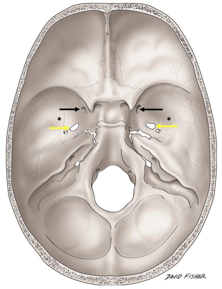
Anatomical Diagram: Superior view (internal aspect) of the human skull base. This medical illustration highlights the bony landmarks and foramina of the anterior, middle, and posterior cranial fossae. Black arrows indicate the bilateral foramina rotundum, while yellow arrows point to the bilateral foramina ovale. Asterisks (*) denote unusual bilateral variant foramina situated approximately 3.1 cm lateral to the standard midline foramina, near the sphenotemporal suture. The diagram clearly depicts the sphenoid bone, temporal bone, and occipital bone transitions, illustrating the spatial relationship between neurovascular exit points and the infratemporal fossa roof. This schematic is designed for osteological study and surgical anatomy education, focusing on anatomical variations within the middle cranial fossa that may be relevant during skull base procedures or diagnostic imaging interpretation.
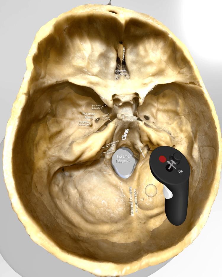
This image displays a highly detailed, three-dimensional digital model of the human skull base, viewed from a superior (internal) perspective. The model is presented within a virtual reality (VR) interface, indicated by the presence of a VR hand controller over the posterior region. Key anatomical landmarks and foramina across the three cranial fossae are clearly labeled. In the anterior cranial fossa, the crista galli and cribriform plate are marked. The middle cranial fossa shows annotations for the foramen rotundum, foramen ovale, foramen spinosum, and the carotid canal. Central landmarks include the sloping clivus and the large, centrally located foramen magnum. Within the posterior cranial fossa, the internal acoustic meatus, jugular foramen, and internal occipital protuberance are identified. This anatomical diagram serves as an educational tool for medical students and neuroanatomy trainees to study the complex spatial relationships of the skull base, including the pathways for cranial nerves and major vasculature.
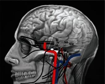
This medical illustration presents a lateral projection of the human cranial region, serving as an anatomical diagram of the skull base, brain, and neurovascular structures. The image overlays a detailed skull rendering with the cerebral hemispheres, emphasizing cortical gyri and sulci. A key feature is the schematic mapping of the twelve cranial nerves (labeled I–XII in Roman numerals), showing their approximate pathways as they exit the brainstem and navigate through skull base foramina. Major vascular structures are highlighted in red (arterial, including the internal carotid artery) and blue (venous), demonstrating their close spatial proximity to the cranial nerves near the zygomatic arch and mandible. The diagram includes orientation labels for 'Anterior' and 'Lateral' perspectives. It is designed for clinical education in neuroanatomy and skull base surgery, illustrating the complex topographic relationships essential for diagnostic imaging and surgical planning in procedures such as microvascular decompression or tumor resection.
mandible anatomy labeled mental foramen
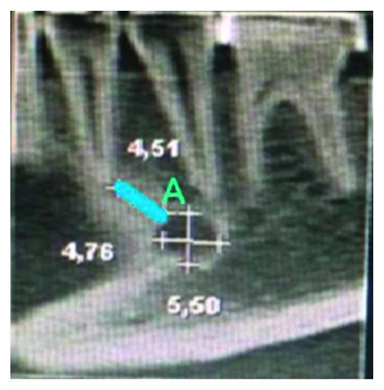
This diagnostic image shows a sagittal view of the human mandible, likely obtained via Cone Beam Computed Tomography (CBCT). The image focuses on the mandibular anatomy and the spatial relationship between the dental roots and the mental foramen (MF). Several lower teeth are visible, characterized by their radiopaque enamel and dentin, with roots extending into the alveolar bone. The mental foramen is identified as a well-defined, ovoid radiolucent area located inferior to the premolar root apices. Quantitative analysis is superimposed on the image: a blue line labeled 'A' measures the vertical distance from the superior cortical border of the mental foramen to the apex of the adjacent tooth root. Numerical annotations (4.51, 4.76, and 5.50) indicate precise morphometric measurements in millimeters for height, length, and distance to anatomical landmarks. This visual is intended for dental and maxillofacial education, specifically regarding preoperative planning for implants or local anesthesia to avoid neurovascular injury.
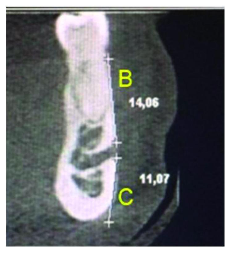
This diagnostic image is a coronal cross-section of a human mandible obtained via Computed Tomography (CT), specifically used for dentomaxillofacial morphometric analysis. The image highlights the vertical position of the mental foramen (MF) relative to local anatomical landmarks. Two linear measurements are annotated: 'Distance B' (labeled 'B') measures 14.06 mm from the superior cortical border of the mental foramen to the alveolar crest; 'Distance C' (labeled 'C') measures 11.07 mm from the superior cortical border of the mental foramen to the inferior base of the mandible. The mental foramen is visible as a radiolucent opening on the vestibular (buccal) aspect of the cortical bone. This type of imaging is clinically significant for preoperative planning in oral surgery, dental implantology, and endodontics to avoid neurovascular injury to the mental nerve. The image clearly demonstrates the cross-sectional anatomy of the mandibular body, including the dense outer cortical bone and internal trabecular patterns.
temporal bone parts petrous mastoid squamous anatomy
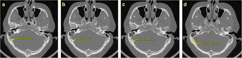
This diagnostic image consists of a series of four high-resolution axial computerized tomography (CT) scans (labeled a–d) of the human skull base, displayed in a bone window. The sequence demonstrates the surgical anatomy of the temporal bone, specifically highlighting the mastoid emissary vein (MEV) and its associated bony canal. In images a, b, and c, yellow arrows trace the course of the mastoid emissary vein as it traverses the mastoid part of the temporal bone. Image d, designated as 'Slice B', identifies the mastoid emissary foramen on the outer cortex. Key landmarks visible across all slices include the petrous and squamous parts of the temporal bone, the mastoid air cells, the sphenoid bone with the sella turcica, and the occipital bone. Annotations in frame d use Greek letters gamma (γ) and delta (δ) to indicate specific measurement points on the inner and outer cortical surfaces, respectively. This visual serves as an anatomical guide for preoperative planning in retrosigmoid approaches to the posterior cranial fossa, helping to avoid intraoperative hemorrhage from emissary veins.
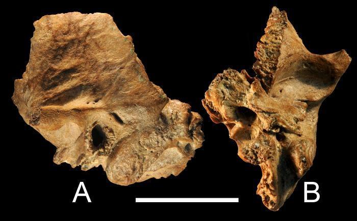
This osteological clinical specimen displays two views of the Xujiayao 15 left temporal bone, representing late archaic human anatomy. View A (lateral) shows the squamous and mastoid portions, highlighting a well-preserved external auditory canal and mastoid process. Key anatomical features include the absence of the zygomatic process and minor chips along the squamous suture. Antero-superiorly, the specimen shows evidence indicative of a prior sutural bone. View B (inferior) provides a perspective of the petrous portion and the skull base surface. This view demonstrates that the medial petrous tip is missing, while the tympanic bone and the inferior aspect of the auditory canal remain largely intact. The specimen exhibits minor osseous changes, specifically Grade 1 External Auditory Exostoses (EAE) characterized by small knobs of bone and surface irregularities along the posterior and posteroinferior tympanic bone, suggesting chronic inflammation or environmental exposure.
Complete University Practical Guide: Maxilla, Mandible, Skull Base & Temporal Bone
HOW TO PRESENT A BONE TO AN EXAMINER - GENERAL FRAMEWORK
1. MAXILLA
Identification & Type
- Paired bone; forms the upper jaw
- Type: Irregular, pneumatic (contains maxillary sinus/antrum of Highmore)
Orientation
- Alveolar process (tooth-bearing margin) downward
- Orbital surface facing upward
- Anterior/facial surface facing you
- The frontal process goes upward toward the nose
Parts of the Maxilla (4 Processes + 1 Body)
| Part | What it does |
|---|---|
| Body | Central mass; contains the maxillary sinus |
| Frontal process | Goes up to articulate with frontal bone |
| Zygomatic process | Goes laterally to meet zygomatic bone |
| Alveolar process | Contains upper teeth sockets (8 per side) |
| Palatine process | Goes medially to form anterior 3/4 of hard palate |
Four Surfaces of the Body
- Anterior (facial) surface - faces forward
- Posterior (infratemporal) surface - faces the infratemporal fossa; has the posterior superior alveolar foramina
- Superior (orbital) surface - forms floor of orbit; carries the infraorbital groove/canal
- Medial (nasal) surface - forms lateral wall of nasal cavity; has the maxillary hiatus
Key Foramina and Their Contents
| Foramen | Location | Contents |
|---|---|---|
| Infraorbital foramen | Anterior surface, ~1 cm below orbital margin, in line with pupil | Infraorbital nerve (V2 branch), infraorbital artery & vein |
| Posterior superior alveolar foramina | Infratemporal (posterior) surface | Posterior superior alveolar nerve & vessels (branch of V2) |
| Anterior superior alveolar canal | Within anterior wall of maxillary sinus | Anterior superior alveolar nerve & vessels |
| Incisive canal | Anterior part of palate (when palatine processes of both maxillae meet) | Nasopalatine nerve (from V2), terminal branch of greater palatine artery |
Maxillary Sinus (Antrum of Highmore)
- Largest paranasal sinus
- Drains via maxillary hiatus into the middle meatus of the nasal cavity
- The hiatus is placed high on the medial wall - poor drainage when upright
- Floor is related to roots of upper molar teeth (especially 1st molar)
- Clinical point: Dental extractions can create an oroantral fistula
Articulations (9 bones)
Muscle Attachments
- Masseter (zygomatic process origin)
- Orbicularis oris, levator labii superioris, levator anguli oris (anterior surface)
- Buccinator (alveolar process)
Potential Viva Questions - Maxilla
- Le Fort I: Horizontal above tooth apices (floats the palate)
- Le Fort II: Pyramidal, through nasal bones, orbital floor, zygomaticomaxillary suture
- Le Fort III: Craniofacial dysjunction - separates whole face from skull
2. MANDIBLE
Identification & Type
- Single unpaired bone (only movable bone of the skull)
- Type: Irregular bone
- Ossification: Intramembranous (body) + endochondral (head/condyle via Meckel's cartilage)
Orientation
- Alveolar arch (tooth-bearing) upward
- Mental protuberance facing you anteriorly
- The two rami going posteriorly and upward
- The base (lower border) forming the inferior margin
Parts of the Mandible
Body
- Has a base (lower border) and alveolar part (upper, carries 16 teeth)
- Two parts meet at midline symphysis menti (in adults, this is fused)
- Mental protuberance - midline bony prominence anteriorly
- Mental tubercles - lateral to the protuberance
- Oblique line - runs from the ramus anteriorly onto the body; attachments for depressors of lower lip
- Mylohyoid line - on medial surface; attachment for mylohyoid muscle (floor of mouth)
- Genial tubercles (mental spines) - midline on the inner surface; upper pair = genioglossus; lower pair = geniohyoid
- Digastric fossa - on either side of midline, inner surface of base; attachment for anterior belly of digastric
- Sublingual fossa - above mylohyoid line = sublingual salivary gland
- Submandibular fossa - below mylohyoid line = submandibular salivary gland
Ramus
- Quadrangular flat plate
- Has coronoid process (anterior, triangular, for temporalis muscle)
- Has condylar process (posterior) = neck + head (condyle)
- Condyle participates in the temporomandibular joint
- Mandibular notch (sigmoid notch) - between coronoid and condylar processes; masseteric nerve and vessels pass through
- Angle of mandible - junction of posterior and inferior borders of ramus (antigonial notching in acromegaly)
- Pterygoid fovea - depression on front of condylar neck; attachment for lateral pterygoid
Key Foramina and Their Contents
| Foramen | Location | Contents |
|---|---|---|
| Mental foramen | Lateral surface of body, below and between premolar roots; at midpoint between upper and lower border | Mental nerve (branch of inferior alveolar nerve, V3), mental artery & vein |
| Mandibular foramen | Medial surface of ramus, guarded anteriorly by lingula | Inferior alveolar nerve (V3), inferior alveolar artery & vein |
Lingula
- Triangular bony projection just anterosuperior to mandibular foramen
- Gives attachment to the sphenomandibular ligament
- This is the landmark for inferior alveolar nerve block (dental injection)
Articulations
- Temporal bone (at temporomandibular joint) - ONLY joint of mandible
- No other bone articulations (all others via teeth/alveolar processes)
Muscle Attachments
| Surface/Process | Muscle |
|---|---|
| Coronoid process | Temporalis |
| Lateral surface of ramus | Masseter |
| Medial surface of ramus | Medial pterygoid |
| Pterygoid fovea (neck) | Lateral pterygoid |
| Genial tubercles (upper) | Genioglossus |
| Genial tubercles (lower) | Geniohyoid |
| Mylohyoid line | Mylohyoid |
| Digastric fossa | Anterior belly of digastric |
Potential Viva Questions - Mandible
3. SKULL BASE (Base of Skull)
Identification & Overview
- The floor of the cranial cavity
- Divided into 3 fossae: Anterior, Middle, and Posterior
- Formed by 5 bones: Frontal, Ethmoid, Sphenoid, Temporal, Occipital
Orientation
- Internal (endocranial) surface - faces upward, contains the 3 fossae at different levels
- External (exocranial) surface - faces downward, forms the roof of the nasopharynx, infratemporal fossa, etc.
- The anterior fossa is the highest level; posterior fossa is the lowest
Skull Base Diagram (Internal View)
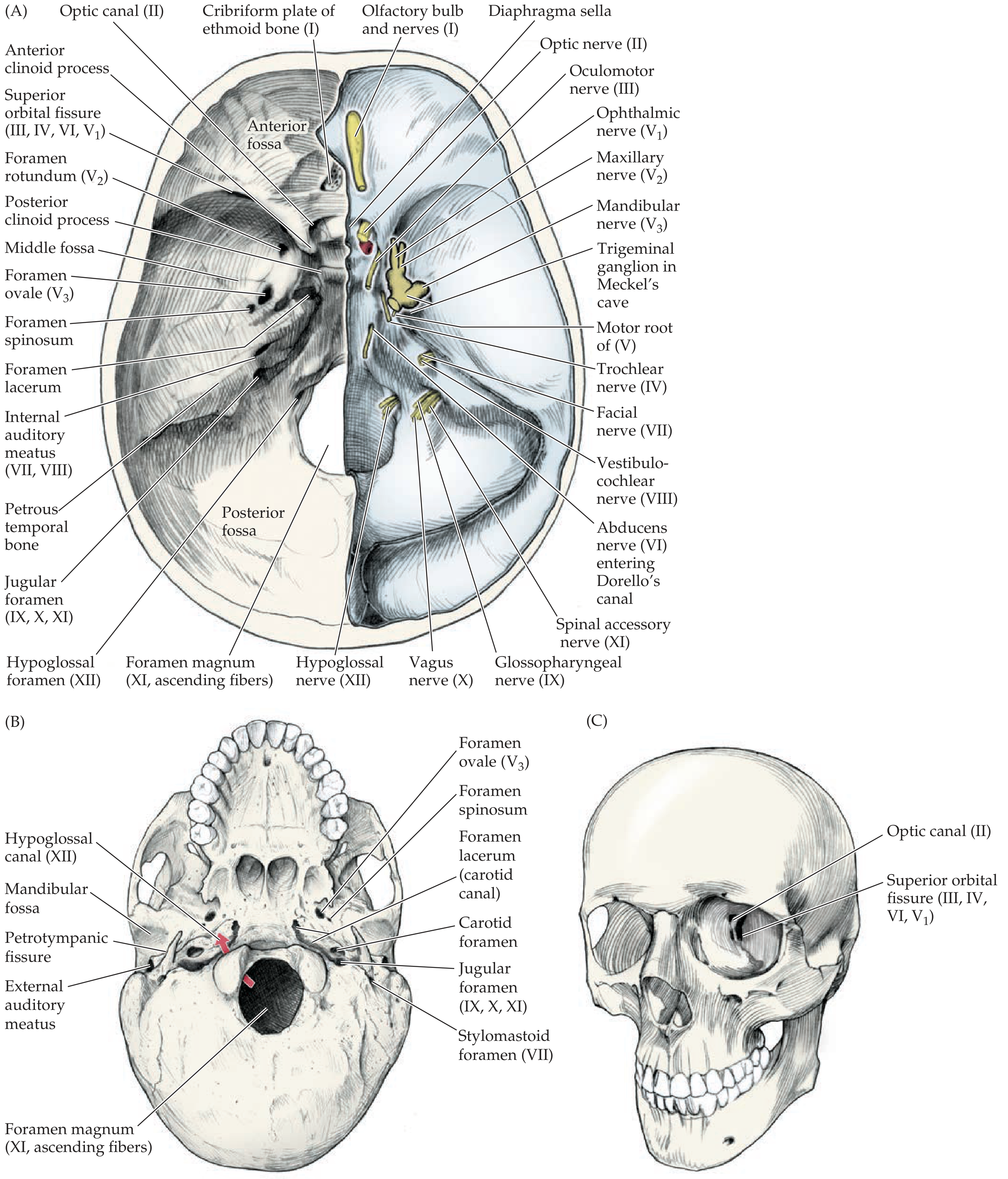
ANTERIOR CRANIAL FOSSA
- Anterior/lateral: Frontal bone
- Posterior: Lesser wings of sphenoid (forming the sphenoidal ridges/anterior clinoid processes)
- Floor: Orbital plates of frontal, cribriform plate of ethmoid, planum sphenoidale
Foramina of Anterior Fossa
| Foramen | Bone | Contents |
|---|---|---|
| Cribriform plate foramina (numerous small holes) | Ethmoid | Olfactory nerve filaments (CN I); anterior ethmoidal nerve and vessels |
| Foramen cecum | Between frontal and ethmoid | Emissary vein connecting nasal veins to superior sagittal sinus (in children) |
| Optic canal (strictly posterior border/middle fossa entry) | Sphenoid, lesser wing | Optic nerve (CN II), ophthalmic artery |
- Crista galli - midline vertical crest of ethmoid; falx cerebri attaches here
- Foramen cecum - small, anterior to crista galli; usually a blind pit
MIDDLE CRANIAL FOSSA
- Anterior: Lesser wings of sphenoid, anterior clinoid processes
- Posterior: Superior border of petrous temporal + dorsum sellae
- Floor: Greater wings of sphenoid, squamous temporal, petrous temporal anterior surface
- Sella turcica - the Turkish saddle; houses the pituitary gland
- Anterior clinoid processes - attachment for tentorium cerebelli
- Cavernous sinus - lateral to sella turcica; contains CN III, IV, V1, V2, VI and internal carotid artery
Foramina of Middle Cranial Fossa
| Foramen | Bone | Contents |
|---|---|---|
| Optic canal | Between roots of lesser wing of sphenoid | CN II (optic nerve), ophthalmic artery (branch of ICA) |
| Superior orbital fissure | Between greater and lesser wings of sphenoid | CN III (oculomotor), CN IV (trochlear), CN VI (abducens), CN V1 (ophthalmic), ophthalmic veins, sympathetic fibers |
| Foramen rotundum | Greater wing of sphenoid | CN V2 (maxillary nerve) |
| Foramen ovale | Greater wing of sphenoid | CN V3 (mandibular nerve), accessory meningeal artery, lesser petrosal nerve (occasionally) |
| Foramen spinosum | Greater wing of sphenoid, near spine of sphenoid | Middle meningeal artery & vein, meningeal branch of V3 |
| Foramen lacerum | Junction of sphenoid, temporal, and occipital | Mostly cartilage in life - internal carotid artery traverses it (superior opening); lesser petrosal nerve; meningeal branches |
| Carotid canal | Petrous temporal | Internal carotid artery with its sympathetic plexus, caroticotympanic nerves |
| Hiatus of facial canal (hiatus for greater petrosal nerve) | Anterior surface of petrous temporal | Greater superficial petrosal nerve (branch of CN VII) |
| Hiatus for lesser petrosal nerve | Anterior surface of petrous temporal (lateral to above) | Lesser petrosal nerve (branch of CN IX - Jacobson's nerve) |
| Arcuate eminence | Petrous temporal | Overlies the superior semicircular canal |
POSTERIOR CRANIAL FOSSA
- Anterior: Clivus (dorsum sellae + basilar occipital), petrous temporal
- Posterior/floor: Occipital bone
- Roof: Tentorium cerebelli (dural fold)
- Clivus - sloping surface formed by dorsum sellae + basilar part of occipital; pons and medulla rest here
- Internal occipital protuberance - midline, attachment for falx cerebri, tentorium cerebelli; confluence of sinuses (torcular Herophili)
- Groove for transverse sinus - horizontal
- Groove for sigmoid sinus - S-shaped, leads to jugular foramen
Foramina of Posterior Cranial Fossa
| Foramen | Bone | Contents |
|---|---|---|
| Internal acoustic meatus (IAM) | Petrous temporal | CN VII (facial), CN VIII (vestibulocochlear), labyrinthine artery |
| Jugular foramen | Between petrous temporal and occipital | CN IX (glossopharyngeal), CN X (vagus), CN XI (accessory); sigmoid sinus becomes internal jugular vein; inferior petrosal sinus |
| Hypoglossal canal | Occipital (condylar part) | CN XII (hypoglossal nerve), meningeal branch of ascending pharyngeal artery |
| Foramen magnum | Occipital bone | Medulla oblongata becoming spinal cord, meninges, vertebral arteries, anterior and posterior spinal arteries, spinal roots of CN XI (ascending), apical ligament of dens |
| Condylar canal | Behind each occipital condyle | Emissary vein (connects sigmoid sinus to sub-occipital venous plexus) |
Potential Viva Questions - Skull Base
- Anterior: Frontal (orbital plates), ethmoid (cribriform plate), sphenoid (lesser wings)
- Middle: Sphenoid (body + greater wings), temporal (squamous + petrous)
- Posterior: Occipital mainly; also petrous temporal, small sphenoid (dorsum sellae/clivus)
4. TEMPORAL BONE
Identification & Type
- Paired bone (left and right)
- Type: Irregular, pneumatic (contains mastoid air cells)
- The most complex bone in the skull
Orientation
- Hold it with the external acoustic meatus (ear canal opening) facing you
- Mastoid process pointing downward and posteriorly
- Styloid process pointing downward anteriorly (in front of mastoid)
- Petrous part points anteromedially like a wedge/pyramid
Four Parts of the Temporal Bone
1. Squamous Part
- Large, flat, convex plate forming the upper anterior temporal bone
- Contributes to lateral wall of cranium
- Zygomatic process projects anteriorly from its lower surface, forming the zygomatic arch with the temporal process of the zygomatic bone
- Inferior surface = mandibular fossa + articular tubercle (for TMJ)
- On medial (inner) surface: groove for middle temporal artery
- Suprameatal (Macewen's) triangle - posterosuperior to external acoustic meatus; surgical landmark for mastoid antrum (used in cortical mastoidectomy)
- Temporal line = suprameatal crest = marks lower limit of temporalis muscle origin
2. Petrous Part ("Pyramid")
- Wedge-shaped dense bone housing the inner ear organs
- Directed anteromedially; its apex points toward the body of the sphenoid
- Anterior surface faces middle cranial fossa; features:
- Arcuate eminence (over superior semicircular canal)
- Tegmen tympani (roof of middle ear)
- Hiatus for greater petrosal nerve (Facial nerve branch)
- Hiatus for lesser petrosal nerve (CN IX branch)
- Posterior surface faces posterior cranial fossa:
- Internal acoustic meatus - CN VII, VIII, labyrinthine artery
- Subarcuate fossa (for a dural fold, larger in neonates)
- Aperture of vestibular aqueduct (endolymphatic duct)
- Inferior surface (base of skull):
- Stylomastoid foramen (between styloid and mastoid processes)
- Carotid canal (internal carotid artery)
- Jugular fossa (forms roof of jugular foramen with occipital bone)
- Mastoid canaliculus (Arnold's nerve = auricular branch of CN X)
- Petrous canaliculus (Jacobson's nerve = tympanic branch of CN IX)
- Superior border: groove for superior petrosal sinus
- Posterior border: groove for inferior petrosal sinus; sigmoid sinus groove
3. Tympanic Part
- C-shaped curved plate of bone
- Forms the anterior wall, floor, and part of posterior wall of the external acoustic meatus (bony EAC)
- Medially: tympanic sulcus - a groove where the fibrous annulus of the tympanic membrane attaches
- Anteroinferiorly: contributes to the posterior boundary of the mandibular fossa
- Sutures formed in EAC:
- Tympanosquamous suture (anterior)
- Tympanomastoid suture (posterior) - inferior aspect is landmark for identifying facial nerve as it exits stylomastoid foramen
4. Styloid Part
- Long, slender, pointed process projecting downward and forward
- Three muscles (all involved in swallowing): stylopharyngeus (CN IX), stylohyoid (CN VII), styloglossus (CN XII)
- Two ligaments: stylohyoid ligament, stylomandibular ligament
- "Stylopharyngeus Stylohyoid Styloglossus - All swallowing muscles"
Key Foramina of the Temporal Bone
| Foramen | Location | Contents |
|---|---|---|
| External acoustic meatus | Tympanic part | Air (ear canal to tympanic membrane) |
| Internal acoustic meatus | Posterior surface of petrous part | CN VII, CN VIII, labyrinthine artery |
| Stylomastoid foramen | Between styloid and mastoid processes (inferior surface) | Facial nerve (CN VII) exits skull here; stylomastoid artery enters |
| Carotid canal | Inferior surface of petrous part | Internal carotid artery with caroticotympanic nerves and sympathetic plexus |
| Jugular foramen | Between petrous temporal and occipital | CN IX, X, XI; sigmoid sinus → IJV; inferior petrosal sinus |
| Mastoid foramen | Mastoid part, near occipitomastoid suture | Mastoid emissary vein (connects sigmoid sinus to occipital veins); meningeal branch of occipital artery |
| Petrotympanic fissure (Glaserian fissure) | Between petrous and tympanic parts, visible on inferior surface | Chorda tympani (branch of CN VII) exits here; anterior tympanic artery |
| Hiatus for greater petrosal nerve | Anterior surface of petrous | Greater superficial petrosal nerve (preganglionic parasympathetic for lacrimal, nasal glands) |
| Hiatus for lesser petrosal nerve | Anterior surface of petrous (lateral to above) | Lesser petrosal nerve (preganglionic parasympathetic for parotid gland) |
| Mastoid canaliculus | Lateral wall of jugular fossa | Auricular branch of vagus (Arnold's nerve) |
| Aperture of vestibular aqueduct | Posterior surface of petrous | Endolymphatic duct |
| Cochlear canaliculus | Inferior surface near apex | Perilymphatic duct |
Sutures of the Temporal Bone
| Suture | Articulates with |
|---|---|
| Squamosal suture | Parietal bone |
| Sphenosquamous suture | Greater wing of sphenoid |
| Parietomastoid suture | Parietal bone |
| Occipitomastoid suture | Occipital bone |
| Tympanosquamous suture | Internal suture within temporal bone |
| Petrotympanic fissure | Internal (petrous and tympanic parts) |
The Mastoid Process
- Projects inferiorly from the mastoid part
- Contains mastoid air cells (pneumatized in adults)
- Develops after birth (absent at birth - that's why the facial nerve is superficial in neonates)
- Mastoid process develops as SCM muscle grows and pulls the bone down (2nd year of life)
- Sigmoid sinus grooves the inner surface
- Attached muscles: SCM (main), splenius capitis, longissimus capitis
- McEwen's (Macewen's/Suprameatal) Triangle - posterosuperior to EAC; lies over the mastoid antrum; entry point in cortical mastoidectomy
Pterion (Relevant to Temporal Bone)
- Formed by 4 bones meeting in an H-shape: frontal + parietal + greater wing of sphenoid + squamous temporal
- The squamous temporal is involved; thinnest part of skull
- Middle meningeal artery runs just deep to it
- Clinical: epidural hematoma after trauma here
Potential Viva Questions - Temporal Bone
QUICK COMPARISON TABLE FOR ALL BONES
| Feature | Maxilla | Mandible | Temporal Bone | Skull Base |
|---|---|---|---|---|
| Type | Irregular, pneumatic | Irregular | Irregular, pneumatic | Composite |
| Paired? | Yes (paired) | No (unpaired) | Yes (paired) | N/A |
| Joint | Immovable (sutures) | TMJ (synovial) | TMJ | N/A |
| Key foramen | Infraorbital | Mental, Mandibular | Stylomastoid, IAM | Foramen magnum |
| Key nerve exiting | V2 (infraorbital) | V3 (mental, IA) | VII (facial) | Multiple CNs |
| Sinus | Maxillary sinus | None | Mastoid air cells | Cavernous sinus |
| Key clinical | Le Fort fractures, sinusitis | Dental blocks, TMD | Facial palsy, CSOM | Skull base tumors, meningitis |
MASTER FORAMEN SUMMARY TABLE (for the Examiner)
| Foramen | Bone/Fossa | Contents |
|---|---|---|
| Cribriform plate | Ethmoid / ACF | CN I (olfactory filaments), anterior ethmoidal n. & vessels |
| Optic canal | Sphenoid / ACF-MCF border | CN II, ophthalmic artery |
| Superior orbital fissure | Sphenoid / MCF | CN III, IV, VI, V1, ophthalmic veins |
| Foramen rotundum | Sphenoid / MCF | CN V2 (maxillary) |
| Foramen ovale | Sphenoid / MCF | CN V3 (mandibular), accessory meningeal artery, lesser petrosal nerve |
| Foramen spinosum | Sphenoid / MCF | Middle meningeal artery & vein, nervus spinosus (meningeal V3) |
| Foramen lacerum | Sphenoid-temporal-occipital junction | Fibrocartilage; ICA transits its roof |
| Carotid canal | Petrous temporal / MCF-base | ICA + sympathetic plexus |
| Hiatus for greater petrosal n. | Petrous temporal / MCF | Greater petrosal nerve (CN VII branch) |
| Internal acoustic meatus | Petrous temporal / PCF | CN VII, CN VIII, labyrinthine artery |
| Jugular foramen | Petrous-occipital / PCF | CN IX, X, XI; sigmoid sinus → IJV; inferior petrosal sinus |
| Hypoglossal canal | Occipital / PCF | CN XII |
| Foramen magnum | Occipital / PCF | Spinal cord, vertebral arteries, anterior & posterior spinal arteries, spinal CN XI |
| Stylomastoid foramen | Temporal (inferior) | CN VII (exit), stylomastoid artery |
| Petrotympanic fissure | Temporal (inferior) | Chorda tympani (CN VII branch), anterior tympanic artery |
| Mental foramen | Mandible body | Mental nerve (V3 branch), mental artery & vein |
| Mandibular foramen | Mandible ramus (medial) | Inferior alveolar nerve & vessels (V3) |
| Infraorbital foramen | Maxilla (anterior surface) | Infraorbital nerve (V2), infraorbital artery & vein |
TOP EXAMINER TIPS
-
Always identify left vs. right when presenting a paired bone - state your reasoning ("this is the left temporal bone because the mastoid process is on this side and the petrous apex points in this direction...")
-
Use anatomical terms - say "anterior surface" not "front side," "superior" not "top," "alveolar process" not "tooth area"
-
Foramina questions are favorites - always know the foramen, its bone, fossa, and contents (nerve + artery + vein where applicable)
-
Clinical correlations score marks - mention Le Fort for maxilla, dental blocks for mandible, facial nerve palsy for temporal bone, extradural hematoma for pterion/middle meningeal artery
-
Embryology gets bonus marks - the mandible is primarily intramembranous; the petrous temporal is endochondral (from otic capsule); the maxilla is intramembranous
-
Mnemonic for cranial fossae foramina:
- ACF: "Cribriform plate = CN I, Optic canal = CN II"
- MCF: "Round = V2, Oval = V3, Spinosum = MMA, SOF = III, IV, VI, V1"
- PCF: "IAM = VII + VIII, Jugular = IX + X + XI, Hypoglossal = XII, Magnum = cord + vertebrals"
Generate images of each and every bone and give me orientation accordingly. If possible give video related to it
Detailed anatomical illustration of the human Maxilla bone showing all surfaces and features. Show anterior (facial) surface with infraorbital foramen labeled, frontal process going upward, zygomatic process going laterally, alveolar process with tooth sockets at bottom, palatine process extending medially. Also show the posterior/infratemporal surface with posterior superior alveolar foramina. Label all key structures: infraorbital foramen, infraorbital groove, orbital surface (floor of orbit), anterior nasal spine, nasal notch, incisive fossa, canine fossa, canine eminence, zygomatic process, frontal process, alveolar process, palatine process, maxillary sinus (antrum of Highmore) shown in cross-section view. Include orientation arrows showing superior, inferior, anterior, posterior. Medical textbook style, white background, clean professional anatomical drawing with clear labels and leader lines.
Detailed anatomical illustration of the human Mandible bone showing both lateral/external view and medial/internal view side by side. External/lateral view: label mental protuberance, mental tubercles, mental foramen, oblique line, body of mandible, base of mandible, angle of mandible, ramus, coronoid process, condylar process (head and neck), mandibular notch (sigmoid notch), pterygoid fovea. Internal/medial view: label mandibular foramen, lingula, mylohyoid groove, mylohyoid line, genial tubercles (mental spines - upper and lower), digastric fossa, sublingual fossa, submandibular fossa. Include orientation arrows. Show alveolar part at top with tooth sockets. Medical textbook style anatomical drawing, white background, clear professional labels with leader lines, educational illustration quality.
Detailed anatomical illustration of the internal (endocranial/superior) view of the Skull Base showing all three cranial fossae. Clearly divide and label: ANTERIOR CRANIAL FOSSA (highest level, frontal color), MIDDLE CRANIAL FOSSA (intermediate level, different color), POSTERIOR CRANIAL FOSSA (lowest level, third color). Label all foramina with their contents: Cribriform plate (CN I), Foramen cecum, Optic canal (CN II, ophthalmic artery), Superior orbital fissure (CN III IV VI V1), Foramen rotundum (V2), Foramen ovale (V3), Foramen spinosum (middle meningeal artery), Foramen lacerum, Carotid canal, Internal acoustic meatus (CN VII VIII), Jugular foramen (CN IX X XI), Hypoglossal canal (CN XII), Foramen magnum (spinal cord vertebral arteries). Label key structures: crista galli, sella turcica, dorsum sellae, clivus, anterior clinoid processes, posterior clinoid processes, petrous temporal bone, arcuate eminence, tegmen tympani. Medical textbook style, color-coded fossae, white background, professional anatomical illustration with clear labels.
Detailed anatomical illustration of the human Temporal Bone showing multiple views. Show: (1) Lateral external view with squamous part, zygomatic process, external acoustic meatus, mastoid process, styloid process, suprameatal triangle (Macewen's triangle) labeled, tympanic part, squamosal suture, parietomastoid suture. (2) Inferior view showing stylomastoid foramen, carotid canal, jugular fossa, petrotympanic fissure (Glaserian fissure), mandibular fossa, articular tubercle, mastoid canaliculus, tympanic sulcus. (3) Medial/petrous view showing internal acoustic meatus, arcuate eminence, tegmen tympani, hiatus for greater petrosal nerve, hiatus for lesser petrosal nerve, apex of petrous, groove for superior petrosal sinus. Label all four parts: Squamous part, Petrous part, Tympanic part, Styloid part, Mastoid part. Include orientation arrows. Medical textbook style anatomical drawing, white background, clean professional labels with leader lines.
temporal bone anatomy orientation practical examination YouTube video
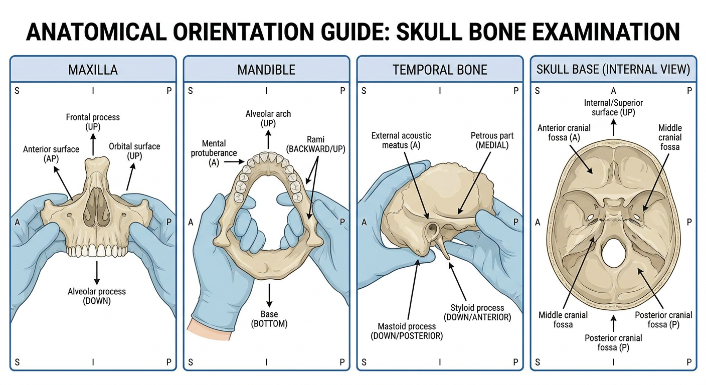
Anatomical orientation guide illustration showing how to correctly hold and orient each skull bone for examination. Four panels side by side: Panel 1 - Maxilla: show hands holding the maxilla with alveolar process (tooth sockets) pointing DOWN, orbital surface facing UP, frontal process going UP, anterior surface facing the examiner. Arrows and labels showing correct orientation. Panel 2 - Mandible: show hands holding mandible with alveolar arch pointing UP, mental protuberance facing examiner anteriorly, rami going backward and upward, base at bottom. Panel 3 - Temporal bone: show holding with external acoustic meatus (ear canal) facing examiner, mastoid process pointing downward-posteriorly, styloid process pointing downward-anteriorly, petrous part pointing medially. Panel 4 - Skull base: show internal/superior surface facing up showing all three fossae. Each panel has clear orientation labels (S=Superior I=Inferior A=Anterior P=Posterior). Medical education style, clean white background, professional illustration.
mandible maxilla skull base anatomy university practical viva YouTube
BONE IMAGES WITH ORIENTATION GUIDE
1. MAXILLA - Labeled Anatomy
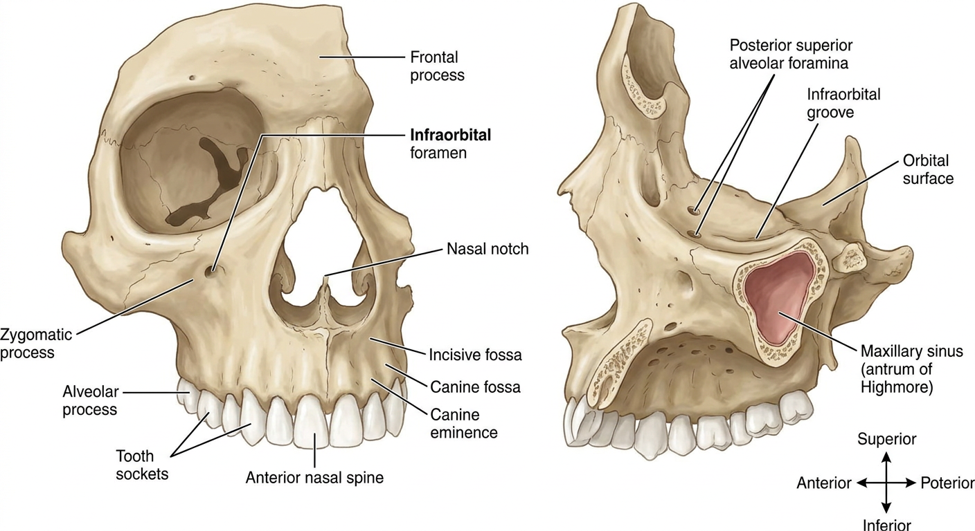
How to Orient the Maxilla in Front of the Examiner
- Alveolar process (tooth sockets) → pointing DOWNWARD
- Frontal process (nasal side) → pointing UPWARD
- Anterior/facial surface → facing YOU (the examiner)
- Orbital plate → facing UPWARD and backward
- Palatine process → going medially (toward the opposite side)
2. MANDIBLE - Labeled Anatomy
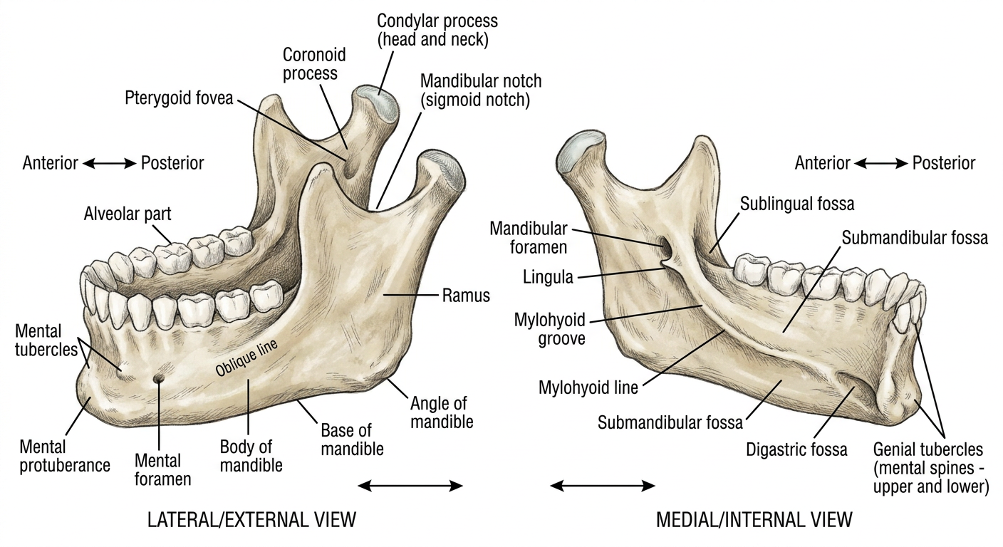
How to Orient the Mandible in Front of the Examiner
- Alveolar arch (tooth sockets) → pointing UPWARD
- Mental protuberance (chin bump) → facing the EXAMINER anteriorly
- Two rami → going POSTERIORLY and UPWARD
- Base (inferior border) → forming the BOTTOM
- Coronoid processes → the flat anterior projections at top of rami
- Condylar processes (condyles) → the posterior projections at top of rami
3. SKULL BASE - Internal View with All Foramina
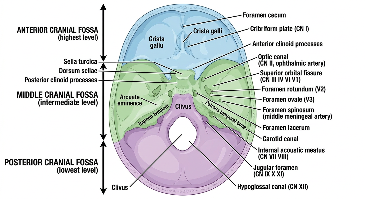
How to Orient the Skull Base
- Internal (endocranial) surface → facing UPWARD toward you
- Anterior fossa → at the FRONT (highest level)
- Middle fossa → MIDDLE (two wing-like depressions on either side of sella turcica)
- Posterior fossa → at the BACK (lowest, largest)
- Crista galli → at the very front midline
- Foramen magnum → at the very back midline
4. TEMPORAL BONE - All Views
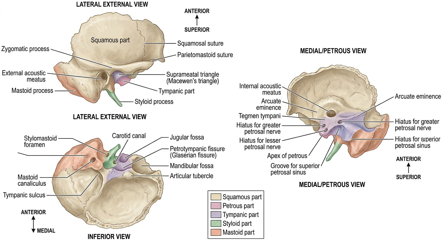
How to Orient the Temporal Bone
- External acoustic meatus (ear canal opening) → facing LATERALLY toward the examiner
- Mastoid process → pointing DOWNWARD and POSTERIORLY
- Styloid process → pointing DOWNWARD and ANTERIORLY (in front of mastoid)
- Petrous part/apex → pointing ANTEROMEDIALLY (like a wedge going inward)
- Squamous part → flat, curving UPWARD and ANTERIORLY
- Zygomatic process → projecting ANTERIORLY from below the squamous part
5. ORIENTATION GUIDE - All Four Bones Together
VIDEO RESOURCES
QUICK ORIENTATION CHEAT SHEET
| Bone | Key "up" structure | Key "facing you" surface | How to identify side |
|---|---|---|---|
| Maxilla | Frontal process | Anterior (facial) surface | Frontal process is nasal side; check infraorbital foramen position |
| Mandible | Alveolar arch | Mental protuberance | Oblique line runs from ramus onto body on each side |
| Temporal | Squamous part | External acoustic meatus | Mastoid process and EAM are both on the same (lateral) side |
| Skull Base | Crista galli (front) | Internal (endocranial) surface up | Crista galli is anterior; foramen magnum is posterior |