
Write a short note on chronic cholecystitis Refer Robbins
Chronic Cholecystitis
Definition and Epidemiology
Pathogenesis
Morphology
Gross
- The gallbladder may be contracted, normal in size, or enlarged.
- The serosa is usually smooth and glistening but may be dulled by subserosal fibrosis; dense fibrous adhesions may represent sequelae of prior acute inflammation.
- On sectioning, the wall is variably thickened with an opaque gray-white appearance; marked subepithelial and subserosal fibrosis is present.
- The lumen contains green-yellow mucoid bile and usually stones.
- The mucosa itself is generally preserved.
Microscopic
- In mild cases: scattered lymphocytes, plasma cells, and macrophages in the mucosa and subserosal fibrous tissue.
- In more advanced cases: marked subepithelial and subserosal fibrosis with mononuclear cell infiltration.
- Rokitansky-Aschoff sinuses - outpouchings of mucosal epithelium through the gallbladder wall - may be quite prominent and can contain bile pigment stones.
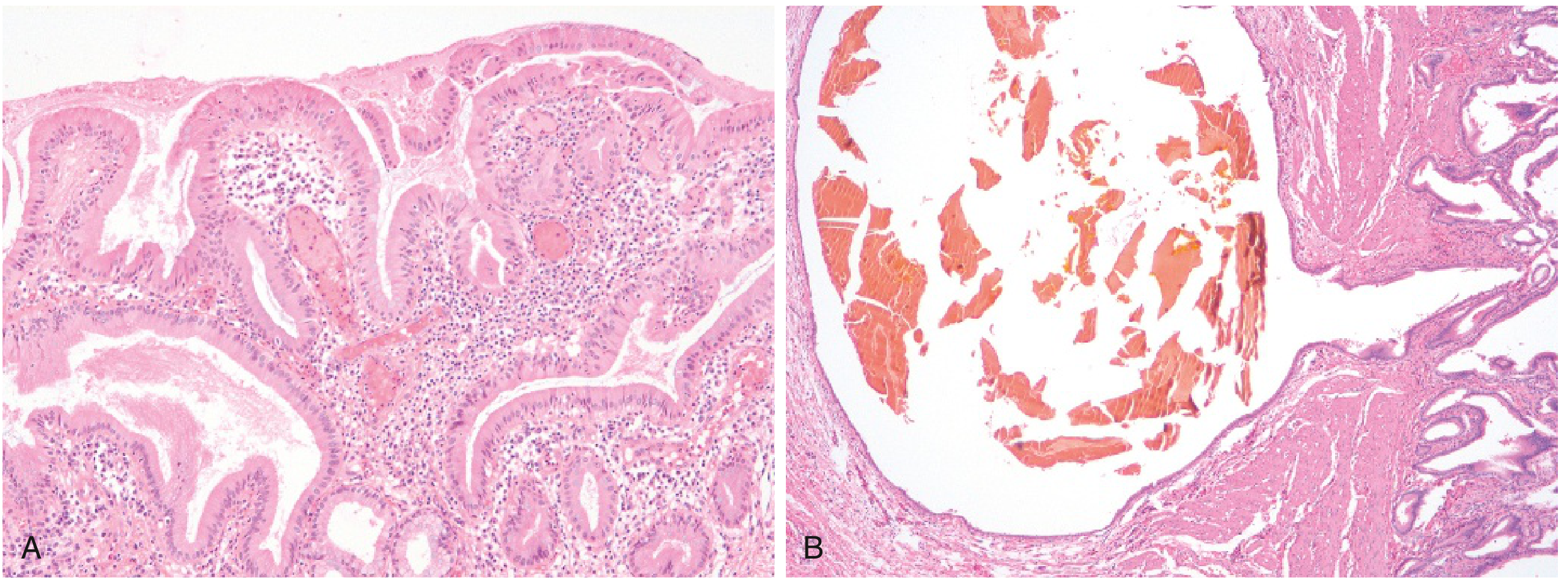
Special Variants
| Variant | Key Feature |
|---|---|
| Porcelain gallbladder | Extensive dystrophic calcification of the gallbladder wall |
| Hyalinizing cholecystitis | Complete replacement of wall and mucosa by dense fibrosis, with or without calcification; associated with increased risk of gallbladder carcinoma |
| Xanthogranulomatous cholecystitis | Massively thickened, shrunken, nodular wall; triggered by rupture of Rokitansky-Aschoff sinuses; foamy lipid-laden xanthoma cells accumulate in wall |
| Hydrops of gallbladder | Atrophic, chronically obstructed, often dilated gallbladder containing only clear secretions |
| IgG4-related sclerosing cholecystitis | A manifestation of IgG4-related fibrosing disease |
- Robbins, Cotran & Kumar Pathologic Basis of Disease, p. 813
Clinical Features
- Recurrent attacks of steady epigastric or right upper quadrant pain
- Nausea and vomiting
- Intolerance for fatty foods
- It is ultimately a pathologic diagnosis based on examination of the resected gallbladder.
Complications
- Bacterial superinfection with cholangitis or sepsis
- Gallbladder perforation leading to local abscess formation or diffuse peritonitis
- Biliary-enteric (cholecystoenteric) fistula - with drainage of bile into adjacent organs, entry of air and bacteria into the biliary tree, and potentially gallstone-induced intestinal obstruction (ileus)
- Aggravation of preexisting cardiac, pulmonary, renal, or hepatic conditions
- Gallbladder carcinoma - the most feared long-term complication
Give me a hard drawn diagram for above question
Hand-drawn medical diagram style sketch on white paper showing chronic cholecystitis, like a student's notebook drawing. Include: 1. A cross-section diagram of a thickened gallbladder wall with labels pointing to: - Thickened fibrotic wall - Rokitansky-Aschoff sinuses (mucosal outpouchings through the wall) - Chronic inflammatory infiltrate (lymphocytes, plasma cells, macrophages) - Gallstone in lumen - Subepithelial and subserosal fibrosis - Preserved mucosa 2. A small inset box showing microscopic histology sketch with: - Mucosal epithelium - Lymphocyte infiltration - Rokitansky-Aschoff sinus with bile 3. A summary box listing variants: - Porcelain gallbladder (calcification) - Hyalinizing cholecystitis - Xanthogranulomatous cholecystitis (xanthoma cells) - Hydrops of gallbladder 4. Clinical features listed on the side: - RUQ pain (recurrent) - Nausea, vomiting - Fat intolerance Title at top: "CHRONIC CHOLECYSTITIS - Robbins" All in pencil/pen sketch style with neat handwritten labels and arrows, like a medical student's diagram notes
400, message: Got more than 4194304 bytes when reading: b'data: {"candidates": [{"content": {"role": "model","parts": [{"inlineData": {"mimeType": "image/png"...'.
We encountered an error while processing your request. Please try again or reach out to us for help.
Give me a hand drawn diagram
Hand-drawn pencil sketch diagram of chronic cholecystitis on white paper, medical student notebook style. Show a cross-section of the gallbladder with labeled arrows pointing to: thickened fibrotic wall, gallstones in lumen, Rokitansky-Aschoff sinuses (mucosal outpouchings), chronic inflammatory cells (lymphocytes and plasma cells), subepithelial fibrosis, and preserved mucosa. Include a small inset microscopy sketch showing lymphocyte infiltration and a Rokitansky-Aschoff sinus containing bile. Add handwritten labels and neat pencil arrows throughout. Title at top reads "Chronic Cholecystitis"
400, message: Got more than 4194304 bytes when reading: b'data: {"candidates": [{"content": {"role": "model","parts": [{"inlineData": {"mimeType": "image/png"...'.
We encountered an error while processing your request. Please try again or reach out to us for help.