
Reading File
I can see this is a BPT Biochemistry exam paper from P P Savani University. Let me clarify what kind of help you need before diving in.
Asking for Preferences
What would you like me to help you with for this exam paper?
I'll answer all questions thoroughly. Let me search the medical library for the key topics simultaneously.
Finding Sources
Finding Sources
Finding Sources
Finding Sources
Finding Sources
Finding Sources
Finding Sources
Finding Sources
I have found good textbook sources. Now let me read the key sections for comprehensive answers.
Reading File
Reading File
Reading File
I have enough excellent textbook source material. Now let me compile thorough answers for all questions.Here are thorough answers to all questions in your P P Savani University BPT Biochemistry paper:
SPPT1070 BIOCHEMISTRY - Complete Answers
SECTION - I
Q-1 (Essay Questions) [10 Marks]
Q-1(i): Hierarchical Levels of Chromosome Packaging with Diagram - Why Essential for Eukaryotic Cells
Eukaryotic cells must pack approximately 2 meters of DNA into a nucleus only ~6 micrometers in diameter. This requires a multi-level hierarchical compaction system.
Level 1 - Nucleosome (Beads on a String)
- DNA wraps around a core of 8 histone proteins (octamer: 2 each of H2A, H2B, H3, H4)
- ~147 base pairs of DNA wrap around each nucleosome
- Adjacent nucleosomes are connected by linker DNA (~20-80 bp) associated with H1 histone
- This reduces the DNA length ~7-fold
- In this state, chromatin looks like "beads on a string" under electron microscopy
Level 2 - 30 nm Fibre (Solenoid)
- Nucleosomes coil and fold further, assisted by histone H1, to form a 30 nm chromatin fibre
- This provides ~40-fold compaction
- Called heterochromatin (inactive/condensed) or euchromatin (active/open)
Level 3 - Loops and Scaffold
- The 30 nm fibre organizes into loop domains (~50,000 bp each) anchored to a protein scaffold
- Creates a 300 nm fibre (radial loop structure)
Level 4 - Coiled Coils / Condensed Chromosome
- Loops further condense and coil into 700-900 nm structures
- At metaphase, fully condensed chromosome (~1,400 nm) is visible
Diagram
DNA double helix (2 nm)
      ↓
Nucleosomes - "beads on a string" (11 nm)
      ↓
30 nm chromatin fibre (solenoid)
      ↓
300 nm loop domains (radial loops)
      ↓
700 nm condensed fibre
      ↓
Metaphase chromosome (1,400 nm)
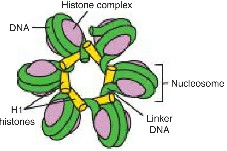
Why Essential for Eukaryotic Cells?
- Physical fit - Without packaging, 2 m of DNA cannot fit in a 6 µm nucleus
- Gene regulation - Euchromatin (loose) = active transcription; heterochromatin (tight) = gene silencing. Histone modifications (acetylation, methylation) control which genes are active
- DNA replication and repair - Organized packaging ensures orderly duplication
- Cell division - Condensed chromosomes are needed for accurate segregation during mitosis/meiosis
- Protection - Packaging protects DNA from physical damage and random transcription
(Source: Langman's Medical Embryology; Kaplan & Sadock's Comprehensive Textbook of Psychiatry)
Q-1(ii): Differentiate Glycogenesis, Glycogenolysis, and Gluconeogenesis as Components of Carbohydrate Metabolism
These three pathways collectively maintain blood glucose homeostasis.
Glycogenesis (Glycogen Synthesis)
| Feature | Detail |
|---|---|
| Definition | Synthesis of glycogen from glucose |
| Location | Liver and skeletal muscle |
| Trigger | High blood glucose, high insulin/glucagon ratio (fed state) |
| Key enzyme | Glycogen synthase (rate-limiting) |
| Starting substrate | Glucose → Glucose-6-phosphate → Glucose-1-phosphate → UDP-glucose → Glycogen |
| Branching enzyme | Amylo-(1,4→1,6)-transglucosylase creates α-1,6 branches |
| Purpose | Store glucose in times of plenty |
| Regulation | Insulin activates; glucagon/epinephrine inhibit via covalent phosphorylation |
Key steps:
- Glucose → Glucose-6-phosphate (hexokinase/glucokinase)
- Glucose-6-P → Glucose-1-phosphate (phosphoglucomutase)
- Glucose-1-P + UTP → UDP-glucose (UDP-glucose pyrophosphorylase)
- UDP-glucose added to glycogen chain by glycogen synthase
- Branching enzyme creates branch points every 8-12 glucose residues
Glycogenolysis (Glycogen Breakdown)
| Feature | Detail |
|---|---|
| Definition | Breakdown of glycogen to release glucose |
| Location | Liver (releases glucose to blood); Muscle (uses glucose locally) |
| Trigger | Fasting, exercise; high glucagon/epinephrine (catabolic state) |
| Key enzyme | Glycogen phosphorylase (rate-limiting) |
| Products | Glucose-1-phosphate (mostly) + free glucose from branch points |
| Debranching enzyme | Removes α-1,6 branches (releasing free glucose) |
| Regulation | Glucagon and epinephrine activate via cAMP → PKA → phosphorylates phosphorylase kinase → activates phosphorylase |
Key steps:
- Glycogen phosphorylase cleaves α-1,4 bonds releasing Glucose-1-phosphate
- Debranching enzyme removes branch points (releases free glucose)
- Glucose-1-P → Glucose-6-P (phosphoglucomutase)
- In liver: Glucose-6-P → Glucose (glucose-6-phosphatase) → released into blood
- In muscle: No glucose-6-phosphatase → glucose-6-P enters glycolysis directly
Gluconeogenesis (New Glucose Formation)
| Feature | Detail |
|---|---|
| Definition | Synthesis of glucose from non-carbohydrate precursors |
| Location | Primarily liver (90%), kidney cortex (10%) |
| Trigger | Prolonged fasting, starvation, intense exercise |
| Precursors | Lactate, pyruvate, glucogenic amino acids, glycerol |
| Key enzymes | Pyruvate carboxylase, PEPCK, Fructose-1,6-bisphosphatase, Glucose-6-phosphatase |
| Purpose | Maintain blood glucose during prolonged fasting when glycogen is depleted |
| Regulation | Glucagon, cortisol, glucocorticoids stimulate; insulin suppresses |
Key bypass reactions (irreversible glycolysis steps bypassed):
- Pyruvate → Oxaloacetate (pyruvate carboxylase, requires biotin)
- OAA → PEP (PEPCK, requires GTP)
- Fructose-1,6-bisphosphate → Fructose-6-phosphate (fructose-1,6-bisphosphatase)
- Glucose-6-phosphate → Glucose (glucose-6-phosphatase)
Comparison Table
| Feature | Glycogenesis | Glycogenolysis | Gluconeogenesis |
|---|---|---|---|
| Process | Synthesis of glycogen | Breakdown of glycogen | Synthesis of new glucose |
| Precursor | Glucose | Glycogen | Lactate, amino acids, glycerol |
| Net result | Glucose stored | Glucose released | Glucose released |
| Organ | Liver + muscle | Liver + muscle | Liver + kidney |
| State | Fed (post-meal) | Short fasting/exercise | Prolonged fasting |
| Key stimulus | Insulin | Glucagon/epinephrine | Glucagon/cortisol |
| Energy cost | Requires ATP/UTP | Releases energy | Requires ATP (energy costly) |
(Source: Biochemistry, 8th ed, Lippincott Illustrated Reviews; Basic Medical Biochemistry - A Clinical Approach, 6e)
Q-2 (Short Notes - Any Two) [10 Marks]
Q-2(i): Key Differences Between Direct ELISA and Sandwich ELISA with Diagram
ELISA (Enzyme-Linked Immunosorbent Assay) is a heterogeneous immunoassay technique where one reactant is bound to a solid phase (microtiter well, magnetic particle, or plastic bead), which facilitates separation of bound and free labelled reactants.
Direct ELISA
- Principle: Antigen is coated directly onto the solid phase. A single enzyme-labelled antibody binds directly to the antigen. No secondary antibody is used.
- Steps:
- Coat microtiter plate with antigen
- Block non-specific binding
- Add enzyme-labelled primary antibody
- Wash away unbound antibody
- Add substrate → color development proportional to antigen amount
- Advantages: Faster, fewer steps, no cross-reactivity from secondary antibody
- Disadvantages: Low sensitivity; primary antibody must be labelled (expensive); no signal amplification
Sandwich ELISA
- Principle: A capture antibody is first coated onto the solid phase. Antigen from sample binds to it. Then a second enzyme-labelled detection antibody binds to a different epitope on the same antigen, forming an antibody-antigen-antibody "sandwich."
- Steps:
- Coat plate with capture antibody
- Add sample (antigen binds to capture antibody)
- Wash
- Add enzyme-labelled detection antibody
- Wash
- Add substrate → colour develops
- Measure optical density
- Advantages: High sensitivity (signal amplification possible); high specificity (two antibodies); can detect very low antigen concentrations
- Disadvantages: Requires two antibodies to different epitopes; more steps; more expensive
Diagrams
DIRECT ELISA:
 ___________Plate___________
|  Ag  Ag  Ag  Ag  Ag  Ag  |
|  |   |   |   |   |   |   |
| Ab* Ab* Ab* Ab* Ab* Ab*  |  (* = enzyme-labelled)
 ---------------------------
         ↓ + Substrate
         Color proportional to antigen

SANDWICH ELISA:
 ___________Plate___________
|  Ab  Ab  Ab  Ab  Ab  Ab  |  (capture antibody)
|  |   |   |   |   |   |   |
|  Ag  Ag  Ag  Ag  Ag  Ag  |  (antigen from sample)
|  |   |   |   |   |   |   |
| Ab* Ab* Ab* Ab* Ab* Ab*  |  (detection antibody + enzyme)
 ---------------------------
         ↓ + Substrate
         Color - high sensitivity
Key Differences Table
| Feature | Direct ELISA | Sandwich ELISA |
|---|---|---|
| Antibodies used | 1 (enzyme-labelled) | 2 (capture + detection) |
| Antigen on plate? | Yes, directly coated | No - captured by antibody |
| Sensitivity | Lower | Higher |
| Specificity | Moderate | High (dual antibody) |
| Steps | Fewer | More |
| Signal amplification | No | Yes (possible) |
| Cost | Lower | Higher |
| Best use | Detecting antibodies; simple antigen quantification | Detecting antigens in complex samples (e.g., serum) |
(Source: Tietz Textbook of Laboratory Medicine, 7th Edition)
Q-2(ii): Protein Metabolism and Significance of Post-Translational Modifications
Protein Metabolism
Protein metabolism includes synthesis (anabolism), digestion, and degradation (catabolism) of proteins in the body.
Digestion of dietary proteins:
- Stomach: Pepsin (works at pH 1.5-2) cleaves peptide bonds; HCl denatures proteins
- Pancreatic enzymes: Trypsin, Chymotrypsin, Elastase (endopeptidases); Carboxypeptidases A and B (exopeptidases)
- Intestinal brush border: Aminopeptidases, Dipeptidases
- Final products: Free amino acids, di- and tripeptides absorbed via active transport (Na+-dependent)
Amino acid catabolism:
- Transamination: Amino group transferred to alpha-ketoglutarate → glutamate (aminotransferases, requires B6/pyridoxal phosphate)
- Oxidative deamination: Glutamate → alpha-ketoglutarate + NH4+ (glutamate dehydrogenase in liver mitochondria)
- Urea cycle: NH4+ is detoxified to urea in liver → excreted by kidneys
- Carbon skeletons enter as pyruvate, acetyl-CoA, TCA intermediates → energy or gluconeogenesis
Nitrogen balance:
- Positive (growth, pregnancy, anabolism)
- Negative (starvation, illness, catabolism)
- Neutral (normal adults)
Post-Translational Modifications (PTMs)
PTMs are covalent modifications of proteins that occur after the ribosome has completed translation. They greatly expand the functional diversity of the proteome (>20,000 genes → >1 million protein forms).
| Modification | Group Added | Enzyme | Significance |
|---|---|---|---|
| Phosphorylation | Phosphate group | Kinases/Phosphatases | Signal transduction, enzyme activation/inhibition (e.g., glycogen phosphorylase) |
| Glycosylation | Sugar moieties | Glycosyltransferases | Protein folding, cell-cell recognition, blood group antigens, immune function |
| Acetylation | Acetyl group | Acetyltransferases | Histone modification → gene regulation; protein stability |
| Ubiquitination | Ubiquitin protein | E1, E2, E3 ligases | Targets protein for proteasomal degradation; controls protein turnover |
| Hydroxylation | Hydroxyl group | Hydroxylases (require Vit C) | Collagen cross-linking and stability (deficient in scurvy) |
| Carboxylation | Carboxyl group | Carboxylases (require Vit K) | Activation of clotting factors II, VII, IX, X |
| Methylation | Methyl group | Methyltransferases | Histone regulation, gene expression |
| Disulfide bond formation | S-S bonds | PDI in ER | Protein tertiary structure and stability (e.g., insulin, immunoglobulins) |
| Cleavage (proteolytic) | Removal of peptide | Proteases | Converts zymogens to active enzymes (e.g., trypsinogen → trypsin; proinsulin → insulin) |
| Lipidation | Lipid group | Lipid transferases | Membrane anchoring (e.g., Ras protein) |
Clinical Significance of PTMs:
- Abnormal phosphorylation: cancer signalling (e.g., BCR-ABL in CML)
- Defective glycosylation: lysosomal storage diseases, CDG syndromes
- Abnormal ubiquitination: neurodegenerative diseases (Parkinson's, Alzheimer's)
- Defective hydroxylation: scurvy (Vitamin C deficiency)
- Defective carboxylation: bleeding disorders (Vitamin K deficiency)
Q-2(iii): β-Oxidation vs ω-Oxidation of Fatty Acids - Pathway, Location, and Energy Yield
β-Oxidation (Beta-Oxidation)
Definition: The primary pathway for fatty acid degradation, occurring at the beta (second) carbon of the fatty acid chain.
Location: Mitochondrial matrix (main site); Peroxisomes (for very long chain fatty acids >22C)
Preparation steps:
- Fatty acid activated to Fatty acyl-CoA by acyl-CoA synthetase (in cytoplasm, uses 2 ATP equivalents)
- Transport into mitochondria via carnitine shuttle (carnitine acyltransferase I on outer membrane, rate-limiting step)
One cycle of β-oxidation (4 reactions):
- Oxidation - FAD-dependent: Acyl-CoA → Trans-Enoyl-CoA (produces FADH2)
- Hydration - Water added: Trans-Enoyl-CoA → L-3-Hydroxyacyl-CoA
- Oxidation - NAD+-dependent: Hydroxyacyl-CoA → 3-Ketoacyl-CoA (produces NADH)
- Thiolysis - CoA added: 3-Ketoacyl-CoA → Acetyl-CoA + shortened Acyl-CoA (by 2 carbons)
Each cycle produces: 1 FADH2 + 1 NADH + 1 Acetyl-CoA
Energy yield for palmitic acid (C16:0):
- 7 cycles of β-oxidation → 7 FADH2 + 7 NADH + 8 Acetyl-CoA
- 7 FADH2 × 1.5 ATP = 10.5 ATP
- 7 NADH × 2.5 ATP = 17.5 ATP
- 8 Acetyl-CoA × 10 ATP (via TCA) = 80 ATP
- Total = 108 ATP - 2 ATP (activation) = 106 net ATP
Regulation: Malonyl-CoA inhibits carnitine acyltransferase I (prevents futile cycle when fatty acid synthesis is active)
ω-Oxidation (Omega-Oxidation)
Definition: Minor pathway of fatty acid oxidation occurring at the omega (last, methyl) carbon.
Location: Smooth endoplasmic reticulum (microsomal fraction) - NOT mitochondria
Pathway:
- Terminal methyl (-CH3) group → hydroxylated to omega-hydroxy fatty acid (by cytochrome P450, requires NADPH and O2)
- Omega-hydroxy fatty acid → oxidized to omega-aldehyde (by alcohol dehydrogenase)
- Omega-aldehyde → oxidized to dicarboxylic acid (by aldehyde dehydrogenase)
- Dicarboxylic acid can undergo β-oxidation from both ends
Energy yield: Low; no direct FADH2/NADH; primarily a detoxification/metabolic route
Significance:
- Minor pathway under normal conditions (<5% of fatty acid oxidation)
- Becomes important when β-oxidation is impaired (e.g., Zellweger syndrome - peroxisomal disorder)
- Generates dicarboxylic acids excreted in urine (dicarboxylic aciduria) - diagnostic marker
- Important for metabolism of certain drugs and xenobiotics
- Medium-chain fatty acids are good substrates
Comparison Table
| Feature | β-Oxidation | ω-Oxidation |
|---|---|---|
| Site of oxidation | Beta (2nd) carbon | Omega (last) carbon |
| Organelle/Location | Mitochondria (mainly); Peroxisomes (VLCFA) | Smooth ER (microsomes) |
| Pathway type | Major pathway | Minor pathway |
| Products | Acetyl-CoA, FADH2, NADH | Dicarboxylic acids |
| Energy yield | High (e.g., 106 ATP from palmitate) | Low (minor energy) |
| Clinical role | Primary energy source from fats | Backup when β-oxidation fails; dicarboxylic aciduria |
| Substrates | All even-chain fatty acids | Medium-chain FA mainly |
| Enzyme system | Acyl-CoA dehydrogenase, enoyl hydratase, etc. | Cytochrome P450 (CYP4A), alcohol/aldehyde dehydrogenase |
Q-3 (Very Short Notes - Any Five) [15 Marks - 3 marks each]
Q-3(i): Role of Essential Minerals in Maintaining Physiological Homeostasis
Essential minerals are inorganic elements required in small amounts that cannot be synthesized by the body.
Macrominerals (required >100 mg/day):
| Mineral | Physiological Role | Homeostasis Function |
|---|---|---|
| Calcium (Ca2+) | Bone/teeth structure, muscle contraction, blood clotting, nerve transmission, second messenger | PTH, calcitonin, Vitamin D regulate blood Ca2+ (8.5-10.5 mg/dL) |
| Phosphorus (P) | Bone/teeth, ATP/ADP/AMP, nucleic acids, phospholipids, phosphorylation reactions | Regulated with Ca2+; PTH increases renal excretion |
| Sodium (Na+) | Major extracellular cation; osmotic pressure, nerve impulse, fluid balance | Aldosterone/ADH/ANP control; maintains ECF volume |
| Potassium (K+) | Major intracellular cation; resting membrane potential (-70mV), cardiac rhythm | Aldosterone regulates; critical for cardiac function |
| Magnesium (Mg2+) | Cofactor for >300 enzymes (ATP-Mg complex), DNA/RNA synthesis, protein synthesis | Bone reservoir; regulated by kidneys |
| Chloride (Cl-) | Osmotic balance, gastric HCl production, HCO3- exchange in RBC | Follows sodium; acid-base balance |
Microminerals (trace elements):
| Mineral | Role |
|---|---|
| Iron (Fe) | Hemoglobin (O2 transport), myoglobin, cytochromes, enzyme cofactors |
| Zinc (Zn) | Cofactor for >200 enzymes (carbonic anhydrase, alkaline phosphatase, insulin storage), wound healing, immunity |
| Iodine (I) | Thyroid hormone (T3/T4) synthesis - controls basal metabolic rate |
| Copper (Cu) | Ceruloplasmin, cytochrome oxidase, superoxide dismutase |
| Selenium (Se) | Glutathione peroxidase - antioxidant defense |
| Fluoride (F) | Dental enamel hardening, bone strength |
| Manganese (Mn) | Mitochondrial superoxide dismutase, arginase (urea cycle) |
Homeostatic significance:
- Electrolytes (Na+, K+, Ca2+) maintain resting membrane potentials and cardiac rhythm
- Ca2+ and phosphate regulate bone remodeling (parathyroid-calcitonin axis)
- Trace elements are essential cofactors without which key enzymatic reactions fail
Q-3(ii): Role of Osmotic Pressure in Nutrient Absorption
Osmotic pressure is the pressure exerted by a solution to prevent the flow of water across a semipermeable membrane due to differences in solute concentration (governed by van't Hoff's law: π = iCRT).
Role in nutrient absorption in the GI tract:
-
Driving water absorption: After active transport of nutrients (glucose, amino acids) into enterocytes, the intracellular osmolarity rises. Water follows by osmosis from the intestinal lumen into the cells (and then into portal capillaries), carrying dissolved nutrients along with it (solvent drag).
-
Paracellular transport: Osmotic gradients drive water through tight junctions between enterocytes, taking small dissolved molecules (electrolytes, small nutrients) with it.
-
Glucose and Na+ coupled transport: Na+/glucose cotransporter (SGLT1) moves glucose into cells; intraluminal osmolarity in the fed state drives water absorption which concentrates nutrients and enhances passive absorption.
-
Colloidal osmotic pressure (oncotic pressure): Plasma proteins (albumin) in blood capillaries create a colloid osmotic pressure (~25 mmHg) that draws absorbed nutrients and water from interstitial fluid into capillaries. Hypoalbuminemia → edema and impaired nutrient uptake.
-
Hyperosmolar vs. hypoosmolar solutions: Hyperosmolar solutions (e.g., high-sugar drinks) draw water into the gut lumen (osmotic diarrhea), impairing nutrient absorption. Isotonic solutions (ORS - oral rehydration salts, 245 mOsm/L) optimize absorption.
-
Lymphatic absorption of fats: Dietary fats (chylomicrons) are too large for capillary absorption; they enter lacteals (lymphatics) where lower oncotic pressure allows entry. The hydrostatic pressure gradient drives lymph flow.
Clinical relevance:
- Oral rehydration therapy (ORS) exploits Na+-glucose cotransport and osmotic water absorption
- Osmotic diarrhea (lactulose, sorbitol, Mg2+ salts) results from non-absorbable osmoles drawing water into gut
- Hypoalbuminemia (liver disease, malnutrition) impairs fluid return to capillaries → ascites and edema
Q-3(iii): Well-Labeled Diagrams of tRNA and mRNA with Major Role in Protein Synthesis
mRNA (Messenger RNA)
Structure:
5'----[5' Cap (7-methylguanosine)]--[5' UTR]--[AUG]--[CODING SEQUENCE (codons)]--[STOP codon]--[3' UTR]--[Poly-A tail (AAAA...)]----3'
- 5' Cap: 7-methylguanosine added post-transcriptionally; protects mRNA, aids ribosome binding
- 5' UTR: Untranslated region; contains Kozak sequence for ribosome recognition
- Start codon AUG: Codes for methionine; initiates translation
- Coding sequence: Series of triplet codons (each = 3 nucleotides = 1 amino acid)
- Stop codons: UAA, UAG, UGA - terminates translation
- 3' UTR: Regulatory sequences, stability elements
- Poly-A tail: 100-200 adenine nucleotides; protects mRNA from degradation, aids nuclear export and translation
Role in protein synthesis: mRNA is the template that carries genetic information from DNA to ribosome. Each codon on mRNA specifies a particular amino acid (or start/stop signal).
tRNA (Transfer RNA)
Structure - Cloverleaf secondary structure:
                    [Acceptor Stem]
                   3'-CCA-OH (amino acid attachment site)
                   5'-end
                    |
             [TΨC Arm] --- [D (Dihydrouridine) Arm]
                    |
               [Variable loop]
                    |
             [Anticodon Arm]
                   |||
              [Anticodon] (3 bases complementary to mRNA codon)
Key features:
- ~73-93 nucleotides; L-shaped 3D structure
- Acceptor stem: 3'-CCA-OH end where specific amino acid is attached by aminoacyl-tRNA synthetase
- Anticodon loop: 3 nucleotide sequence that base-pairs with complementary codon on mRNA (antiparallel, anticodon)
- TΨC arm: Binds to ribosome (50S subunit)
- D arm: Contains dihydrouridine; binds aminoacyl-tRNA synthetase
Role in protein synthesis:
- tRNA is the adaptor molecule - it decodes mRNA and brings the correct amino acid to the ribosome
- Aminoacyl-tRNA synthetase charges tRNA with the correct amino acid (using ATP → AMP + PPi)
- During translation: charged tRNA enters the A site of the ribosome; anticodon base-pairs with mRNA codon; peptide bond forms; tRNA moves to P site, then E site, then exits
Ribosomal sites:
- A site (Aminoacyl): New charged tRNA enters
- P site (Peptidyl): Growing peptide chain attached to tRNA
- E site (Exit): Uncharged tRNA exits the ribosome
Q-3(iv): Basic Principle of Basal Metabolic Rate (BMR) and Impact on Energy Expenditure
Definition: BMR is the minimum amount of energy required to maintain vital body functions at rest, in a post-absorptive state (12-18 hours after last meal), at a comfortable temperature, free from physical/mental stress.
What it represents:
- Energy needed for: heartbeat, breathing, circulation, body temperature maintenance, brain function, kidney filtration, cell repair/maintenance
- Represents ~60-75% of total daily energy expenditure (TDEE)
Measurement:
- Direct calorimetry: Measures heat produced by the body in an insulated chamber
- Indirect calorimetry (standard): Measures O2 consumed and CO2 produced; uses respiratory quotient (RQ = CO2 produced/O2 consumed)
- Harris-Benedict equation: Estimates BMR from height, weight, age, sex
- Males: BMR = 66 + (13.7 × weight kg) + (5 × height cm) - (6.8 × age)
- Females: BMR = 655 + (9.6 × weight) + (1.8 × height) - (4.7 × age)
Factors affecting BMR (and hence energy expenditure):
| Factor | Effect |
|---|---|
| Body surface area | Larger surface area → higher BMR (more heat loss) |
| Age | BMR decreases with age (loss of lean muscle mass) |
| Sex | Males > Females (more lean muscle mass) |
| Thyroid hormones (T3/T4) | Major regulator; hyperthyroidism ↑ BMR 50-100%; hypothyroidism ↓ BMR 30-40% |
| Lean muscle mass | More muscle = higher BMR (muscle is metabolically active) |
| Body temperature/fever | Every 1°C rise in temp → 13% increase in BMR |
| Nutritional state | Starvation/prolonged fasting → ↓ BMR (metabolic adaptation) |
| Sympathetic nervous system | Catecholamines (adrenaline) ↑ BMR |
| Pregnancy/growth | ↑ BMR due to increased tissue synthesis |
| Climate | Cold climate → ↑ BMR (thermogenesis); hot → slight increase |
Impact on energy expenditure:
- Total Daily Energy Expenditure (TDEE) = BMR + Physical Activity + Thermic Effect of Food (TEF)
- BMR is the largest component (~60-75%)
- Individuals with high BMR burn more calories at rest → less tendency for obesity
- Reduced BMR (hypothyroidism, starvation) → weight gain and fatigue
- Clinical use: BMR measurement guides nutritional support in ICU patients, guides weight management programs
Q-3(v): Biochemical Events Involved in Muscle Contraction
Muscle contraction at the molecular level is explained by the sliding filament theory and the cross-bridge cycle.
Key Proteins
- Thick filaments: Myosin (heavy chain with globular head - ATPase activity)
- Thin filaments: Actin (F-actin), Tropomyosin, Troponin complex (TnT, TnI, TnC)
- Titin: Elastic protein connecting myosin to Z-disk
- Sarcomere: Functional unit of muscle (Z-disk to Z-disk)
Biochemical Steps of Contraction
1. Excitation-Contraction Coupling:
- Motor nerve action potential → acetylcholine released at neuromuscular junction
- ACh binds nicotinic receptors → depolarization of muscle membrane (sarcolemma)
- Depolarization spreads along T-tubules → triggers L-type Ca2+ channels (DHP receptors)
- DHP receptor activates ryanodine receptor (RyR) on sarcoplasmic reticulum → Ca2+ floods into cytoplasm (from ~0.1 µM to ~10 µM)
2. Activation of Thin Filament:
- Ca2+ binds Troponin C (TnC) → conformational change
- Troponin-I releases actin inhibition; Tropomyosin shifts, exposing myosin-binding sites on actin
3. Cross-Bridge Cycle (ATP-dependent):
- Rigor state: Myosin head tightly bound to actin (no ATP)
- ATP binding: ATP binds myosin head → myosin releases actin
- ATP hydrolysis: ATP → ADP + Pi; myosin head cocks to high-energy (90°) position
- Weak binding: Cocked myosin head weakly attaches to next actin binding site
- Power stroke: Pi released → strong bond formed; myosin head swings to 45° → actin filament slides (thin filament pulled toward center → sarcomere shortens = contraction)
- ADP release: ADP released; myosin returns to rigor state
- Cycle repeats as long as Ca2+ and ATP are available
4. Relaxation:
- Motor nerve stimulation stops → Ca2+ pumped back into SR by SERCA pump (ATP-dependent)
- Cytoplasmic Ca2+ falls → Ca2+ dissociates from TnC
- Tropomyosin moves back to block actin binding sites
- Cross-bridges detach → muscle relaxes
Energy Sources for Contraction:
- Immediate: Phosphocreatine (CP) donates phosphate to ADP → ATP (creatine kinase)
- Short-term: Glycolysis (anaerobic) → ATP + lactate
- Long-term: Oxidative phosphorylation (aerobic) - fats and glucose via TCA + ETC
Q-3(vi): Essential vs. Non-Essential Amino Acids with Their Roles
Amino acids are the building blocks of proteins. Of the 20 standard amino acids:
- Essential amino acids (EAA): Cannot be synthesized by the human body (or in adequate amounts) - must be obtained from diet
- Non-essential amino acids (NEAA): Can be synthesized endogenously from metabolic precursors
Essential Amino Acids (9)
Mnemonic: PVT TIM HALL (or "Any Help In Learning These Little Molecules Proves Truly Valuable")
| Amino Acid | Role |
|---|---|
| Phenylalanine | Precursor for tyrosine, dopamine, epinephrine, norepinephrine, melanin, thyroid hormones |
| Valine | Branched-chain AA (BCAA); muscle energy, tissue repair; glucogenic |
| Tryptophan | Precursor for serotonin (5-HT), melatonin, NAD+ (niacin/B3); antidepressant mood |
| Threonine | Serine synthesis precursor; collagen synthesis; immune function |
| Isoleucine | BCAA; hemoglobin synthesis; muscle metabolism, glucogenic + ketogenic |
| Methionine | SAM (S-adenosylmethionine) - methyl group donor for methylation reactions; cysteine synthesis; one-carbon metabolism |
| Histidine | Histamine precursor (allergy/immune); hemoglobin; essential in infants; semi-essential in adults |
| Leucine | BCAA; stimulates protein synthesis (mTOR pathway); purely ketogenic; muscle sparing |
| Lysine | Collagen hydroxylation crosslinks; carnitine synthesis (fat metabolism); histone modification |
Conditionally essential AA (essential during illness/growth): Arginine, Cysteine, Glutamine, Tyrosine, Proline, Glycine
Non-Essential Amino Acids (11)
| Amino Acid | Synthesis Source | Role |
|---|---|---|
| Alanine | Transamination of pyruvate | Glucose-alanine cycle; gluconeogenesis |
| Aspartate | Transamination of OAA | Urea cycle, purine synthesis, pyrimidine synthesis |
| Asparagine | From aspartate | Protein glycosylation, N-glycosylation |
| Glutamate | Transamination of alpha-ketoglutarate | Neurotransmitter; nitrogen donor/acceptor; urea cycle |
| Glutamine | From glutamate | Nitrogen transport in blood; fuel for enterocytes/lymphocytes; purine/pyrimidine synthesis |
| Glycine | From serine | Heme synthesis (porphyrin), creatine, bile acids, collagen; inhibitory neurotransmitter |
| Proline | From glutamate | Collagen structure (hydroxyproline after PTM); wound healing |
| Serine | From 3-phosphoglycerate | Phospholipid synthesis, sphingolipids, one-carbon metabolism (via serine hydroxymethyltransferase) |
| Tyrosine | From phenylalanine | Dopamine, epinephrine, norepinephrine, melanin, T3/T4 thyroid hormones |
| Cysteine | From methionine + serine | Glutathione (antioxidant), coenzyme A, taurine, disulfide bonds |
| Arginine | From citrulline (urea cycle) | Urea cycle; NO synthesis; creatine synthesis; conditionally essential |
Clinical Significance:
- Phenylketonuria (PKU): Deficiency of phenylalanine hydroxylase → toxic phenylalanine accumulation → intellectual disability
- Homocystinuria: Methionine metabolism defect
- Maple syrup urine disease: BCAA (Leu, Ile, Val) catabolism defect
- Deficiency of Trp → pellagra (niacin deficiency) in low-protein diets (maize-based)
Q-3(vii): Classification of Vitamins into Fat-Soluble and Water-Soluble with Physiological Functions
Vitamins are organic micronutrients required in small amounts for normal metabolism that cannot be synthesized in adequate amounts by the body.
FAT-SOLUBLE VITAMINS (ADEK)
Stored in liver and adipose tissue; absorbed with dietary fats; require bile for absorption; toxicity possible with excess.
| Vitamin | Active Form | Sources | Physiological Functions | Deficiency |
|---|---|---|---|---|
| Vitamin A (Retinol) | Retinal, Retinoic acid | Liver, egg yolk, dairy, beta-carotene (carrots, yellow veg) | 1) Vision - 11-cis retinal in rhodopsin (night vision) 2) Epithelial differentiation/integrity 3) Immune function 4) Bone growth 5) Retinoic acid - gene expression | Night blindness, xerophthalmia, Bitot's spots, keratomalacia; increased infections |
| Vitamin D (Calciferol) | 1,25-dihydroxycholecalciferol (calcitriol) | Sunlight (D3), fortified milk, fish liver oil | 1) Intestinal Ca2+ and phosphate absorption 2) Bone mineralization 3) Renal Ca2+ reabsorption 4) Muscle function 5) Immune modulation | Rickets (children), Osteomalacia (adults), hypocalcemia, tetany |
| Vitamin E (Tocopherol) | α-Tocopherol | Vegetable oils, nuts, seeds, wheat germ | 1) Antioxidant - protects cell membranes from lipid peroxidation 2) Protects RBCs from hemolysis 3) Maintains nerve function (myelin) 4) Immune function | Hemolytic anemia (esp. premature infants), peripheral neuropathy, ataxia |
| Vitamin K | Menaquinone (K2), Menadione (K3) | Leafy greens (K1), gut bacteria (K2) | 1) Carboxylation of glutamate residues (gamma-carboxylation) in clotting factors II, VII, IX, X and proteins C and S 2) Bone metabolism (osteocalcin) | Bleeding tendency, prolonged PT; hemorrhagic disease of newborn |
WATER-SOLUBLE VITAMINS (B complex + C)
Not stored significantly; excreted in urine; daily intake required; toxicity rare (except B6, niacin).
| Vitamin | Active Form | Key Physiological Functions | Deficiency Disease |
|---|---|---|---|
| B1 (Thiamine) | Thiamine pyrophosphate (TPP) | Coenzyme for pyruvate dehydrogenase, alpha-ketoglutarate dehydrogenase, transketolase (HMP shunt); carbohydrate metabolism | Beriberi (wet = cardiac; dry = neurological), Wernicke-Korsakoff syndrome (alcoholics) |
| B2 (Riboflavin) | FMN, FAD | Electron carrier in oxidative phosphorylation (ETC); fatty acid beta-oxidation; amino acid catabolism | Ariboflavinosis: angular stomatitis, glossitis, corneal vascularization, photophobia |
| B3 (Niacin) | NAD+, NADP+ | Electron carrier in >400 redox reactions; glycolysis, TCA cycle, beta-oxidation | Pellagra (4 Ds: Diarrhea, Dermatitis, Dementia, Death) |
| B5 (Pantothenic acid) | Coenzyme A (CoA) | Central role in metabolism - CoA carrier; fatty acid synthesis and oxidation; TCA (acetyl-CoA); steroid synthesis | Rare; "burning feet" syndrome in deficiency |
| B6 (Pyridoxine) | Pyridoxal phosphate (PLP) | Transamination; decarboxylation; neurotransmitter synthesis (GABA, serotonin, dopamine); heme synthesis; glycogenolysis | Peripheral neuropathy, dermatitis, glossitis, microcytic anemia, irritability |
| B7 (Biotin) | Biocytin | Carboxylation reactions: pyruvate carboxylase, acetyl-CoA carboxylase (FA synthesis), propionyl-CoA carboxylase | Rare; dermatitis, alopecia, neurological symptoms; caused by raw egg white consumption (avidin inhibits biotin) |
| B9 (Folate/Folic acid) | Tetrahydrofolate (THF) | One-carbon metabolism; nucleotide synthesis (purines, thymidine); amino acid interconversion; critical in pregnancy for neural tube development | Megaloblastic anemia, neural tube defects (spina bifida), glossitis |
| B12 (Cobalamin) | Methylcobalamin, Adenosylcobalamin | Methylation of homocysteine → methionine; myelin synthesis (neurological function); propionate metabolism; requires intrinsic factor for absorption | Megaloblastic anemia + subacute combined degeneration of spinal cord (neurological = unique to B12) |
| Vitamin C (Ascorbic acid) | Ascorbate | 1) Collagen synthesis - hydroxylation of proline and lysine (requires Fe2+) 2) Antioxidant 3) Iron absorption (reduces Fe3+ → Fe2+) 4) Carnitine synthesis 5) Tyrosine metabolism 6) Immune function (neutrophil activity) | Scurvy: bleeding gums, poor wound healing, perifollicular hemorrhages, corkscrew hairs, joint pain |
Summary of key exam tips:
- Fat-soluble: ADEK - stored in liver, need bile, can cause toxicity
- Water-soluble: B1, B2, B3, B5, B6, B7, B9, B12, C - excreted in urine, deficiency faster
- The B vitamins primarily act as enzyme cofactors in metabolic pathways
*All answers based on Lippincott's Illustrated Biochemistry 8th Ed, Basic Medical Biochemistry 6e, Tietz Textbook of Laboratory Medicine 7th Ed, Langman's Medical Embryology, Guyton & Hall Medical Physiology.*All questions have been answered in full above. Here's a quick navigation guide:
Quick Navigation Guide
| Question | Topic | Marks |
|---|---|---|
| Q1(i) | Chromosome Packaging - 5 levels of compaction, diagram, why essential | 10 |
| Q1(ii) | Glycogenesis vs Glycogenolysis vs Gluconeogenesis - full comparison table | 10 |
| Q2(i) | Direct ELISA vs Sandwich ELISA - diagrams + difference table | Short note |
| Q2(ii) | Protein metabolism + Post-translational modifications table | Short note |
| Q2(iii) | β-Oxidation vs ω-Oxidation - pathway, location, energy yield | Short note |
| Q3(i) | Essential minerals - macrominerals and trace elements | 3 |
| Q3(ii) | Osmotic pressure in nutrient absorption | 3 |
| Q3(iii) | tRNA and mRNA structure and role in protein synthesis | 3 |
| Q3(iv) | BMR - principle, Harris-Benedict equation, factors | 3 |
| Q3(v) | Muscle contraction - cross-bridge cycle, Ca2+ role | 3 |
| Q3(vi) | Essential vs non-essential amino acids - full tables | 3 |
| Q3(vii) | Fat-soluble (ADEK) vs water-soluble vitamins (B+C) - functions and deficiencies | 3 |
Key tips for your exam:
- For Q1, attempt only one of the two essay options
- For Q2, attempt only two of the three short notes
- For Q3, attempt only five of the seven very short notes
- Draw neat labeled diagrams wherever asked (nucleosome, ELISA, tRNA cloverleaf, sarcomere) - they carry marks
- For the fatty acid oxidation question, clearly state the location (mitochondria vs ER) as that is a key differentiator
This is a shared conversation. Sign in to Orris to start your own chat.