
Apoptosis
Reading File
Now let me search the medical library for comprehensive apoptosis content in parallel.
Finding Sources
Searching PubMed
apoptosis mechanisms pathways
Robbins Pathology and the GI/Immunology textbooks have strong apoptosis sections. Let me read those in parallel.
Reading File
Reading File
Reading File
Excellent content from Robbins and GI textbook. Let me get more detail from Robbins (the main source) and also fetch the apoptosis diagram image.
Reading File
Loading Image
I have rich content. Let me now get the Robbins pathway details (the truncated section) and the second Robbins diagram.
Reading File
Loading Image
I now have comprehensive, well-illustrated content from Robbins & Kumar Basic Pathology. Here is the full, detailed answer:
Apoptosis
Apoptosis is a pathway of programmed cell death in which cells activate enzymes that degrade their own nuclear DNA and nuclear/cytoplasmic proteins. The term comes from the Greek "falling off" - fragments called apoptotic bodies bud off from the dying cell, are rapidly phagocytosed by macrophages, and are cleared without triggering inflammation. This distinguishes apoptosis fundamentally from necrosis.
Morphological Features
In H&E-stained sections, apoptotic cells show:
- Chromatin condensation (pyknosis) → karyorrhexis (nuclear fragmentation)
- Cell shrinkage and cytoplasmic budding
- Formation of membrane-bound apoptotic bodies containing organelles and cytosol
- No inflammatory response - even widespread apoptosis may be histologically undetectable because fragments are cleared so rapidly
At the molecular level, DNA is cleaved into nucleosome-sized fragments (DNA laddering on gel electrophoresis - a classic diagnostic feature).
Causes of Apoptosis
Physiologic Apoptosis
| Condition | Mechanism |
|---|---|
| Embryogenesis / tissue patterning | Loss of growth factor signaling |
| Turnover of proliferative tissues (intestinal epithelium, lymphocytes) | Loss of survival signals |
| Involution of hormone-dependent tissues (e.g., endometrium) | Reduced hormonal survival signals |
| End of immune responses (leukocyte decline) | Loss of activation stimulus |
| Elimination of self-reactive lymphocytes | Both mitochondrial & death receptor pathways |
Pathologic Apoptosis
- DNA damage - radiation, cytotoxic drugs → activation of proapoptotic BH3-only proteins
- Misfolded protein accumulation (ER stress) → BH3-only protein activation
- Viral infections - viral proteins activate proapoptotic molecules; cytotoxic T lymphocytes kill infected cells via caspase activation
Mechanisms: Two Converging Pathways
Both pathways ultimately activate caspases - cysteine proteases that cleave after aspartic acid residues. They work in a cascade (initiator caspases activate executioner caspases).
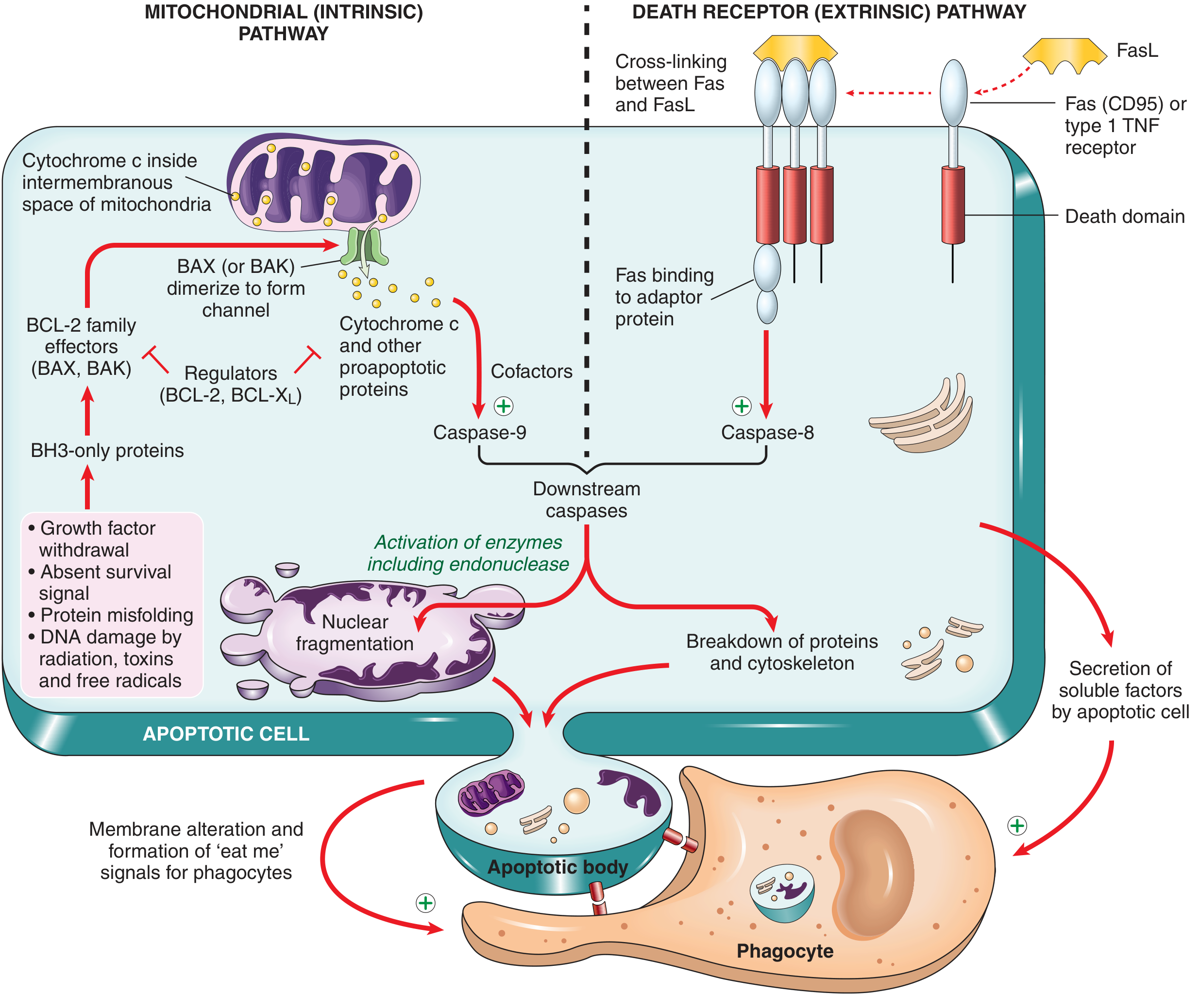
FIG. 1.12 from Robbins & Kumar Basic Pathology - Mechanisms of Apoptosis
1. Mitochondrial (Intrinsic) Pathway
This is the dominant pathway in most physiologic and pathologic apoptosis.
Key molecular players:
| Protein | Family | Role |
|---|---|---|
| BCL-2, BCL-X | BCL-2 anti-apoptotic | Maintain mitochondrial membrane integrity; keep BAX/BAK inactive |
| BAX, BAK | BCL-2 pro-apoptotic effectors | Dimerize and insert into mitochondrial membrane to form channels |
| BH3-only proteins (BIM, PUMA, NOXA, BID, BAD) | BCL-2 sensors | Activated by stress; shift balance toward BAX/BAK |
Sequence of events:
- Stress signals (DNA damage, growth factor withdrawal, protein misfolding) → activate BH3-only proteins
- BH3-only proteins inhibit BCL-2/BCL-X
Land directly activate BAX/BAK - BAX/BAK dimerize and insert into the mitochondrial outer membrane, forming pores
- Cytochrome c (and other pro-apoptotic proteins) leak into the cytosol
- Cytochrome c + APAF-1 + dATP → forms the apoptosome (wheel-shaped heptameric complex)
- Apoptosome recruits and activates caspase-9 (initiator caspase)
- Caspase-9 activates executioner caspases 3 and 7
2. Death Receptor (Extrinsic) Pathway
Triggered by ligand binding to death receptors on the cell surface - members of the TNF receptor superfamily containing a cytoplasmic "death domain."
Key receptors and ligands:
- Fas (CD95) + FasL (expressed on activated T lymphocytes)
- TNFR1 + TNF
Sequence of events:
- FasL cross-links Fas receptors on target cells
- Fas death domains recruit adaptor proteins (FADD)
- Adaptor proteins recruit and activate caspase-8 (initiator caspase)
- Caspase-8 directly activates executioner caspases 3 and 7
- (Caspase-8 can also cleave BID → truncated tBID → amplifies via the mitochondrial pathway)
Roles: Elimination of self-reactive lymphocytes; cytotoxic T lymphocyte-mediated killing of target cells.
Terminal Phase (Common to Both Pathways)
Executioner caspases 3 and 7 cleave numerous substrates:
- Endonucleases → nuclear DNA fragmentation (nucleosome-sized)
- Cytoskeletal proteins → cell shrinkage and blebbing
- Nuclear lamins → nuclear fragmentation
- Proteins involved in membrane asymmetry → phosphatidylserine flip
Clearance of Apoptotic Cells
Apoptotic cells send "eat-me" signals to phagocytes:
- Phosphatidylserine flip: normally confined to the inner membrane leaflet; in apoptosis it flips to the outer leaflet where macrophage receptors recognize it
- Secretion of soluble chemoattractants that recruit phagocytes
Macrophages engulf apoptotic bodies efficiently - no cell contents are released, so no inflammatory response is generated. This is a defining functional difference from necrosis.
Comparison: Apoptosis vs. Necrosis
| Feature | Apoptosis | Necrosis |
|---|---|---|
| Mechanism | Programmed, enzyme-driven | Passive, pathologic |
| Cell size | Shrinks | Swells |
| Nucleus | Pyknosis → karyorrhexis | Karyolysis or pyknosis |
| Membrane | Intact until phagocytosis | Disrupted early |
| Inflammation | None | Yes (DAMPs released) |
| Apoptotic bodies | Yes | No |
| Energy requirement | Yes (ATP-dependent) | No |
Other Related Cell Death Pathways
- Necroptosis: TNF-induced, features of both necrosis and apoptosis
- Pyroptosis: Inflammasome-driven, releases IL-1, causes fever and inflammation
- Ferroptosis: Iron-dependent, involves lipid peroxidation
- Anoikis: Apoptosis triggered by loss of cell-matrix attachment
Clinical Relevance
- Cancer: Escape from apoptosis is a hallmark of cancer. BCL-2 overexpression (t(14;18) translocation in follicular lymphoma) is the paradigm example. Drugs like venetoclax (BCL-2 inhibitor) exploit this pathway therapeutically.
- Autoimmunity: Defective Fas/FasL signaling → failure to eliminate self-reactive lymphocytes → autoimmune lymphoproliferative syndrome (ALPS)
- Neurodegeneration: Excessive neuronal apoptosis contributes to Alzheimer's, Parkinson's, ALS
- Ischemia/reperfusion injury: Apoptosis occurs alongside necrosis in infarcted tissue
Sources: Robbins & Kumar Basic Pathology; Sleisenger & Fordtran's Gastrointestinal and Liver Disease; Cellular and Molecular Immunology (Abbas)
Recent reviews for further reading: Apoptosis in Cancer Biology and Therapy (Moyer et al., Annu Rev Pathol, 2025) | Comprehensive Overview of Apoptosis Signaling (Mustafa et al., Cells, 2024)
This is a shared conversation. Sign in to Orris to start your own chat.