
Leukocyte circulation and migration to tissues
Reading File
This is a broad topic, so I'll run parallel searches across headings and paragraphs to get comprehensive coverage.
Finding Sources
Finding Sources
Finding Sources
Excellent - found a dedicated chapter. Let me read the main sources in parallel.
Reading File
Reading File
Good content so far. Let me read more of the Cellular and Molecular Immunology chapter and the Robbins content on leukocyte migration, plus fetch the key diagram images.
Reading File
Reading File
Loading Image
Excellent content and a great diagram. Now let me read more of the Cellular & Molecular Immunology chapter covering chemokines, T lymphocyte migration, and B lymphocyte migration.
Reading File
Loading Image
I now have rich content from the main chapter. Let me read a bit more to cover T cell recirculation, effector T cell migration, and B cell migration sections.
Reading File
I see I'm now in the references section. Let me go back and get the key content on T cell and B cell migration, chemokines, and leukocyte extravasation - that critical content was in the offset 1450-1650 range but got truncated. Let me read the most important part more carefully.
Reading File
I now have comprehensive content from both Cellular and Molecular Immunology and Robbins & Cotran. I have enough information to write an extremely thorough answer. Let me now compile everything.
Leukocyte Circulation and Migration to Tissues
A defining feature of the immune system - unlike any other organ system - is the continuous, regulated movement of its cellular components through blood, into tissues, and back into circulation. This process serves three core purposes:
- Delivery of myeloid leukocytes (neutrophils, monocytes) to sites of infection or tissue injury to eliminate pathogens and clear debris
- Delivery of naive lymphocytes from bone marrow/thymus to secondary lymphoid organs, where they encounter antigen and differentiate
- Delivery of effector lymphocytes from secondary lymphoid organs back to peripheral tissues where infection is occurring
- Cellular and Molecular Immunology, p. 133
Overview of Key Concepts
| Term | Definition |
|---|---|
| Homing | Migration of a leukocyte out of blood into a particular tissue |
| Recruitment | Movement from blood into tissues (general term) |
| Recirculation | Repeated homing of lymphocytes to secondary lymphoid organs and return to blood |
| Extravasation / Diapedesis | Transmigration of leukocytes through endothelium |
| Chemotaxis | Directional locomotion along a chemical concentration gradient |
Step 1: Overview - The Multistep Adhesion Cascade
Leukocyte migration from blood into tissue is not a single event - it is a sequential, multi-step process orchestrated by adhesion molecules and chemokines. The steps are:
- Tethering and rolling - mediated by selectins
- Integrin activation by endothelium-bound chemokines
- Stable (firm) adhesion - mediated by integrins
- Transmigration (diapedesis) through endothelium - mediated by PECAM-1 (CD31)
- Chemotaxis through extracellular matrix toward the inflammatory site
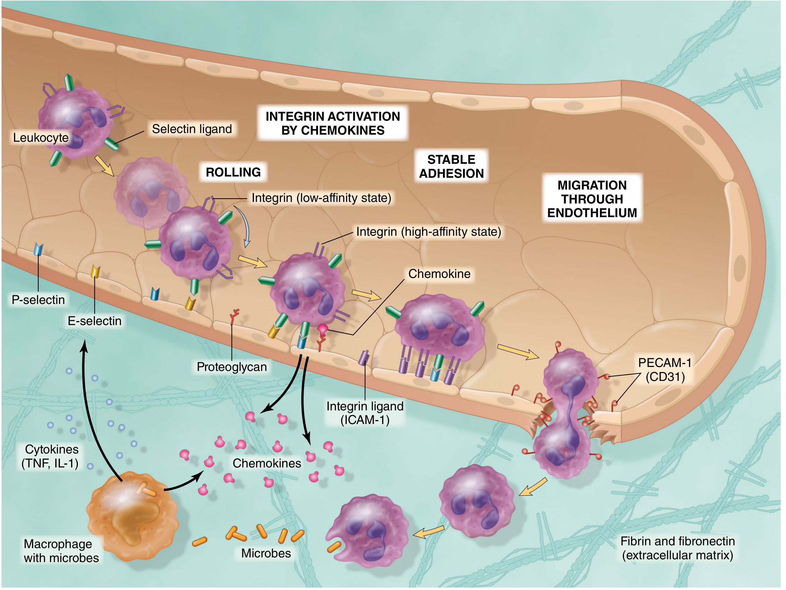
Fig. 3.4 from Robbins & Cotran - The multistep process of leukocyte migration through blood vessels. Selectins mediate rolling; chemokines activate integrins; integrins mediate firm adhesion; CD31/PECAM-1 mediates transmigration.
Step 2: Margination
Under normal blood flow, erythrocytes occupy the central laminar stream, pushing leukocytes to the periphery. During inflammation, vasodilation causes stasis of blood flow, increasing margination - leukocytes redistribute to a peripheral position along the vessel wall. This allows them to sense endothelial signals and begin rolling.
- Robbins & Cotran, p. 85
Step 3: Rolling - Selectins
Rolling is the first contact between a leukocyte and inflamed endothelium. It is mediated by selectins, which form low-affinity, fast-dissociating bonds with their carbohydrate ligands.
The Three Selectins
| Selectin | Location | Key Ligand(s) |
|---|---|---|
| P-selectin (CD62P) | Endothelium (Weibel-Palade bodies), platelets | PSGL-1 (sialyl Lewis X) |
| E-selectin (CD62E) | Activated endothelium | PSGL-1, E-selectin ligand-1 |
| L-selectin (CD62L) | Leukocytes | PNAd, MAdCAM-1 on HEVs |
- P-selectin is mobilized rapidly (within minutes) from endothelial Weibel-Palade bodies to the cell surface in response to histamine and thrombin
- E-selectin is newly synthesized over 1-2 hours in response to TNF and IL-1
- L-selectin sits at the tips of leukocyte microvilli for efficient capture
The carbohydrate ligands for P- and E-selectin are sialylated oligosaccharides (notably the tetrasaccharide sialyl Lewis X). P-selectin glycoprotein ligand 1 (PSGL-1) is post-translationally modified to display this structure as the major P-selectin counter-receptor.
Because these interactions have a fast off-rate, the leukocyte binds, detaches, and binds again - creating the characteristic rolling behavior visualized on intravital microscopy.
- Cellular and Molecular Immunology, p. 137-138
- Robbins & Cotran, p. 85
Step 4: Integrin Activation by Chemokines
Rolling slows the leukocyte sufficiently for it to encounter chemokines displayed on the endothelial surface. These chemokines are produced at the site of infection/injury and bind tightly to heparan sulfate proteoglycans on the endothelial surface - essentially presenting a high local concentration to rolling leukocytes.
Chemokines activate leukocytes via 7-transmembrane G-protein-coupled receptors (GPCRs), triggering intracellular signaling that converts leukocyte integrins from a low-affinity to a high-affinity conformation. This "inside-out" signaling is the key switch that transforms rolling into firm adhesion.
- Cellular and Molecular Immunology, p. 143-148
Key Chemokines in Leukocyte Recruitment
| Chemokine | Receptor | Target Cell | Key Function |
|---|---|---|---|
| CXCL8 (IL-8) | CXCR1, CXCR2 | Neutrophils | Neutrophil recruitment |
| CXCL1 (GROα) | CXCR2 | Neutrophils | Neutrophil recruitment |
| CCL2 (MCP-1) | CCR2 | Monocytes | Monocyte/mixed leukocyte recruitment |
| CCL19/CCL21 | CCR7 | T cells, DCs | Migration to parafollicular zones of lymph nodes |
| CXCL13 | CXCR5 | B cells | Migration to B cell follicles |
| CCL25 (TECK) | CCR9 | Lymphocytes | Recruitment to intestine |
| CCL27 (CTACK) | CCR10 | T cells | Recruitment to skin |
| CCL11 (Eotaxin) | CCR3 | Eosinophils, basophils, Th2 | Eosinophil recruitment |
Step 5: Firm Adhesion - Integrins
Integrins are heterodimeric transmembrane proteins (one α + one β chain) whose cytoplasmic tails interact with cytoskeletal components (talin, vinculin, actin). Their extracellular domains bind ligands on endothelial cells (IgSF family members - ICAMs).
Key Integrins in Leukocyte Adhesion
| Integrin | Also Known As | CD Designation | Endothelial Ligand |
|---|---|---|---|
| αLβ2 | LFA-1 | CD11a/CD18 | ICAM-1 (CD54), ICAM-2 |
| αMβ2 | Mac-1, CR3 | CD11b/CD18 | ICAM-1 |
| α4β1 | VLA-4 | CD49d/CD29 | VCAM-1 |
| α4β7 | LPAM-1 | - | MAdCAM-1 (gut HEVs) |
Mechanism of firm adhesion:
-
TNF and IL-1 (from tissue macrophages sensing microbes) induce endothelial expression of VCAM-1 and ICAM-1
-
Chemokines activate rolling leukocytes → inside-out signaling → integrins switch to high-affinity state
-
High-affinity integrin + upregulated ICAM-1/VCAM-1 = firm, stable adhesion
-
The leukocyte stops rolling, its cytoskeleton reorganizes, and it spreads out on the endothelial surface
-
Robbins & Cotran, p. 85-86; Cellular and Molecular Immunology, p. 139-141
Step 6: Transmigration (Diapedesis)
After firm adhesion, chemokines stimulate leukocytes to migrate through interendothelial junctions - a process called transmigration or diapedesis. This occurs mainly in postcapillary venules.
Key molecule: CD31 (PECAM-1, platelet-endothelial cell adhesion molecule), expressed homotypically on both leukocytes AND endothelial cell junctions. Homotypic CD31-CD31 interactions facilitate passage through the junctional space.
Two routes of transmigration:
- Paracellular - squeezing between endothelial cells (predominant route)
- Transcellular - movement through the cytoplasm of individual endothelial cells
After crossing the endothelium, leukocytes pierce the basement membrane by secreting collagenases. Once through, the basement membrane is repaired and becomes continuous again.
- Robbins & Cotran, p. 86; Cellular and Molecular Immunology, p. 148
Step 7: Chemotaxis in the Tissue
Once in the extravascular space, leukocytes migrate toward the inflammatory stimulus along a chemical gradient (chemotaxis). Chemoattractants include:
| Type | Examples |
|---|---|
| Bacterial products | N-formyl-methionyl peptides (e.g., fMet-Leu-Phe) |
| Complement | C5a |
| Lipid mediators | Leukotriene B4 (LTB4) |
| Chemokines | CXCL8 (IL-8), CCL2 (MCP-1) |
All bind to 7-transmembrane G-protein-coupled receptors → second messenger activation → actin polymerization at the leading edge, myosin contraction at the rear → directional locomotion.
- Robbins & Cotran, p. 86
Leukocyte Adhesion Deficiencies (LADs)
Clinical proof that this cascade is essential:
| Disease | Defect | Consequence |
|---|---|---|
| LAD type 1 | Absent/reduced β2 integrins (LFA-1, Mac-1) | Failure of firm adhesion; recurrent bacterial infections, absent pus |
| LAD type 2 | Absent leukocyte ligands for E- and P-selectins (fucosylation defect) | Failure of rolling; recurrent infections |
- Cellular and Molecular Immunology, p. 371
T Lymphocyte Migration and Recirculation
Naive T Cell Recirculation Through Lymph Nodes
Naive T cells must patrol secondary lymphoid organs to find their cognate antigen. They enter lymph nodes via specialized high endothelial venules (HEVs) - postcapillary venules lined by plump, cuboidal endothelial cells found in the T cell zones.
The adhesion cascade for naive T cells at HEVs uses distinct molecules compared to myeloid cell extravasation at sites of inflammation:
| Step | Molecule on T cell | Ligand on HEV |
|---|---|---|
| Rolling | L-selectin (CD62L) | PNAd (GlyCAM-1, CD34 on lymph node HEV; MAdCAM-1 on gut Peyer's patches) |
| Integrin activation | CCR7 | CCL19/CCL21 displayed on HEV surface |
| Firm adhesion | LFA-1 (αLβ2) | ICAM-1 |
| Transmigration | PECAM-1, CD99 | - |
After entering the lymph node parenchyma, if a T cell does not encounter its antigen, it exits via efferent lymphatics and returns to blood through the thoracic duct - completing the recirculation loop. Exit from lymph nodes requires sphingosine-1-phosphate (S1P) signaling: T cells express the receptor S1PR1, which is attracted by high S1P concentrations in lymph and blood. Retention signals (e.g., CCR7) are downregulated after exit.
- Cellular and Molecular Immunology, p. 159-163
Effector T Cell Migration to Peripheral Tissue
After activation and clonal expansion in secondary lymphoid organs, effector T cells acquire a distinct adhesion molecule and chemokine receptor profile that permits them to exit circulation into peripheral tissues (rather than returning to lymph nodes):
- Downregulate L-selectin (CD62L) and CCR7 (the "lymph node homing" molecules)
- Upregulate PSGL-1, E-selectin ligands, VLA-4, and tissue-specific chemokine receptors
Tissue-specific homing of effector T cells:
-
Gut-homing: Express α4β7 integrin (binds MAdCAM-1 on intestinal HEVs) + CCR9 (binds CCL25/TECK produced in intestinal mucosa) - imprinted by gut dendritic cells that produce retinoic acid
-
Skin-homing: Express CLA (cutaneous lymphocyte antigen) as the E-selectin ligand + CCR4/CCR10 (binding CCL17/CCL27 produced in skin)
-
Cellular and Molecular Immunology, p. 163-167
Memory T Cell Migration
Memory T cells are broadly divided into:
- Central memory T cells (TCM): Express CCR7 + L-selectin → home to lymph nodes (like naive cells); long-lived surveyors
- Effector memory T cells (TEM): Express tissue-homing receptors (lack CCR7/L-selectin) → patrol peripheral non-lymphoid tissues; provide rapid local responses
B Lymphocyte Migration
Naive B Cell Recirculation
Like naive T cells, naive follicular B cells express L-selectin and CCR7, allowing them to enter lymph nodes via HEVs. However, once inside, CCR7 signals direct naive T cells to T cell zones, while B cells upregulate CXCR5 and respond to CXCL13 (BLC) secreted by follicular stromal cells - directing them into B cell follicles.
Plasmablasts and Plasma Cells
Antibody-secreting plasmablasts generated in germinal center reactions downregulate CXCR5 (losing the follicular retention signal) and upregulate:
- CXCR4 → migrating toward bone marrow niches
- CXCR3 → migration to inflamed peripheral tissues
Long-lived plasma cells home to bone marrow survival niches via CXCL12 (SDF-1) signaling through CXCR4.
Memory B Cells
Memory B cells may retain or acquire tissue-specific homing receptors similar to effector memory T cells, allowing them to patrol peripheral tissues and mucosae.
- Cellular and Molecular Immunology, p. 168-171
Summary: Molecules by Step
| Step | Key Molecules |
|---|---|
| Stimulus (infection) | TNF, IL-1, IL-17 → upregulate endothelial adhesion molecules |
| Margination | Blood flow slowing, endothelial activation |
| Rolling | Selectins (P/E/L) + their carbohydrate ligands (sialyl Lewis X, PNAd) |
| Integrin activation | Chemokines (displayed on proteoglycans) → GPCRs → inside-out signaling |
| Firm adhesion | Integrins (LFA-1, VLA-4) + ICAM-1, VCAM-1, MAdCAM-1 |
| Transmigration | PECAM-1 (CD31), CD99; collagenases to cross basement membrane |
| Chemotaxis in tissue | fMet peptides, C5a, LTB4, chemokines → GPCR → actin polymerization |
| Lymphocyte exit from LN | S1P → S1PR1 (S1P gradient toward lymph/blood) |
| Tissue-specific homing | α4β7 + CCR9 (gut); CLA + CCR10/CCR4 (skin); CXCR4 (bone marrow) |
Sources:
- Cellular and Molecular Immunology (Abbas, Lichtman, Pillai), Chapter 3, pp. 132-171
- Robbins & Cotran Pathologic Basis of Disease, Chapter 3, pp. 85-86
This is a shared conversation. Sign in to Orris to start your own chat.