
Lung ultrasound
"lung ultrasound"[MeSH Terms] AND diagnosis
"lung ultrasound" AND "point of care"
Lung Ultrasound (LUS)
Transducer Selection
- High-frequency linear-array (5-10 MHz): preferred for pneumothorax detection (higher resolution and sensitivity: 82% vs. 76% compared with convex transducers)
- Phased-array or curvilinear (lower frequency): preferred when depth is needed (e.g., B-line assessment, pleural effusion, deep consolidation - must reach >15 cm depth to distinguish B-lines from Z-lines)
- Microconvex: versatile for general use
Core Ultrasound Signs
1. The Pleural Line
2. A-Lines (Horizontal Artifacts)
- Equidistant, horizontal, hyperechoic lines parallel to the pleural surface
- Reverberation artifact from reflections of the pleural line
- Seen in normal aerated lung, but also in pneumothorax, PE, COPD, and asthma
- Not specific - their meaning depends on whether lung sliding is present or absent
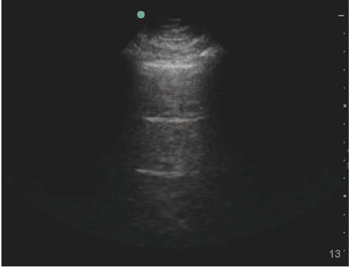
3. B-Lines (Vertical Artifacts - "Comet Tails" / "Lung Rockets")
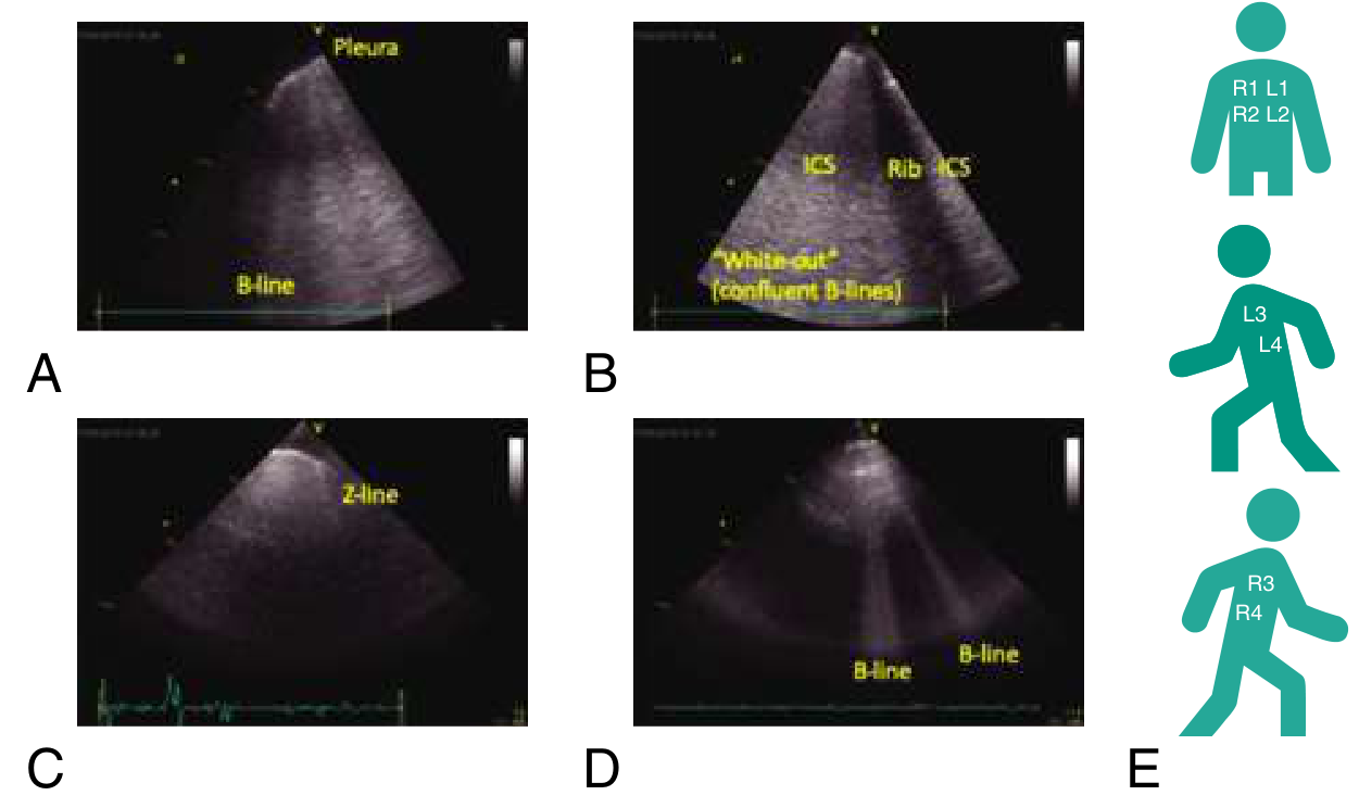
- Discrete, hyperechoic, laser-like vertical rays arising from the pleural surface
- Extend to the bottom of the screen (>15 cm deep) without fading
- Slide with pleural movement
- Arise because thickened intralobular septa near the visceral pleura allow ultrasound waves to penetrate and continuously reverberate
- Presence of B-lines rules out pneumothorax (confirms visceral-parietal pleural apposition)
- 1-2 B-lines per field: normal (especially at bases)
- ≥3 B-lines per intercostal space = abnormal ("pulmonary interstitial syndrome") - indicates interstitial edema, fibrosis, or alveolar disease
- Diffuse, confluent B-lines ("white lung") = severe pulmonary edema or diffuse alveolar hemorrhage
Z-lines vs. B-lines: Z-lines are hyperechoic lines from the pleura that extend <15 cm and are non-diagnostic. B-lines must reach >15 cm depth to count.
M-Mode Patterns
| Finding | M-Mode Pattern | Meaning |
|---|---|---|
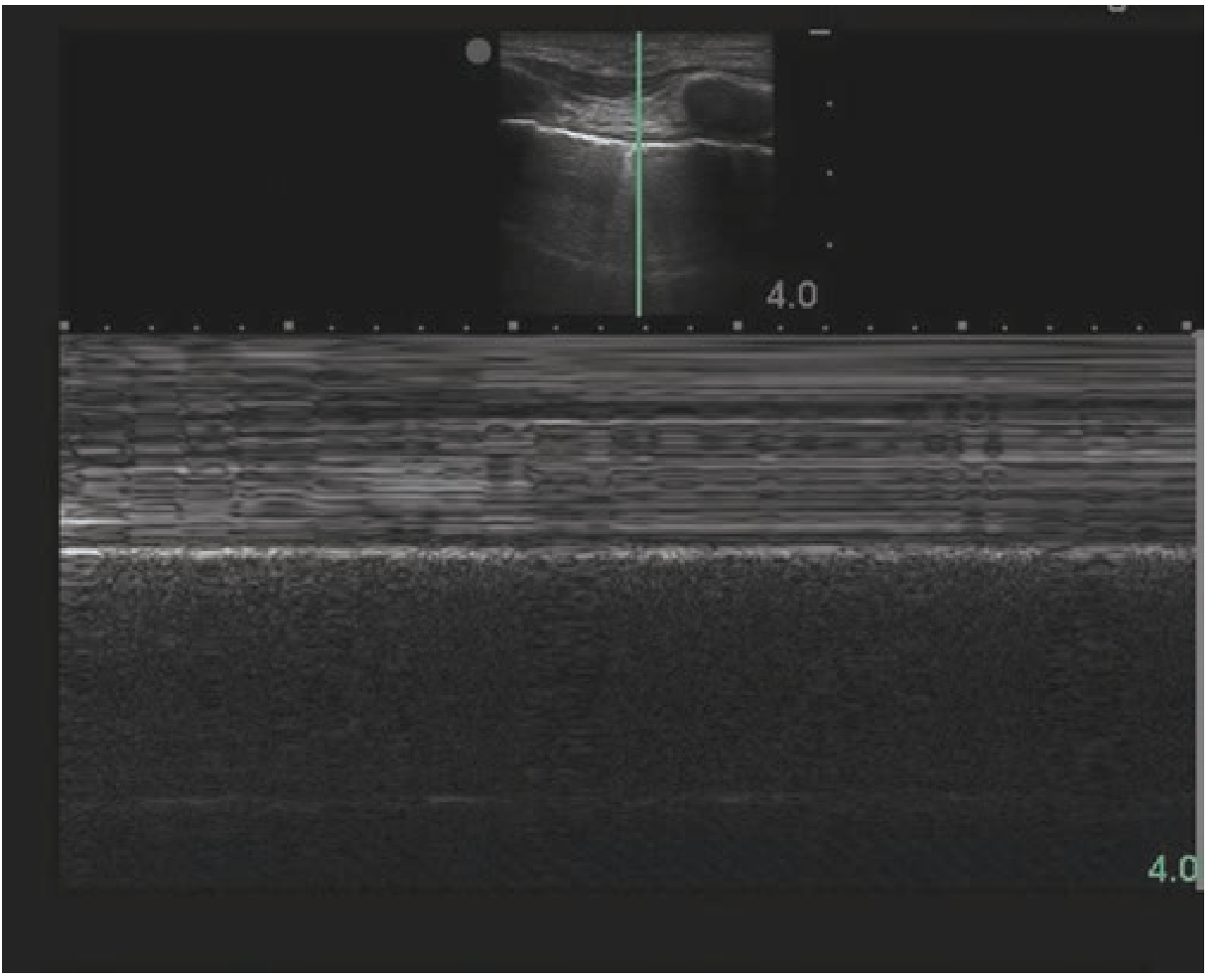
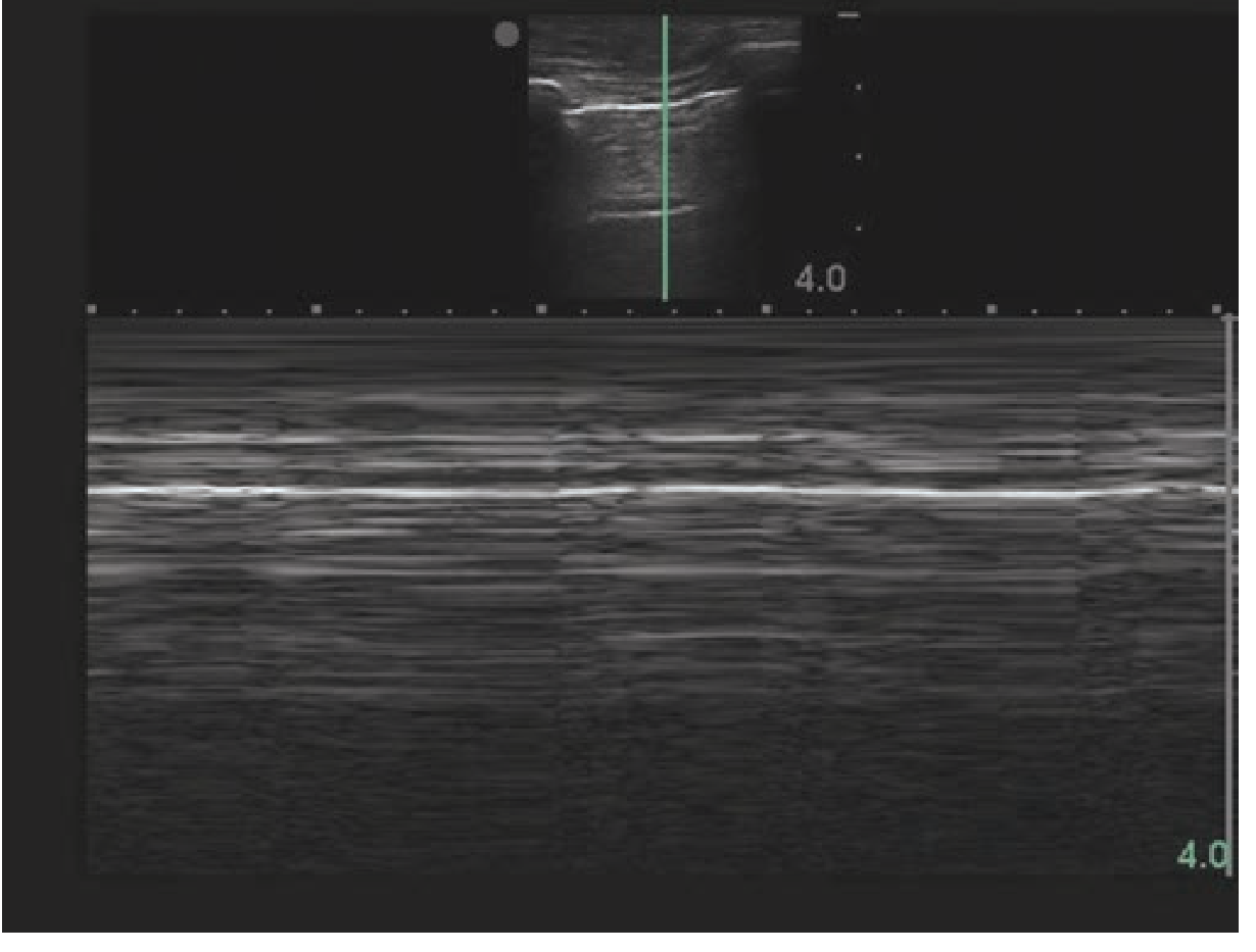
| Normal lung sliding | "Seashore sign" - horizontal lines (chest wall) above, granular pattern below | Pleura are apposed; no pneumothorax |
| Pneumothorax | "Stratosphere sign" / "Barcode sign" - horizontal lines throughout | Air separates pleural layers |
| Lung point | Alternating seashore + barcode | Edge of pneumothorax - 100% specific |
Clinical Applications
Pneumothorax
- Lung sliding absent = key sign; absence is NOT specific to pneumothorax (also seen with pleurodesis, fibrotic adhesions, mainstem intubation, atelectasis, apnea)
- Lung point = boundary between collapsed and aerated lung; 100% specific for pneumothorax; found anteriorly for small, laterally for large pneumothorax in supine patients
- Presence of lung sliding definitively rules out pneumothorax (NPV 100%)
- Presence of B-lines also rules out pneumothorax (confirms pleural apposition)
| Test | Pooled Sensitivity | Pooled Specificity |
|---|---|---|
| Lung ultrasound | 79-95% | 98-99% |
| Chest radiography | 40-52% | 99-100% |
Pulmonary Edema / Acute Heart Failure (AHF)
- The 8-point LUS exam scans 2 anterior + 2 lateral intercostal spaces (ICS) on each hemithorax
- Positive: ≥3 B-lines in at least 1 ICS on each side
- Positive and negative likelihood ratios for LUS equal or outperform all other binary tests for AHF diagnosis
- Speeds ED diagnosis without loss of accuracy
- B-line count change from ED to discharge (not initial count) predicts 30-day death or AHF readmission
- LUS-guided diuretic dosing (RCT feasibility): reduced pulmonary edema at 48 hours
Consolidation / Pneumonia
- Consolidated lung is easily penetrated by ultrasound and appears tissue-like (hepatization)
- Air bronchograms appear as hyperechoic foci within the consolidation coursing parallel to pulmonary arteries
- LUS is useful for triaging children with suspected community-acquired pneumonia
- A 2024 systematic review (PMID 38299193) confirms point-of-care LUS detects pneumonia with clinically useful accuracy
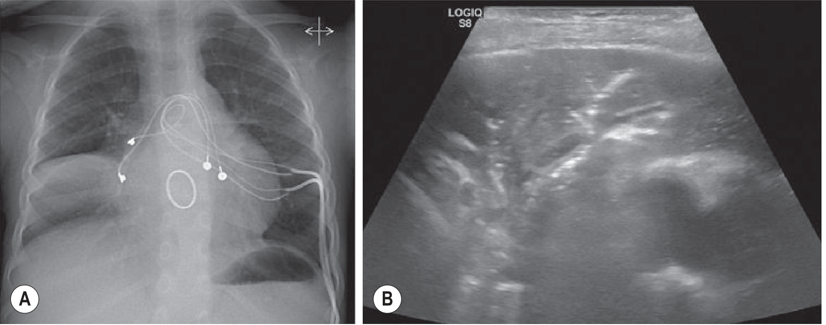
Pleural Effusion
- Arguably best imaging technique for pleural effusion assessment
- Demonstrates thin septa or echogenic debris that CT often misses
- Guides real-time needle placement for diagnostic tap or chest drain
- Superior to CXR for detecting small effusions
ARDS vs. Cardiogenic Pulmonary Edema
- Cardiogenic edema: diffuse, bilateral, homogenous B-lines; spared areas absent
- ARDS/noncardiogenic: focal B-lines with non-homogenous distribution, spared areas present, subpleural consolidations, irregular pleural line, reduced lung sliding
Atelectasis
- Appears as tissue-like opacity similar to consolidation
- No air bronchograms (or only dynamic air bronchograms)
- B-lines may still be visible at the margins
The BLUE Protocol (Bedside Lung Ultrasound in Emergency)
- Anterior lung sliding (present or absent)
- Multiple bilateral B-lines (present or absent)
- Posterior consolidations or effusion (present or absent)
| Profile | Typical Diagnosis |
|---|---|
| A-profile (A-lines + sliding) | Normal or PE/asthma/COPD |
| B-profile (bilateral B-lines) | Pulmonary edema |
| A/B profile (asymmetric) | Pneumonia |
| C-profile (anterior consolidation) | Pneumonia |
| A-profile + no sliding | Pneumothorax |
Limitations
- Cannot visualize central pulmonary structures, mediastinal nodes, or central abscesses (unlike CT)
- Operator-dependent
- Limited in obese patients or those with subcutaneous emphysema
- Cannot assess abnormalities not contacting the visceral pleura (e.g., central lesions)
- In well-resourced settings, does not replace CXR for assessment of tubes, lines, and central thoracic structures (Grainger & Allison's)
Recent Evidence
- PMID 38299193 (Systematic Review, 2024): Point-of-care LUS detects pneumonia with good diagnostic accuracy
- PMID 38205982 (Systematic Review/Meta-Analysis, 2024): Lung ultrasound score in COVID-19 correlates with PaO2/FiO2 ratio, intubation rates, and mortality
Generate a quick reference PDF comparing LUS patterns in pneumonia vs. pulmonary edema vs. pneumothorax
ls -la /tmp/workspace/ 2>/dev/null || echo "workspace not found"
mkdir -p /tmp/workspace/lus-quickref && python3 -c "from reportlab.platypus import SimpleDocTemplate; print('reportlab ok')"
~/lus-quickref/generate_lus_pdf.py
#!/usr/bin/env python3
"""
LUS Quick Reference PDF - Pneumonia vs Pulmonary Edema vs Pneumothorax
Generated with ReportLab
"""

from reportlab.platypus import (
    SimpleDocTemplate, Table, TableStyle, Paragraph, Spacer,
    HRFlowable, KeepTogether
)
from reportlab.lib.styles import getSampleStyleSheet, ParagraphStyle
from reportlab.lib import colors
from reportlab.lib.pagesizes import A4, landscape
from reportlab.lib.units import cm, mm
from reportlab.lib.enums import TA_CENTER, TA_LEFT, TA_RIGHT
from reportlab.platypus.flowables import BalancedColumns
import os

OUTPUT = "/tmp/workspace/lus-quickref/LUS_Quick_Reference.pdf"

# ── Colour palette ──────────────────────────────────────────────────────────
C_NAVY    = colors.HexColor("#1A2B4A")   # header bg
C_BLUE    = colors.HexColor("#2E6DA4")   # section headers
C_LTBLUE  = colors.HexColor("#D6E8F7")   # alternate row / label bg
C_ORANGE  = colors.HexColor("#E8500A")   # pneumothorax accent
C_GREEN   = colors.HexColor("#1E7E34")   # pneumonia accent
C_PURPLE  = colors.HexColor("#5A3E8B")   # pulm edema accent
C_GOLD    = colors.HexColor("#C98B00")   # warnings / notes
C_LIGHTGRAY = colors.HexColor("#F4F6F8")
C_WHITE   = colors.white
C_BLACK   = colors.black

# ── Page setup ───────────────────────────────────────────────────────────────
PAGE = landscape(A4)
MARGIN = 1.5 * cm
doc = SimpleDocTemplate(
    OUTPUT,
    pagesize=PAGE,
    leftMargin=MARGIN, rightMargin=MARGIN,
    topMargin=MARGIN, bottomMargin=MARGIN,
    title="Lung Ultrasound Quick Reference",
    author="Orris Medical",
    subject="LUS patterns: Pneumonia vs Pulmonary Edema vs Pneumothorax"
)

styles = getSampleStyleSheet()

def style(name, **kw):
    s = ParagraphStyle(name, **kw)
    return s

# Custom paragraph styles
S_TITLE = style("Title2",
    fontSize=20, fontName="Helvetica-Bold",
    textColor=C_WHITE, alignment=TA_CENTER, leading=24)

S_SUBTITLE = style("Subtitle2",
    fontSize=10, fontName="Helvetica",
    textColor=colors.HexColor("#BBDDFF"), alignment=TA_CENTER, leading=13)

S_SECTION = style("SectionHead",
    fontSize=11, fontName="Helvetica-Bold",
    textColor=C_WHITE, alignment=TA_CENTER, leading=14)

S_BODY = style("Body2",
    fontSize=8.5, fontName="Helvetica",
    textColor=C_BLACK, leading=12)

S_BODY_BOLD = style("BodyBold",
    fontSize=8.5, fontName="Helvetica-Bold",
    textColor=C_BLACK, leading=12)

S_SMALL = style("Small",
    fontSize=7.5, fontName="Helvetica",
    textColor=colors.HexColor("#333333"), leading=10)

S_SMALL_BOLD = style("SmallBold",
    fontSize=7.5, fontName="Helvetica-Bold",
    textColor=C_BLACK, leading=10)

S_CELL = style("Cell",
    fontSize=8, fontName="Helvetica",
    textColor=C_BLACK, leading=11, leftIndent=3)

S_CELL_BOLD = style("CellBold",
    fontSize=8, fontName="Helvetica-Bold",
    textColor=C_BLACK, leading=11, leftIndent=3)

S_CELL_WHITE = style("CellWhite",
    fontSize=8.5, fontName="Helvetica-Bold",
    textColor=C_WHITE, leading=11, alignment=TA_CENTER)

S_TICK = style("Tick",
    fontSize=8, fontName="Helvetica",
    textColor=C_BLACK, leading=11, leftIndent=6)

S_NOTE = style("Note",
    fontSize=7.5, fontName="Helvetica-Oblique",
    textColor=colors.HexColor("#444444"), leading=10, leftIndent=3)

S_WARN = style("Warn",
    fontSize=7.5, fontName="Helvetica-Bold",
    textColor=C_GOLD, leading=10, leftIndent=3)

S_FOOTER = style("Footer",
    fontSize=7, fontName="Helvetica",
    textColor=colors.HexColor("#777777"), alignment=TA_CENTER, leading=9)

def P(text, s=S_BODY): return Paragraph(text, s)
def SP(h=4): return Spacer(1, h)

# ── Helper: coloured header cell ─────────────────────────────────────────────
def hdr(text, bg, color=C_WHITE, size=9):
    s = ParagraphStyle("hdr_tmp", fontSize=size, fontName="Helvetica-Bold",
                       textColor=color, alignment=TA_CENTER, leading=size+3)
    return Paragraph(text, s)

# ────────────────────────────────────────────────────────────────────────────
# PAGE HEADER BANNER
# ────────────────────────────────────────────────────────────────────────────
header_data = [[
    P("🫁  LUNG ULTRASOUND  ·  QUICK REFERENCE CARD", S_TITLE),
]]
header_table = Table(header_data, colWidths=[PAGE[0] - 2*MARGIN])
header_table.setStyle(TableStyle([
    ("BACKGROUND",  (0,0), (-1,-1), C_NAVY),
    ("TOPPADDING",  (0,0), (-1,-1), 10),
    ("BOTTOMPADDING",(0,0),(-1,-1), 10),
    ("LEFTPADDING", (0,0), (-1,-1), 8),
    ("RIGHTPADDING",(0,0), (-1,-1), 8),
    ("ROUNDEDCORNERS", [6,6,6,6]),
]))

subtitle_data = [[
    P("Pneumonia   ·   Pulmonary Edema   ·   Pneumothorax   |   Pattern Recognition at the Bedside", S_SUBTITLE),
]]
subtitle_table = Table(subtitle_data, colWidths=[PAGE[0] - 2*MARGIN])
subtitle_table.setStyle(TableStyle([
    ("BACKGROUND",  (0,0), (-1,-1), C_BLUE),
    ("TOPPADDING",  (0,0), (-1,-1), 5),
    ("BOTTOMPADDING",(0,0),(-1,-1), 5),
]))

# ────────────────────────────────────────────────────────────────────────────
# MAIN COMPARISON TABLE
# ────────────────────────────────────────────────────────────────────────────

# Column widths (landscape A4 usable ~25 cm)
USABLE = PAGE[0] - 2*MARGIN
C0 = USABLE * 0.17   # Feature
C1 = USABLE * 0.28   # Pneumonia
C2 = USABLE * 0.28   # Pulm Edema
C3 = USABLE * 0.27   # Pneumothorax

def cell(txt, bold=False, italic=False, color=C_BLACK, size=8):
    fname = "Helvetica-Bold" if bold else ("Helvetica-Oblique" if italic else "Helvetica")
    s = ParagraphStyle("_c", fontSize=size, fontName=fname, textColor=color, leading=11, leftIndent=4)
    return Paragraph(txt, s)

def bullet_list(items, color=C_BLACK, size=8):
    lines = "".join(f"• {i}<br/>" for i in items)
    s = ParagraphStyle("_bl", fontSize=size, fontName="Helvetica",
                       textColor=color, leading=11.5, leftIndent=4)
    return Paragraph(lines, s)

PNE_C = colors.HexColor("#E8F5E9")   # pneumonia row bg
OED_C = colors.HexColor("#EDE7F6")   # oedema row bg
PTX_C = colors.HexColor("#FFF3E0")   # pneumothorax row bg

HEADER_ROW = [
    hdr("Feature", C_NAVY, size=9),
    hdr("Pneumonia", C_GREEN, size=9),
    hdr("Pulmonary Edema", C_PURPLE, size=9),
    hdr("Pneumothorax", C_ORANGE, size=9),
]

rows = [HEADER_ROW]

data_rows = [
    (
        "Lung Sliding",
        ["Usually PRESENT", "May be ABSENT over consolidation"],
        ["Usually PRESENT", "May be absent in severe cases"],
        ["ABSENT (key sign)", "NPV 100% when present → rules out PTX"],
    ),
    (
        "A-Lines\n(horizontal artifacts)",
        ["Absent or obscured", "Replaced by B-lines or consolidation"],
        ["Absent (replaced by B-lines)"],
        ["PRESENT with absent sliding", "= diagnostic combination for PTX"],
    ),
    (
        "B-Lines\n(vertical artifacts)",
        ["Focal / regional", "Non-homogenous distribution", "Localised to affected lobe/segment"],
        ["Diffuse bilateral ≥3 per ICS", "Homogenous distribution", "May fuse into 'white lung'"],
        ["ABSENT (rules out PTX when present)", "B-lines confirm pleural apposition"],
    ),
    (
        "Pleural Line\nAppearance",
        ["Irregular, fragmented", "Subpleural micro-consolidations", "Pleural line thickened"],
        ["Smooth, regular", "Normal pleural line"],
        ["Normal but STATIONARY", "No shimmer/glide sign"],
    ),
    (
        "Consolidation",
        ["YES — tissue-like hepatization", "Air bronchograms present", "Parallel to pulmonary arteries"],
        ["Absent (unless severe ARDS overlap)"],
        ["Absent", "(collapsed lung may be seen only with large PTX)"],
    ),
    (
        "Air Bronchograms",
        ["Static or dynamic air bronchograms", "Static = obstruction", "Dynamic = patent airway"],
        ["Absent"],
        ["Absent"],
    ),
    (
        "Pleural Effusion",
        ["May be present (parapneumonic)", "Anechoic or complex with septations"],
        ["Often bilateral, anechoic", "Corresponds to heart failure severity"],
        ["Absent"],
    ),
    (
        "M-Mode Pattern",
        ["Seashore sign may be lost over consolidation"],
        ["Seashore sign present (if sliding intact)"],
        ["Stratosphere / Barcode sign", "Lung point: 100% specific for PTX"],
    ),
    (
        "Distribution",
        ["Focal / lobar", "Posterior > anterior", "Usually unilateral"],
        ["Bilateral, symmetric", "Dependent > non-dependent"],
        ["Anterior in supine", "Small: anterior MCL", "Large: extends laterally"],
    ),
    (
        "Lung Point",
        ["Absent"],
        ["Absent"],
        ["PRESENT — 100% specific", "= edge of pneumothorax", "Not always found in large PTX"],
    ),
    (
        "Key Differentiator",
        ["Irregular pleural line +\nfocal B-lines +\nconsolidation with air bronchograms"],
        ["Diffuse bilateral B-lines\n(≥3 per ICS bilateral)\nSmooth pleural line\nNo consolidation"],
        ["Absent sliding +\nA-lines +\nLung point (confirms)"],
    ),
    (
        "BLUE Protocol\nProfile",
        ["C-profile (anterior consolidation)", "A/B profile (asymmetric B-lines)", "A-profile + posterior consolidation"],
        ["B-profile (bilateral B-lines)"],
        ["A-profile + absent sliding"],
    ),
    (
        "Performance\n(vs CXR)",
        ["Sens 85-93%, Spec 88-95%\n(paediatric CAP)", "Superior to CXR in resource-limited settings"],
        ["LR+ and LR− outperform\nall other binary tests\nfor AHF in ED"],
        ["Sens 79-95%, Spec 98-99%\n(meta-analysis)\nvs CXR Sens 40-52%"],
    ),
]

for feature, pne, oed, ptx in data_rows:
    row = [
        cell(feature, bold=True, color=C_NAVY, size=8),
        bullet_list(pne, color=colors.HexColor("#1B5E20"), size=8),
        bullet_list(oed, color=colors.HexColor("#311B92"), size=8),
        bullet_list(ptx, color=colors.HexColor("#BF360C"), size=8),
    ]
    rows.append(row)

main_table = Table(rows, colWidths=[C0, C1, C2, C3], repeatRows=1)

# Build row-by-row background styles
bg_styles = [
    # Header row
    ("BACKGROUND", (0,0), (0,0), C_NAVY),
    ("BACKGROUND", (1,0), (1,0), C_GREEN),
    ("BACKGROUND", (2,0), (2,0), C_PURPLE),
    ("BACKGROUND", (3,0), (3,0), C_ORANGE),
    ("TEXTCOLOR",  (0,0), (-1,0), C_WHITE),
    ("FONTNAME",   (0,0), (-1,0), "Helvetica-Bold"),
    ("FONTSIZE",   (0,0), (-1,0), 9),
    ("ALIGN",      (0,0), (-1,0), "CENTER"),
    ("VALIGN",     (0,0), (-1,-1), "TOP"),
    # Alternating row bgs (feature col always light blue)
    ("BACKGROUND", (0,1), (0,-1), C_LTBLUE),
]
for i in range(1, len(rows)):
    bg = C_LIGHTGRAY if i % 2 == 1 else C_WHITE
    bg_styles += [
        ("BACKGROUND", (1,i), (1,i), PNE_C if i % 2 == 1 else colors.HexColor("#F1F8F1")),
        ("BACKGROUND", (2,i), (2,i), OED_C if i % 2 == 1 else colors.HexColor("#F5F0FB")),
        ("BACKGROUND", (3,i), (3,i), PTX_C if i % 2 == 1 else colors.HexColor("#FFF8F0")),
    ]

main_table.setStyle(TableStyle(bg_styles + [
    ("GRID",        (0,0), (-1,-1), 0.4, colors.HexColor("#BBBBCC")),
    ("LINEBELOW",   (0,0), (-1,0), 1.5, C_NAVY),
    ("TOPPADDING",  (0,0), (-1,-1), 5),
    ("BOTTOMPADDING",(0,0),(-1,-1), 5),
    ("LEFTPADDING", (0,0), (-1,-1), 4),
    ("RIGHTPADDING",(0,0),(-1,-1), 4),
    # Thicker left border for each condition column
    ("LINEAFTER",   (0,0), (0,-1), 1.5, C_NAVY),
    ("LINEBEFORE",  (1,0), (1,-1), 1.5, C_GREEN),
    ("LINEBEFORE",  (2,0), (2,-1), 1.5, C_PURPLE),
    ("LINEBEFORE",  (3,0), (3,-1), 1.5, C_ORANGE),
]))

# ────────────────────────────────────────────────────────────────────────────
# BOTTOM LEGEND / NOTES BAR
# ────────────────────────────────────────────────────────────────────────────

legend_items = [
    ("B-line rule:", "Discrete vertical hyperechoic ray from pleura to screen bottom (>15 cm). ≥3 per ICS = abnormal."),
    ("Z-line:", "Hyperechoic vertical line <15 cm — non-diagnostic, do not count as B-lines."),
    ("Lung point:", "100% specific for PTX. Alternating seashore/barcode on M-mode."),
    ("BLUE protocol:", "3 binary signs: anterior sliding · bilateral B-lines · posterior consolidation/effusion."),
    ("Transducer:", "Linear 5-10 MHz for PTX (higher sensitivity). Phased-array/curvilinear for B-lines (need >15 cm depth)."),
    ("Sources:", "Fishman's Pulmonary Diseases · Rosen's Emergency Medicine · Murray & Nadel · Grainger & Allison · Miller's Anesthesia"),
]

def legend_cell(label, text):
    s_lbl = ParagraphStyle("_ll", fontSize=7, fontName="Helvetica-Bold",
                            textColor=C_NAVY, leading=10)
    s_txt = ParagraphStyle("_lt", fontSize=7, fontName="Helvetica",
                            textColor=colors.HexColor("#222222"), leading=10)
    return [Paragraph(label, s_lbl), Paragraph(text, s_txt)]

legend_rows = [legend_cell(lbl, txt) for lbl, txt in legend_items]
# Two columns of legend items
half = len(legend_rows) // 2 + len(legend_rows) % 2
col1_rows = legend_rows[:half]
col2_rows = legend_rows[half:]

def flat_legend_col(items):
    """Flatten legend items into a single-column 2-col table"""
    rows = [[lbl, txt] for lbl, txt in items]
    t = Table(rows, colWidths=[2.8*cm, 9.5*cm])
    t.setStyle(TableStyle([
        ("VALIGN", (0,0), (-1,-1), "TOP"),
        ("TOPPADDING", (0,0), (-1,-1), 1),
        ("BOTTOMPADDING", (0,0), (-1,-1), 1),
        ("LEFTPADDING", (0,0), (-1,-1), 2),
        ("RIGHTPADDING", (0,0), (-1,-1), 2),
    ]))
    return t

legend_outer = Table(
    [[flat_legend_col(col1_rows), flat_legend_col(col2_rows)]],
    colWidths=[(USABLE/2)-4, (USABLE/2)-4]
)
legend_outer.setStyle(TableStyle([
    ("BACKGROUND", (0,0), (-1,-1), colors.HexColor("#EEF3F9")),
    ("BOX",       (0,0), (-1,-1), 0.5, C_BLUE),
    ("LINEAFTER",  (0,0), (0,-1), 0.5, C_BLUE),
    ("TOPPADDING", (0,0), (-1,-1), 5),
    ("BOTTOMPADDING", (0,0), (-1,-1), 5),
    ("LEFTPADDING", (0,0), (-1,-1), 6),
    ("RIGHTPADDING", (0,0), (-1,-1), 6),
]))

legend_header = Table(
    [[hdr("  KEY  ·  DEFINITIONS  ·  SOURCES", C_BLUE, size=8)]],
    colWidths=[USABLE]
)
legend_header.setStyle(TableStyle([
    ("BACKGROUND", (0,0), (-1,-1), C_BLUE),
    ("TOPPADDING", (0,0), (-1,-1), 3),
    ("BOTTOMPADDING", (0,0), (-1,-1), 3),
]))

# ────────────────────────────────────────────────────────────────────────────
# BUILD DOCUMENT
# ────────────────────────────────────────────────────────────────────────────
elements = [
    header_table,
    subtitle_table,
    SP(5),
    main_table,
    SP(6),
    legend_header,
    legend_outer,
]

doc.build(elements)
print(f"PDF created: {OUTPUT}")
print(f"File size: {os.path.getsize(OUTPUT):,} bytes")
cd /tmp/workspace/lus-quickref && python3 generate_lus_pdf.py 2>&1
python3 -c " import pdfplumber with pdfplumber.open('/tmp/workspace/lus-quickref/LUS_Quick_Reference.pdf') as pdf: print(f'Pages: {len(pdf.pages)}') page = pdf.pages[0] print(f'Page size: {round(page.width/72*2.54,1)} x {round(page.height/72*2.54,1)} cm (landscape A4 = ~29.7 x 21 cm)') text = page.extract_text() print('--- First 400 chars ---') print(text[:400] if text else '(no text extracted)') "
LUS Quick Reference
PDF Document · PDF
| Row | What's compared |
|---|---|
| Lung Sliding | Present vs. absent, with NPV note for PTX |
| A-Lines | Role in each pattern |
| B-Lines | Focal/regional vs. diffuse bilateral vs. absent |
| Pleural Line | Irregular + fragmented vs. smooth vs. stationary |
| Consolidation | Hepatization + air bronchograms vs. absent vs. absent |
| Air Bronchograms | Static/dynamic vs. absent vs. absent |
| Pleural Effusion | Parapneumonic vs. bilateral anechoic vs. absent |
| M-Mode Pattern | Seashore loss vs. intact vs. barcode/stratosphere |
| Distribution | Focal/lobar vs. bilateral symmetric vs. anterior-first |
| Lung Point | Absent vs. absent vs. 100% specific |
| Key Differentiator | Summary diagnostic fingerprint per condition |
| BLUE Protocol Profile | C/A-B profile vs. B-profile vs. A + absent sliding |
| Performance vs. CXR | Sensitivity/specificity from meta-analyses |