
Erithroproaesis
I'll look up erythropoiesis from the medical library for you.
Searching sources...
Reading File
Now let me search the medical library for erythropoiesis content in parallel:
Finding Sources
Finding Sources
Good results. Now let me read the most relevant sections in parallel - the Histology textbook (which covers development stages) and the Biochemistry textbook:
Reading File
Reading File
Reading File
Now let me read the histology textbook's dedicated erythropoiesis section and the kidney textbook's normal erythropoiesis section:
Reading File
Reading File
Now let me find the histology textbook's specific erythropoiesis development section with cell stages, and fetch the key diagram:
Finding Sources
Loading Image
Excellent! Now let me find the histology textbook's developmental stages content (pronormoblast through reticulocyte):
Finding Sources
Finding Sources
Now let me read both the normoblastic maturation section from Henry's and the histology plate section:
Reading File
Reading File
I now have rich content. Let me also read the continuation of Henry's normoblastic section for the reticulocyte and the proerythroblast section from histology:
Reading File
Reading File
Let me fetch the hemopoiesis diagram from the histology textbook and then read the proerythroblast section:
Loading Image
Reading File
I now have all the information needed for a comprehensive answer. Let me fetch the RNA/hemoglobin timeline diagram as well:
Loading Image
I now have all the material needed for a thorough answer. Here is the full synthesis:
Erythropoiesis
Erythropoiesis is the process by which mature red blood cells (erythrocytes) are produced from hematopoietic stem cells, primarily in the red bone marrow of adults.
1. Overview and Regulation
The delivery of oxygen to peripheral tissues is tightly regulated, and the circulating red cell mass is the key determinant. Under normal conditions, approximately 1% of circulating erythrocytes is replaced daily - roughly 250 billion cells, or 2.5 to 3 million erythrocytes per second.
The control is based on a classic negative feedback loop:
- Decreased oxygen delivery to tissues (hypoxia) is sensed by peritubular interstitial cells of the kidney
- The kidney releases erythropoietin (EPO) - a 30.4 kDa glycoprotein hormone
- EPO binds to specific EPO receptors (EPO-R) on bone marrow erythroid progenitors, stimulating proliferation and maturation
- As red cell mass rises, oxygen delivery improves, and EPO production falls
Additional cofactors required: iron, vitamin B12, and folic acid.
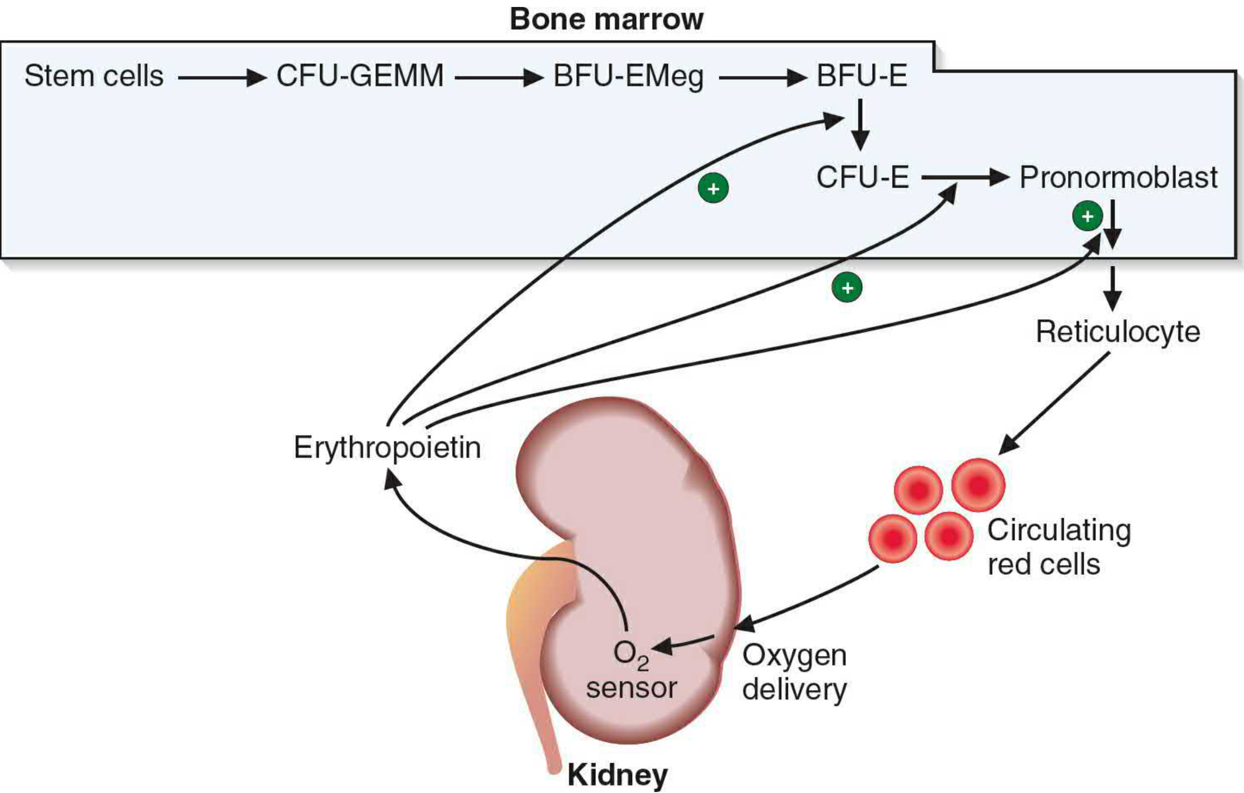
2. Progenitor Cell Hierarchy (Pre-microscopic stages)
These stages are not visible by light microscopy but are identified by colony assays:
| Cell | Notes |
|---|---|
| Hematopoietic Stem Cell (HSC) | Self-renewing multipotent progenitor |
| CMP (Common Myeloid Progenitor) | Under EPO, IL-3, IL-4 influence |
| MEP (Megakaryocyte/Erythrocyte Progenitor) | Bipotential; requires GATA-1 transcription factor for erythroid commitment |
| ErP (Erythrocyte Progenitor) | Erythropoietin-sensitive committed progenitor |
| BFU-E (Burst-Forming Unit - Erythroid) | Early committed progenitor |
| CFU-E (Colony-Forming Unit - Erythroid) | More mature progenitor; highly EPO-sensitive |
3. Morphologically Recognizable Stages (Normoblastic Maturation)
The following cells are visible under the light microscope in bone marrow smears:
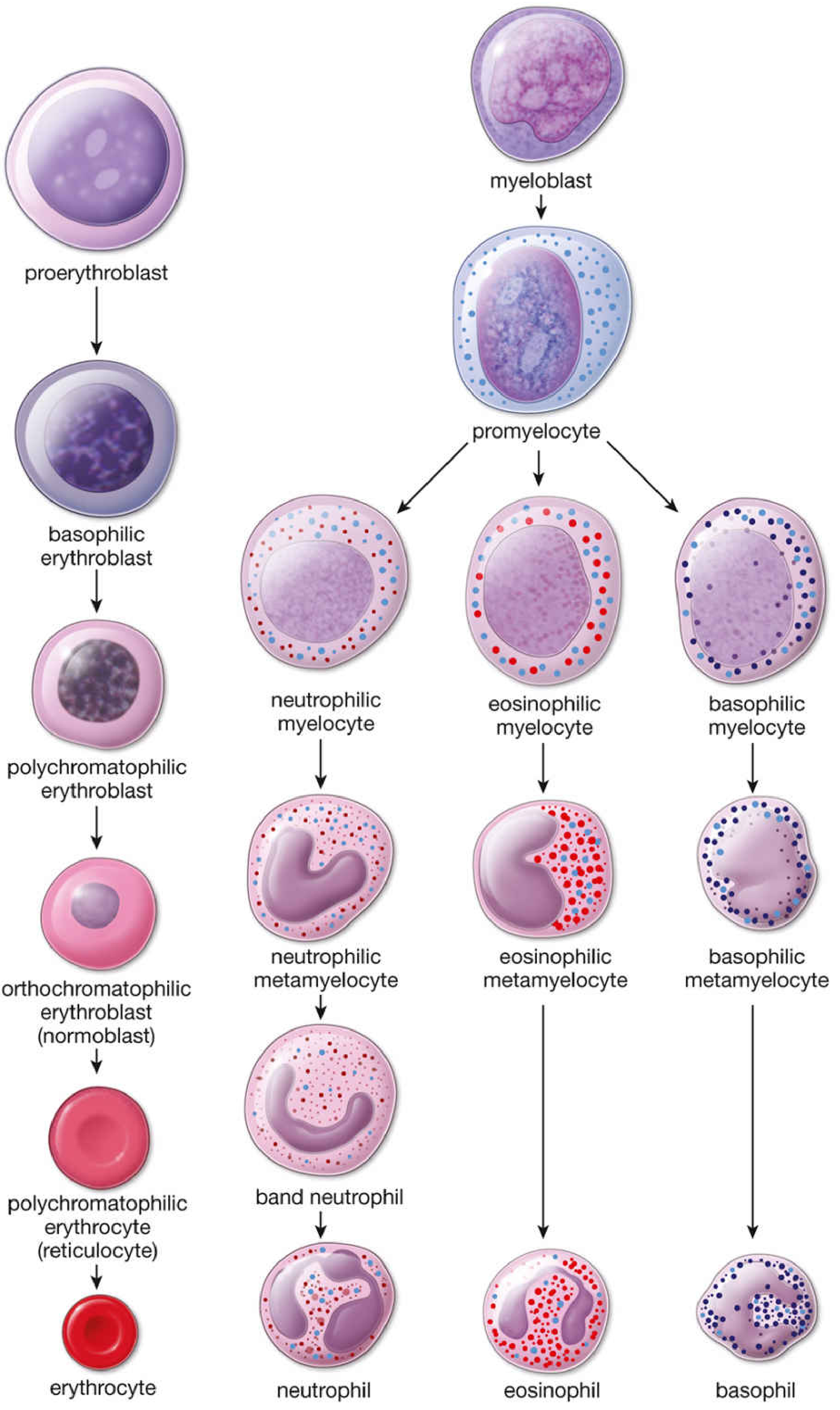
1. Proerythroblast (Pronormoblast)
- Size: 12-20 μm (largest precursor)
- Nucleus: Large, spherical; fine uniform chromatin; 1-2 prominent nucleoli; prominent nuclear membrane
- Cytoplasm: Mildly basophilic (free ribosomes beginning to accumulate); no granules
- Activity: Components for Hgb production begin to accumulate; stage lasts ~24 hours
2. Basophilic Erythroblast (Basophilic Normoblast)
- Size: 10-16 μm; smaller than proerythroblast (arises by mitosis)
- Nucleus: Smaller, progressively heterochromatic; chromatin may appear as "wheel with broad spokes"; nucleoli present but often not visible; parachromatin stains pink
- Cytoplasm: Deeply basophilic due to abundant polyribosomes synthesizing Hgb; cell borders may be irregular with pseudopodia
- Activity: Active Hgb synthesis begins; highest RNA content; stage lasts ~24 hours
3. Polychromatophilic Erythroblast (Polychromatophilic Normoblast)
- Size: Slightly smaller than basophilic erythroblast
- Nucleus: Occupies ~half the cell area; coarse heterochromatin in checkerboard pattern; no visible nucleoli
- Cytoplasm: Mixed blue (RNA) + pink (Hgb) = gray or lilac appearance (polychromasia); distinct pink and purple regions may be visible
- Activity: This is the last stage capable of mitosis (undergoes 1-2 more divisions); stage lasts ~30 hours
4. Orthochromatophilic Erythroblast (Orthochromatic Normoblast / Normoblast)
- Size: Smaller; lower N/C ratio
- Nucleus: Small and pyknotic (densely condensed, no chromatin pattern visible); mitosis no longer possible
- Cytoplasm: Predominantly eosinophilic/pink due to abundant Hgb; fewer polyribosomes remain
- Activity: Nucleus is extruded (with a small rim of cytoplasm) via cytoplasmic contractions to produce a reticulocyte
5. Reticulocyte (Polychromatophilic Erythrocyte)
- No nucleus (ejected at orthochromatic stage)
- Size: Larger than mature RBCs
- Cytoplasm: Pink-gray; still contains residual ribosomes, mRNA, and mitochondria → can still synthesize Hgb
- Location: Remains in marrow stroma for 1-2 days, then released into blood; circulates for ~1-2 days
- Maturation: Final maturation occurs in the spleen, where remaining ribosomes and mitochondria are removed
- Normal count in peripheral blood: ~0.5-1.5% of red cells
6. Mature Erythrocyte
- Size: ~7-8 μm biconcave disc
- No nucleus, no organelles, no protein synthesis
- Packed with Hgb (~280 million molecules per cell)
- Life span: ~120 days; senescent cells phagocytosed by macrophages in spleen, bone marrow, and liver
4. RNA vs. Hemoglobin Dynamics During Maturation
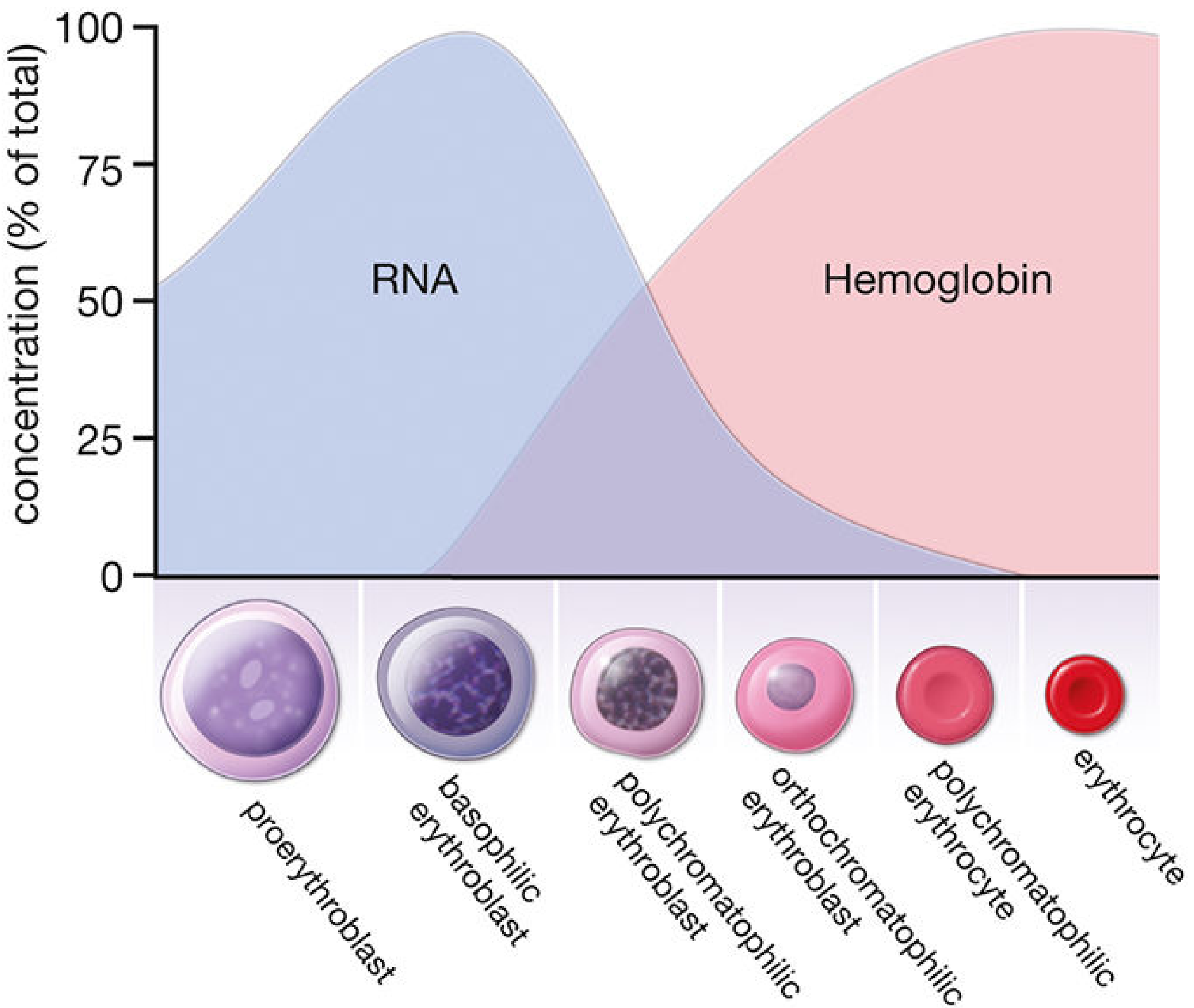
- Early stages (proerythroblast, basophilic erythroblast): High RNA → deep cytoplasmic basophilia
- As maturation proceeds: RNA falls, Hgb rises → polychromasia → eosinophilia
- Key principle: Cytoplasmic color shifts blue → gray/lilac → pink in parallel with RNA→Hgb transition
5. Kinetics
| Parameter | Value |
|---|---|
| Mitotic divisions from pronormoblast | 3-4 divisions over ~3 days |
| Reticulocytes produced per pronormoblast | Up to 16 |
| Time from basophilic erythroblast to circulation | ~1 week |
| Reticulocyte time in marrow | 1-2 days |
| Reticulocyte time in blood | 1-2 days |
| RBC life span | ~120 days |
| Daily RBC production | ~250 billion cells |
6. Erythroblastic Islands
In the marrow, developing erythroid cells cluster around central macrophages in structures called erythroblastic islands. The macrophage supplies iron directly to the developing normoblasts. These islands are usually disrupted when marrow is aspirated, but fragments of macrophage cytoplasm may remain attached to normoblasts (best seen on Prussian blue stain).
7. Sites of Erythropoiesis Across Life
| Period | Site |
|---|---|
| Embryo (yolk sac, 3-8 wks) | Primitive erythropoiesis (nucleated RBCs) |
| Fetus (liver, spleen) | Definitive erythropoiesis |
| After birth | Red bone marrow (axial skeleton in adults) |
| Stress/disease | Extramedullary erythropoiesis (liver, spleen can resume) |
Sources:
- Histology: A Text and Atlas with Correlated Cell and Molecular Biology, pp. 737-746
- Henry's Clinical Diagnosis and Management by Laboratory Methods, pp. 658-659
- Basic Medical Biochemistry: A Clinical Approach (6e), p. 1536
- Brenner and Rector's The Kidney, 2-Volume Set, p. 2465
This is a shared conversation. Sign in to Orris to start your own chat.