
bulbocavernosus and ischiocavernosus muscles exercises
- Bulbocavernosus: surrounds the vaginal opening (females) / base of penis (males); contracts during orgasm and helps with urinary control
- Ischiocavernosus: runs from ischial tuberosity to clitoris/penis; maintains erection by compressing crural veins
- Both are trained primarily through pelvic floor (Kegel) contractions, with some sport-specific add-ons like squats and hip bridges
bulbocavernosus ischiocavernosus muscle exercises pelvic floor
bulbocavernosus ischiocavernosus perineal muscles anatomy diagram
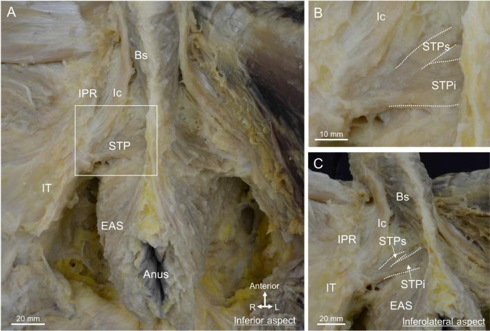
This composite of three clinical photographs displays a detailed anatomical dissection of the male perineum, focusing on the superficial perineal muscles and their spatial relationships. Panel A provides an inferior view, labeling the bulbospongiosus (Bs) centrally, the ischiocavernosus (Ic) running along the ischiopubic ramus (IPR), and the superficial transverse perineal muscle (STP) extending laterally toward the ischial tuberosity (IT). The external anal sphincter (EAS) is shown surrounding the anus. Panel B is a magnified inset of the rectangular area in A, demarcating the division of the STP into a superior bundle (STPs) and an inferior bundle (STPi). Panel C provides an inferolateral aspect with a slight pelvic tilt, illustrating the continuity between the muscle fibers of the bulbospongiosus and the external anal sphincter. The images emphasize the concentration of these muscle groups within the central region of the perineum, providing essential context for understanding pelvic floor anatomy, urogenital support, and surgical landmarks in the male pelvis.
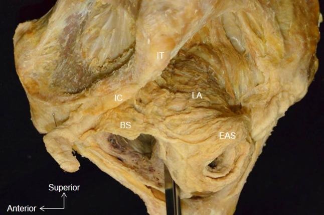
This clinical photograph displays a lateral aspect of a whole pelvis dissection, focusing on the musculature of the pelvic floor and perineum. Key anatomical landmarks are labeled: the bulbospongiosus (BS), external anal sphincter (EAS), ischiocavernosus (IC), ischial tuberosity (IT), and levator ani (LA). The image specifically demonstrates the anatomical continuity between the perineal and anal musculature, showing the muscle bundle of the bulbospongiosus (BS) attaching directly to the lateral surface of the circular external anal sphincter (EAS). Superior to these, the levator ani (LA) is visible, positioned deep to the bony ischial tuberosity (IT). The ischiocavernosus (IC) is situated anterior and superior to the bulbospongiosus. This dissection illustrates the spatial arrangement of the superficial and deep pelvic muscles, highlighting the integrated nature of the pelvic support structures and the sphincteric complex, which is critical for understanding pelvic floor functional anatomy and surgical approaches.
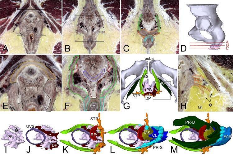
This composite educational graphic details the anatomy of the female perineal body and superficial perineal muscles through high-resolution cross-sectional imaging and 3D reconstructions. Panels A-C provide transverse sections from superior to inferior, illustrating the spatial relationship between the urethra (U), vagina (V), and rectum (R). Panel D indicates the anatomical levels of these sections relative to the pelvis. Panels E, F, and H offer magnified views highlighting muscular attachments and venous plexuses (Vv). Panel G presents an inferior 3D view of the superficial perineal region, identifying the bulbospongiosus (BS), ischiocavernosus (IC), and superficial transverse perineal (STP) muscles flanking the perineal body (PB). Panels I-M display a serial layer-by-layer 3D reconstruction of the perineal body and its complex muscular attachments, including the urethrovaginal sphincter (UVS), deep perineal muscle (DP), rectoperineal muscle (RP), internal anal sphincter (IAS), and the deep (PR-D) and superficial (PR-S) portions of the puborectal muscle. The content demonstrates the integrated structural support of the pelvic floor and the fibrous architecture of the perineal body as a central tendinous point.
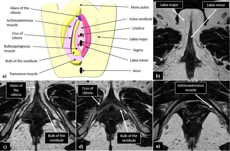
An educational composite featuring a schematic diagram and magnetic resonance imaging (MRI) scans detailing normal female vulvar anatomy. Panel (a) is a color-coded anatomical illustration labeling the mons pubis, labia majora and minora, vulva vestibule, urethra, vagina, clitoris (glans and crus), bulbospongiosus and ischiocavernosus muscles, bulb of the vestibule, and anus. Panels (b) through (e) provide corresponding axial T2-weighted imaging (T2WI) sequences. These radiological images demonstrate the intermediate signal intensity of the labia majora (lateral) and labia minora (medial). Key landmarks visualized in the MRI series include the glans of the clitoris anteriorly, the posterior-medial crus of the clitoris, the bulb of the vestibule, and the ischiocavernosus muscle. The images highlight the high-contrast resolution of MRI for depicting perineal soft tissues, which is essential for the clinical staging of vulvar malignancies. The content is suitable for medical education on pelvic anatomy and radiological interpretation.
pelvic floor muscle training bulbocavernosus ischiocavernosus exercise
pelvic floor exercise erectile dysfunction ejaculation
Bulbocavernosus (Bulbospongiosus) & Ischiocavernosus Muscles - Anatomy and Exercises
Anatomy Overview
| Muscle | Origin | Insertion | Key Function |
|---|---|---|---|
| Ischiocavernosus | Ischial tuberosity & ramus | Crus of penis/clitoris | Forces blood from crura into the erect penis/clitoris; maintains rigidity by compressing crural veins |
| Bulbospongiosus (bulbocavernosus) | Perineal body (+ midline raphe in men) | Bulb of vestibule, perineal membrane, corpus cavernosum | Pumps blood from corpus spongiosum into penile/clitoral body; expels urine/semen; constricts vaginal orifice |
Why Exercise These Muscles?
- Improved erectile rigidity (they raise intracavernous pressure 3-4x above systolic BP during erection)
- Better ejaculatory control and force
- Urinary continence
- Enhanced sexual response in both sexes
- Recovery after prostatectomy or childbirth
Exercise Protocol
1. Core Technique - Kegel (Pelvic Floor Contraction)
- Imagine stopping the flow of urine mid-stream and preventing passing gas simultaneously
- You should feel a lift-and-squeeze inward sensation at the perineum
- The buttocks, inner thighs, and abdomen should NOT contract
- In men: you should see a penile retraction and scrotal lift
- In women: you should feel the vaginal opening draw inward
2. Endurance Holds (Bulbospongiosus + Ischiocavernosus tonic fiber training)
- Contract the pelvic floor muscles to ~50-70% of maximal effort
- Hold for 6-10 seconds
- Fully relax for equal time (6-10 seconds) - the relaxation phase is as important as the contraction
- Repeat 10 reps x 3 sets
- Progress: increase hold duration by 2 seconds each week, up to 30-second holds
3. Quick Flicks (Power/Ejaculatory Control Training)
- Contract the pelvic floor as fast and hard as possible for 1 second
- Fully release immediately
- Repeat 10-15 reps x 3 sets
4. Progressive Resistance Positions
| Stage | Position | Rationale |
|---|---|---|
| 1 | Lying supine (knees bent) | Gravity-assisted; easiest to feel the muscles |
| 2 | Sitting | Increased load from body weight |
| 3 | Standing | Full weight-bearing, most functional |
| 4 | During dynamic movements (squat, stair climb) | Co-contraction with global muscles |
5. Compound Exercises That Recruit These Muscles
- Lie supine, knees bent, feet flat
- Contract pelvic floor first, then lift hips off floor
- Hold 3-5 seconds at the top; lower slowly
- 3 x 10 reps
- Perform a bodyweight squat
- Contract pelvic floor on the way up (exhalation phase)
- This mimics the functional co-contraction pattern
- From all-fours, extend opposite arm and leg
- Maintain pelvic floor contraction throughout
- 3 x 8 each side
6. Ischiocavernosus-Specific Technique (Males)
- Perform a strong pelvic floor contraction, aiming to compress the base/crura
- You should see the penis pulse/lift
- This specifically loads the ischiocavernosus through its functional range
- 10-15 contractions, 2-3 sets
7. Reverse Kegels (Relaxation Training)
- Inhale deeply into the belly and consciously let the perineum descend/bulge outward
- Hold the relaxed/open state for 5-10 seconds
- Exhale and gently re-engage
Training Parameters Summary
| Parameter | Beginner | Intermediate | Advanced |
|---|---|---|---|
| Hold duration | 5-6 sec | 8-10 sec | 15-30 sec |
| Quick flicks per set | 10 | 15 | 20 |
| Sets per session | 3 | 4 | 5 |
| Sessions per day | 2-3 | 2-3 | 2 |
| Weeks to effect | 4-6 weeks | - | 3-6 months |
Important Notes
- Biofeedback (surface EMG or manometric probe) significantly improves motor learning and is used clinically to confirm correct muscle activation
- Quality over quantity: a correctly performed contraction with full relaxation is far more beneficial than many poorly performed ones
- If there is pelvic pain (chronic prostatitis, vaginismus, interstitial cystitis), pelvic floor muscles are often hypertonic - strengthening exercises are contraindicated until relaxation/down-training is achieved first
- If symptoms (erectile dysfunction, incontinence, sexual dysfunction) persist after 3 months of consistent training, referral to a pelvic floor physiotherapist is recommended for individualized assessment including internal examination