
Septo optic dysplasia meaning
Reading File
Now let me search the medical library and web in parallel.
Finding Sources
Finding Sources
Searching the Web
septo-optic dysplasia definition clinical features causes
Good results. Let me read the dedicated sections from the textbooks now.
Reading File
Reading File
Reading File
Reading File
I have comprehensive information from multiple authoritative textbooks. Let me also fetch the optic nerve hypoplasia fundus image since it's directly relevant.
Loading Image
I now have all the information needed for a thorough answer.
Septo-Optic Dysplasia (SOD)
Also called de Morsier syndrome, septo-optic dysplasia is a rare congenital malformation characterized by abnormal development of midline forebrain structures. It is diagnosed when two or more of the following three features are present:
- Optic nerve hypoplasia - underdevelopment of one or both optic nerves/chiasm, causing visual impairment
- Midline brain abnormality - absence (agenesis) of the septum pellucidum, and sometimes the corpus callosum or anterior commissure
- Pituitary/hypothalamic dysfunction - ranging from isolated growth hormone deficiency to panhypopituitarism
- Goldman-Cecil Medicine, p. 561
- Bradley and Daroff's Neurology in Clinical Practice, p. 305
- Grainger & Allison's Diagnostic Radiology, p. 3620
Classic Triad
| Component | What happens |
|---|---|
| Optic nerve hypoplasia | Small optic discs with the characteristic "double ring sign" on fundoscopy; reduced visual acuity, possible nystagmus |
| Absent septum pellucidum | Midline brain septum is missing; frontal horns take on a "box-like" shape on imaging |
| Hypopituitarism | GH deficiency (most common), hypothyroidism, central diabetes insipidus, adrenal insufficiency, or full panhypopituitarism |
Fundus Appearance in Optic Nerve Hypoplasia
The small disc appears as the inner circle of a characteristic "double ring" - the outer ring is a border of choroidal and retinal pigment changes:
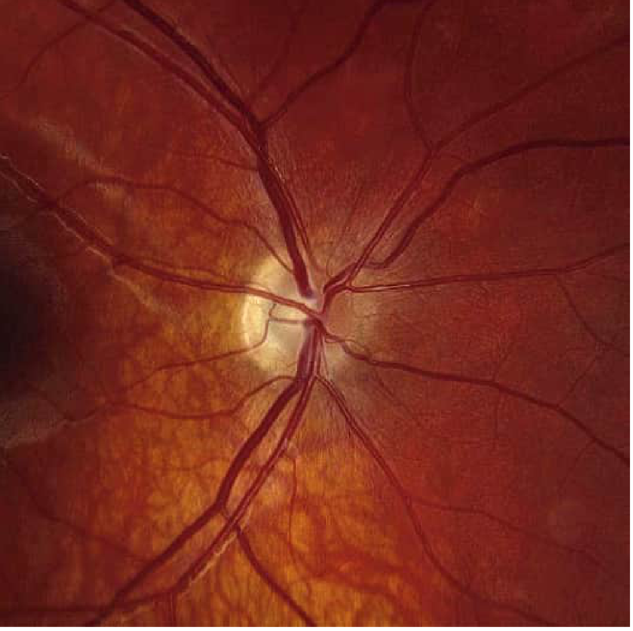
Fundus photo showing optic nerve hypoplasia (ONH) - from Bradley and Daroff's Neurology in Clinical Practice
Genetics and Causes
-
Mutations in the HESX1 gene (chromosome 3p14) have been identified in some cases, though less than 1% of SOD cases carry HESX1 mutations
-
Often sporadic - no familial cases well established
-
Risk factors include:
- Young or teenage maternal age
- Maternal diabetes mellitus
- Maternal use of antiepileptic drugs, quinine, or LSD during pregnancy
- Maternal drug or alcohol abuse in early gestation
-
Goldman-Cecil Medicine, p. 561; Bradley and Daroff's, p. 313
Embryological Basis
SOD arises from defective development of structures sharing a forebrain (prosencephalic) embryological origin. The septum pellucidum, optic nerve, and hypothalamic-pituitary axis all derive from the ventral forebrain, so a single early developmental insult can disrupt all three. It overlaps clinically with semilobar holoprosencephaly.
Associated Features
- Hypoplasia of olfactory bulbs (anosmia or hyposmia) in some cases
- Midline cerebellar defects (rare)
- Hydrocephalus (inconsistently)
- Detachment of the fornix from the corpus callosum
- Rhombencephalosynapsis (aplasia of the cerebellar vermis with fusion of hemispheres) - rare
Clinical Presentation
- Visual impairment - diminished acuity, nystagmus
- Endocrine symptoms - short stature (GH deficiency), hypoglycemia in neonates, polyuria/polydipsia (diabetes insipidus), delayed puberty or precocious puberty
- Seizures are uncommon
- Intellectual development is usually normal
- Ataxia if cerebellum is involved
Neuroimaging (MRI) Findings
-
Absent septum pellucidum with box-like frontal horns
-
Small optic nerves and chiasm
-
Small anterior pituitary gland
-
Ectopic "bright spot" of posterior pituitary on T1-weighted MRI (indicating ectopic posterior pituitary)
-
Grainger & Allison's Diagnostic Radiology, p. 3620
Management
- Hormone replacement for any identified pituitary deficiencies (GH, thyroid, cortisol, ADH as needed)
- Visual and developmental support
- Regular endocrine monitoring throughout childhood
In summary, SOD is not a single defect but a spectrum - some patients have all three features, others only two, and the clinical severity varies widely based on which structures are affected and to what degree.
This is a shared conversation. Sign in to Orris to start your own chat.