
Important clinical features of hepatitis B virus
Reading File
Now let me search the medical library for hepatitis B clinical features, running parallel searches:
Finding Sources
Finding Sources
Searching PubMed
"hepatitis B" clinical features
Good. Now let me read the primary sources - Jawetz Microbiology for clinical features and Harrison's for management, plus Sleisenger for GI/liver perspective:
Reading File
Reading File
Finding Sources
Now let me get the Harrison's and Henry's content, plus the natural history/serology sections from Jawetz:
Finding Sources
Reading File
Excellent content. Let me now fetch the serology image and get Harrison's clinical features content:
Loading Image
Finding Sources
Reading File
Let me now read the Jawetz section on clinical outcomes (chronic hepatitis, cirrhosis, HCC) and the Harrison's section:
Reading File
Finding Sources
Now let me search for Harrison's HBV content and the extrahepatic manifestations:
Finding Sources
Finding Sources
Reading File
Reading File
Reading File
I now have excellent material from multiple authoritative sources. Let me compile a comprehensive answer.
Clinical Features of Hepatitis B Virus (HBV) Infection
1. The Virus and Epidemiology
HBV is a member of the Hepadnaviridae family - a partially double-stranded DNA virus that replicates via reverse transcriptase. It encodes four genes: C (core protein/HBcAg), S (surface antigen/HBsAg - large, middle, and small forms), P (DNA polymerase), and X (unclear function). There are 4 serotypes (adr, adw, ayr, ayw) and 8 genotypes with differing virulence and treatment responses.
- Over 350 million chronic carriers worldwide; ~600,000 deaths/year from HBV-related disease
- 25% of chronic carriers develop chronic active hepatitis
- High viremia: up to 10¹⁰ particles/mL during peak infection
Transmission routes:
- Perinatal (mother-to-newborn during delivery) - most important in endemic regions
- Parenteral (IV drug use, unsterile needles, tattooing, transfusion)
- Sexual contact
- Close household contact
- HBsAg detectable in saliva, semen, vaginal secretions, menstrual fluid - but fecal-oral route NOT documented
High-risk groups: parenteral drug users, hemodialysis patients, healthcare workers, multiply transfused patients, organ transplant recipients, promiscuous individuals, newborns of HBsAg-positive mothers
2. Natural History and Clinical Phases
Incubation period: 50-180 days (mean 60-90 days). Longer with low-dose or non-percutaneous exposure.
Acute HBV Infection
The clinical course varies enormously - from asymptomatic (most common) to fulminant hepatic failure (<1%).
Phases of acute illness:
- Prodrome (1-2 weeks before jaundice): fever, malaise, anorexia, nausea, vomiting, arthralgias, rash (serum sickness-like)
- Icteric phase: jaundice, dark urine, pale stools, right upper quadrant discomfort, hepatomegaly; lasts 2 weeks-3 months
- Convalescent phase: gradual resolution; ALT/AST normalize over 1-2 months
Outcomes of acute infection (age-dependent):
| Age at infection | Risk of chronicity |
|---|---|
| Neonates | ~90% |
| Children 1-5 yr | 25-50% |
| Immunocompetent adults | <5% |
| Immunocompromised adults | Up to 50% |
Most immunocompetent adults (>95%) clear HBsAg spontaneously. Hemodialysis patients have ~50% rate of chronic carriage.
3. Serological Markers - Key to Diagnosis
The chart below shows the classic timeline:
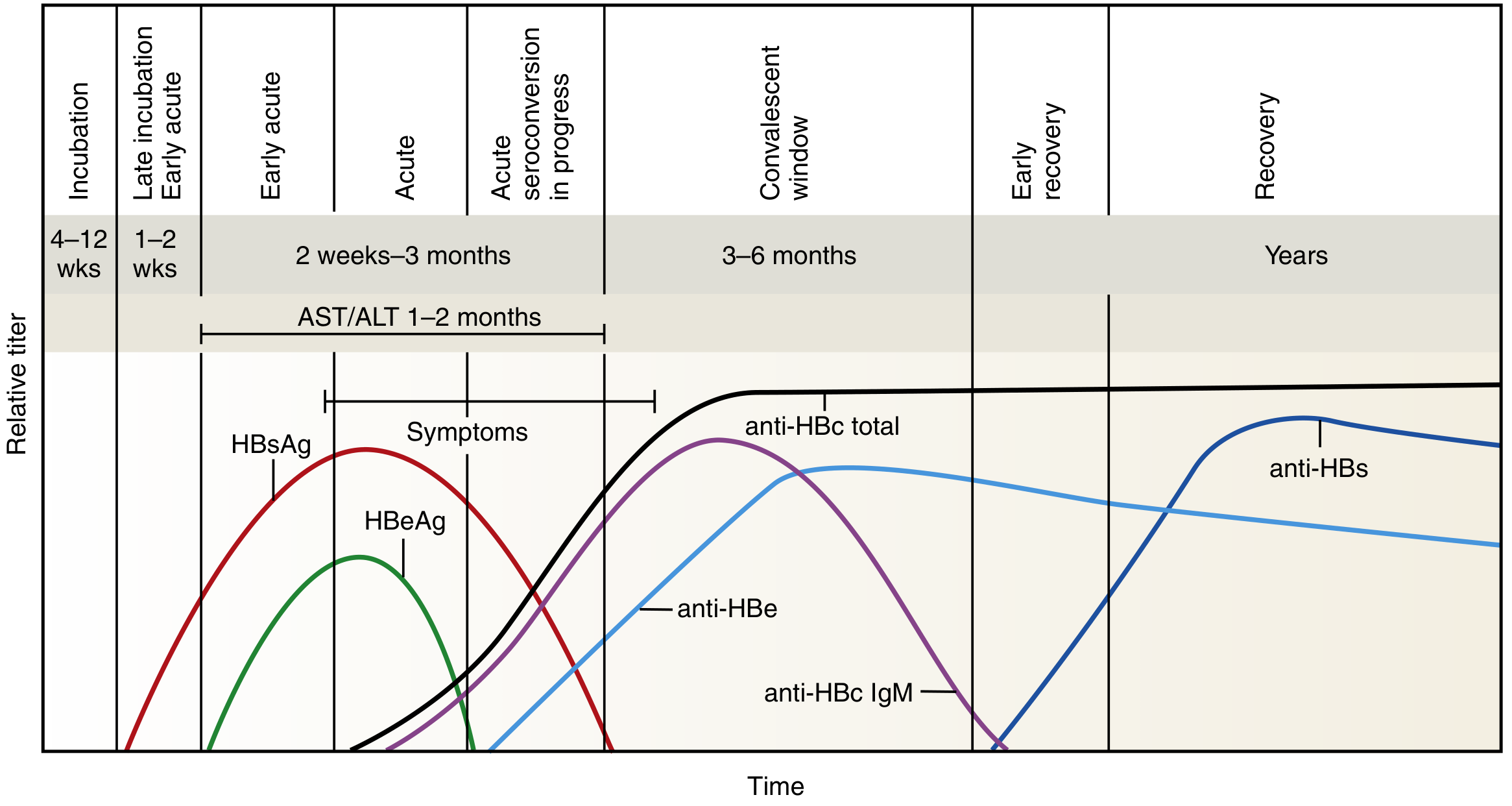
| Marker | Significance |
|---|---|
| HBsAg | First marker to appear (2-6 weeks before symptoms); indicates active infection. Persists >6 months = chronic infection |
| HBeAg | Marker of active viral replication; high infectivity (>10⁹ virions/mL) |
| HBV DNA | Most sensitive and accurate marker of replication (by PCR) |
| IgM anti-HBc | Appears at onset of clinical illness; indicates acute infection or reactivation. "Core window" marker |
| IgG anti-HBc | Persists lifelong; past or chronic infection |
| Anti-HBe | Appears as HBeAg disappears; signals start of resolution |
| Anti-HBs | Protective antibody; indicates recovery or vaccination |
"Core window" period: After HBsAg disappears but before anti-HBs appears, IgM anti-HBc is the only positive marker - critical for diagnosis.
Pre-core mutants: Some patients develop HBeAg-negative chronic hepatitis due to a stop codon mutation at nucleotide 1896, preventing HBeAg production but allowing continued viral replication - an important and aggressive clinical variant.
Chronic carrier definition: HBsAg persisting >6 months, with either HBeAg or anti-HBe present.
4. Phases of Chronic HBV Infection
| Phase | HBsAg | HBeAg | HBV DNA | ALT | Inflammation |
|---|---|---|---|---|---|
| Immune tolerant | + | + | Very high (>10⁶ IU/mL) | Normal | Minimal |
| Immune active (HBeAg+) | + | + | High (>10⁶ IU/mL) | Elevated | Active |
| Inactive carrier | + | - | Low (<10² IU/mL) | Normal | Minimal |
| Immune reactivation (HBeAg-) | + | - | Moderate (>10⁴ IU/mL) | Elevated | Active |
| Resolved | - | - | Negative | Normal | None |
5. Histopathology
Classic features on liver biopsy (relevant in chronic HBV):
- Panlobular mononuclear cell infiltration (lymphocytes predominate)
- Hepatocyte necrosis with ballooning degeneration, cell dropout
- Acidophilic (Councilman/apoptotic) bodies
- Kupffer cell hyperplasia
- Ground-glass hepatocytes (pathognomonic of chronic HBV) - contain HBsAg, identifiable with orcein or aldehyde fuchsin stain
- In severe disease: bridging necrosis (portal-portal, portal-central)
- Cirrhosis in end-stage disease
6. Extrahepatic Manifestations
The pathogenesis involves circulating immune complexes (HBsAg-anti-HBs) depositing in vessel walls with complement activation - serum complement levels are typically low.
| Manifestation | Details |
|---|---|
| Serum sickness-like prodrome | Arthralgias, urticaria, angioedema - in acute HBV (precedes jaundice) |
| Polyarteritis nodosa (PAN) | Up to 30% of PAN patients have HBV; features include fever, rash, abdominal pain, renal disease, mononeuritis multiplex, hypertension, CNS involvement |
| Glomerulonephritis | Membranous GN and MPGN most common; nephrotic syndrome is typical presentation; immune complex + HBV antigen deposition in basement membrane |
| Membranous nephropathy | Especially in children; may resolve after HBeAg seroconversion |
| Cryoglobulinemia | Type II/III; associated vasculitis (purpura, arthralgias, peripheral neuropathy) |
| Papular acrodermatitis (Gianotti-Crosti) | Predominantly in children |
These manifestations can occur without clinically apparent liver disease and may masquerade as independent rheumatologic or nephrologic conditions.
7. Complications of Chronic HBV
- Cirrhosis: 25% of chronic carriers with active disease develop cirrhosis
- Hepatocellular carcinoma (HCC): Major risk - especially in those infected perinatally; HBV is the leading global cause of HCC
- Liver failure: Decompensation with ascites, variceal bleeding, encephalopathy, hepatorenal syndrome
- HBV reactivation: Can occur in immunosuppressed patients (chemotherapy, biologics, steroids) - potentially fatal
- Superinfection with HDV (hepatitis D): Requires HBsAg as its envelope; dramatically worsens prognosis
8. Diagnostic Summary
- HBsAg + IgM anti-HBc = acute infection
- HBsAg + IgG anti-HBc (>6 months) = chronic infection
- Anti-HBs + anti-HBc = past infection with immunity
- Anti-HBs alone = vaccine-induced immunity
- IgM anti-HBc alone = "core window" of acute infection
- HBV DNA = best marker of active replication, used to guide treatment
- Liver biopsy = grades inflammation, stages fibrosis, guides treatment decisions
Sources:
- Jawetz Melnick & Adelberg's Medical Microbiology 28e, Chapter 35
- Harrison's Principles of Internal Medicine 22e (2025)
- Henry's Clinical Diagnosis and Management by Laboratory Methods, Chapter 22
- Sleisenger and Fordtran's Gastrointestinal and Liver Disease, Chapter 79
- Washington Manual of Medical Therapeutics, Chapter 19
Difference between gonorrhea and hepatitis B
Reading File
Now let me search both topics in parallel:
Finding Sources
Finding Sources
Finding Sources
Finding Sources
Reading File
Reading File
Reading File
Reading File
Finding Sources
Now let me get the disseminated gonococcal infection (DGI) section and HBV treatment:
Reading File
Finding Sources
I now have rich material from both diseases across multiple authoritative textbooks. Let me compile the comprehensive comparison.
Gonorrhea vs. Hepatitis B - A Comprehensive Comparison
These are two very different sexually transmitted infections sharing only the route of transmission (sexual contact). Every other aspect - pathogen, organ targeted, clinical course, complications, and treatment - differs fundamentally.
1. Basic Microbiology
| Feature | Gonorrhea | Hepatitis B |
|---|---|---|
| Causative agent | Neisseria gonorrhoeae (bacterium) | Hepatitis B virus (DNA virus, Hepadnaviridae) |
| Type of organism | Gram-negative diplococcus | Partially double-stranded DNA virus; replicates via reverse transcriptase |
| Size/structure | ~0.6-1 µm; aerobic, oxidase-positive | 42 nm Dane particle; envelope (HBsAg), nucleocapsid (HBcAg), genome |
| Key virulence factors | Pili (adhesion), outer membrane proteins (PorA, PorB), IgA protease, LOS | HBsAg (3 forms), HBeAg (replication marker), DNA polymerase (reverse transcriptase), X protein |
2. Epidemiology and Transmission
| Feature | Gonorrhea | Hepatitis B |
|---|---|---|
| Global burden | Second most common bacterial STI (after Chlamydia) | >350 million chronic carriers; ~600,000 deaths/year |
| Primary route | Direct mucosal contact during sex (vaginal, anal, oral) | Sexual contact, percutaneous (IV drugs, needlestick), perinatal (mother-to-newborn) |
| Perinatal transmission | Neonatal conjunctivitis (ophthalmia neonatorum) at delivery | Major route in endemic areas; >90% chronicity if infected at birth |
| Fecal-oral route | No | No |
| Blood/needles | Rare | Major route (up to 10¹⁰ virions/mL in blood) |
| Infectivity in bodily fluids | Semen, vaginal secretions, rectal secretions | Blood, semen, vaginal secretions, saliva, breast milk |
3. Incubation Period
| Gonorrhea | Hepatitis B | |
|---|---|---|
| Range | 2-14 days (typically 2-7 days) | 30-180 days (mean 60-90 days) |
Gonorrhea declares itself within days; HBV remains clinically silent for weeks to months.
4. Primary Organ Targeted
| Gonorrhea | Hepatitis B | |
|---|---|---|
| Primary target | Columnar/transitional mucosal epithelium (genitourinary tract) | Hepatocytes (liver) |
| Entry mechanism | Pili attach to mucosal epithelium → penetrate via endocytosis | Receptor-mediated endocytosis → nucleus via chaperone proteins → cccDNA forms |
5. Clinical Manifestations
Gonorrhea
In men:
- Urethral discharge (initially scant/mucoid, then profuse/purulent) within 2-7 days
- Dysuria, urinary frequency (without urgency)
- Up to 10% remain asymptomatic
- Complications: epididymitis, prostatitis, orchitis, urethral stricture
In women (~50% asymptomatic):
- Mucopurulent cervicitis (most common)
- Vaginal discharge, dysuria, dyspareunia
- Urethritis, Bartholin gland abscess
- Complications: Pelvic Inflammatory Disease (PID) - leading to infertility and ectopic pregnancy
- Gonococcal vaginitis (in prepubertal girls/postmenopausal women)
Other sites:
- Pharyngitis (oral sex)
- Proctitis/anorectal infection (anal sex, or cervical exudate spread in women)
Disseminated Gonococcal Infection (DGI):
- Occurs in 0.5-3% of untreated cases (more common in women)
- Triad: polyarthralgias/migratory arthritis, tenosynovitis, dermatitis (pustular/vesicular skin lesions on distal extremities)
- Septic arthritis (most common in knees, wrists, ankles)
- Rarely: endocarditis, meningitis
Hepatitis B
Acute HBV:
- Prodrome (1-2 weeks): anorexia, nausea, vomiting, fatigue, arthralgias, low-grade fever, myalgias
- Serum sickness-like syndrome (rash, arthralgias) can precede jaundice
- Icteric phase: jaundice, dark urine, clay-colored stools, hepatomegaly (tender), right upper quadrant pain
- Splenomegaly and cervical adenopathy in 10-20%
- ALT/AST elevated for 1-2 months
- Many patients are anicteric (no jaundice)
Chronic HBV (persists >6 months in HBsAg+):
- Phases: immune tolerant → immune active → inactive carrier → immune reactivation
- Chronic hepatitis, cirrhosis, portal hypertension, liver failure
- Hepatocellular carcinoma (HCC) - major long-term risk
Extrahepatic manifestations (immune complex-mediated):
- Polyarteritis nodosa (up to 30% of PAN cases are HBV-related)
- Membranous/membranoproliferative glomerulonephritis (nephrotic syndrome)
- Cryoglobulinemia
- Papular acrodermatitis (Gianotti-Crosti, children)
6. Histopathology
| Gonorrhea | Hepatitis B | |
|---|---|---|
| Key finding | Intense suppurative (neutrophilic) inflammation; gram-negative intracellular diplococci in PMNs | Mononuclear (lymphocytic) infiltration; hepatocyte necrosis; Councilman/apoptotic bodies |
| Pathognomonic feature | Gram-negative intracellular diplococci on Gram stain of discharge | Ground-glass hepatocytes (contain HBsAg; stain with orcein/aldehyde fuchsin) in chronic disease |
| Scarring | Fibrosis/strictures of genitourinary tract; tuboovarian abscesses | Hepatic fibrosis → cirrhosis; bridging necrosis in severe disease |
7. Diagnosis
| Gonorrhea | Hepatitis B | |
|---|---|---|
| First-line test | NAAT (nucleic acid amplification test) - most sensitive | HBsAg (first marker to appear, 2-6 weeks before symptoms) |
| Rapid diagnosis | Gram stain of urethral discharge (high specificity in symptomatic men; ~50% sensitive in women) | IgM anti-HBc (marks acute infection, "core window") |
| Gold standard culture | Modified Thayer-Martin medium (selective); CO₂-enriched atmosphere | PCR for HBV DNA (most accurate replication marker) |
| Chronicity marker | N/A (bacteria cleared or cause complications) | HBsAg persisting >6 months |
| Immunity marker | N/A | Anti-HBs (protective antibody - vaccination or recovery) |
| Replication marker | N/A | HBeAg, HBV DNA |
| Liver assessment | N/A | ALT/AST, liver biopsy (grade inflammation, stage fibrosis) |
8. Treatment
| Gonorrhea | Hepatitis B | |
|---|---|---|
| Acute uncomplicated | Ceftriaxone IM (500 mg single dose; 1g if >150 kg) - first-line | Supportive care; antivirals for severe acute disease |
| Prior options | Fluoroquinolones - NO LONGER recommended (widespread resistance) | No role for antibiotics |
| Chronic/prevention | N/A | Tenofovir (TDF or TAF) or entecavir - preferred oral antivirals; pegylated interferon-alfa (alternative) |
| DGI/complications | Ceftriaxone 1g IV/IM daily | Liver transplant for end-stage liver disease |
| Antibiotic resistance | Growing major problem (esp. azithromycin, fluoroquinolones) | Not applicable (viral) |
| Vaccine | No vaccine available | Highly effective recombinant vaccine available; 3-dose schedule; neonatal immunization recommended |
| Prophylaxis | Neonatal eye prophylaxis (erythromycin) for ophthalmia neonatorum | Neonatal HBsAg + HBIG within 12 hours of birth for HBsAg+ mothers |
9. Summary of Key Differences at a Glance
| Parameter | Gonorrhea | Hepatitis B |
|---|---|---|
| Type of pathogen | Bacterium | Virus |
| Primary organ | Genitourinary tract | Liver |
| Incubation | 2-14 days | 30-180 days |
| Chronicity | None (acute bacterial infection) | Up to 90% (neonates); <5% (adults) |
| Cancer risk | No | Yes - hepatocellular carcinoma |
| Diagnosis | NAAT, Gram stain, culture | Serology (HBsAg, anti-HBc, HBV DNA) |
| Treatment | Antibiotics (ceftriaxone) | Antivirals (tenofovir, entecavir) |
| Vaccine | No | Yes (very effective) |
| Notifiable disease | Yes | Yes |
Sources:
- Harrison's Principles of Internal Medicine 22e (2025), Chapters 135, 161
- Robbins & Kumar Basic Pathology, Chapter 16
- Jawetz Melnick & Adelberg's Medical Microbiology 28e, Chapter 35
- Henry's Clinical Diagnosis and Management by Laboratory Methods, Chapter 22
This is a shared conversation. Sign in to Orris to start your own chat.