I now have all the core content needed. Let me update the todos and compile the comprehensive exam notes.Here are your comprehensive university exam notes for all three topics, sourced from Medical Microbiology 9e and Sherris & Ryan's Medical Microbiology 8e.
🦠 MICROBIOLOGY EXAM NOTES
1. VARICELLA-ZOSTER VIRUS (VZV)
Classification
- Family: Herpesviridae, Subfamily: Alphaherpesvirinae
- HHV-3 (Human Herpesvirus 3)
- Smallest genome of all human herpesviruses (HHVs)
Key Trigger Words (Exam Clue)
Chickenpox, shingles, dermatome, DRG latency, alphaherpesvirus, Oka strain vaccine
Structure & Replication
- Enveloped, dsDNA virus
- Replicates slower and in fewer cell types than HSV
- Productive replication in: human diploid fibroblasts (in vitro), activated T cells, epithelial cells, epidermal cells (in vivo)
- VZV is sequestered into lysosomes and degraded in most cells (via mannose-6-phosphate receptor binding); only terminally differentiated skin cells (lacking this receptor) release infectious virus
- Spreads within the body via cell-to-cell contact
Pathogenesis
Primary Infection (Varicella / Chickenpox):
- Acquired by inhalation - replication begins in tonsils and respiratory mucosa
- Virus spreads via bloodstream and lymphatics to reticuloendothelial system
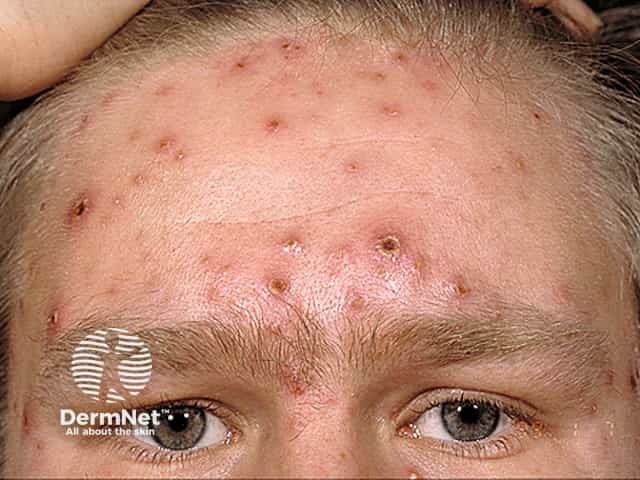
- Secondary viremia spreads virus to skin - forms vesicular rash in successive crops
- VZV infects T cells which home to skin and transfer virus to keratinocytes
- Overcomes IFN-α, produces dermal vesiculopustular rash
CPE: Cowdry type A intranuclear inclusions + syncytia formation
Latency:
- Establishes latency in dorsal root ganglia (DRG), cranial nerve ganglia, and other ganglia
- Unlike HSV: viral RNAs AND specific viral proteins can be detected in latently infected cells
Reactivation (Herpes Zoster / Shingles):
- Occurs when cell-mediated immunity wanes (elderly, immunocompromised)
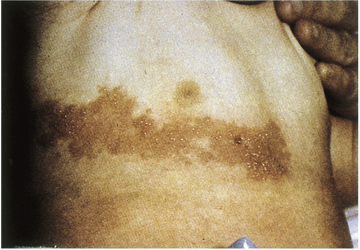
- Virus travels along neuron to skin → vesicular rash in single dermatome
- Typical sites: thoracic dermatome, head (ophthalmic branch of CN V)
- Postherpetic neuralgia (PHN): chronic pain lasting months-years; occurs in ~30%
Immunity
- Cell-mediated immunity is the key control mechanism
- VZV encodes thymidine kinase (like HSV) - basis of antiviral susceptibility
- Immunocompromised patients: risk of dissemination to lungs, brain, liver (potentially fatal)
- Neonates at risk for severe progressive disease
Laboratory Diagnosis
| Test | Notes |
|---|
| PCR / genome detection | Method of choice (VZV labile in transport) |
| Direct fluorescent antibody to membrane antigen (FAMA) | Skin scraping/biopsy |
| Serology (IFA, ELISA) | Screens immunity; antibody levels normally low |
| Culture | NOT routinely done - virus is labile |
Treatment, Prevention, Control
| Drug | Use |
|---|
| Acyclovir (ACV) | Approved for VZV; requires higher doses than for HSV (VZV TK less sensitive) |
| Famciclovir | Better pharmacodynamics |
| Valacyclovir | Better pharmacodynamics |
| VZIg (Varicella-Zoster Immune Globulin) | Post-exposure prophylaxis in high-risk/immunocompromised; NOT effective as therapy for active disease |
Vaccines:
- Varivax: Live attenuated Oka strain - administered after 1 year of age; induces antibody AND cell-mediated immunity
- Shingrix: Adjuvanted subunit vaccine for zoster prevention in adults ≥50
- Medical Microbiology 9e
2. SYPHILIS (Treponema pallidum)
Classification
- Organism: Treponema pallidum subspecies pallidum
- Family: Spirochaetaceae
- Gram negative (does not stain well - outer membrane is antigen-poor)
- Exclusively human pathogen under natural conditions
Key Trigger Words
Chancre, condylomata lata, tabes dorsalis, VDRL/RPR, FTA-ABS, gumma, penicillin G
Transmission & Epidemiology
- Direct sexual contact with active primary/secondary lesion (>50% transmission when lesion present)
- Transplacental (congenital syphilis) - after 4th month of gestation
- Non-genital contact, needle sharing (IDU)
- Late disease is NOT infectious (no blood transmission via modern screening)
- ~6 million new cases annually worldwide; HIV co-infection synergy (syphilitic lesions = portal for HIV)
Pathogenesis
- Spirochete enters via microscopic skin breaks or mucosal passage
- Adheres via adhesin binding to fibronectin and extracellular matrix components
- Multiplies slowly, minimal initial tissue reaction (paucity of outer membrane antigens)
- Basic pathologic lesion: endarteritis - swelling and proliferation of endothelial cells of arterioles
- Endarteritis reduces blood supply → necrotic ulceration of primary lesion
Stages of Disease
PRIMARY SYPHILIS
- Incubation: median 3 weeks (range 3-90 days)
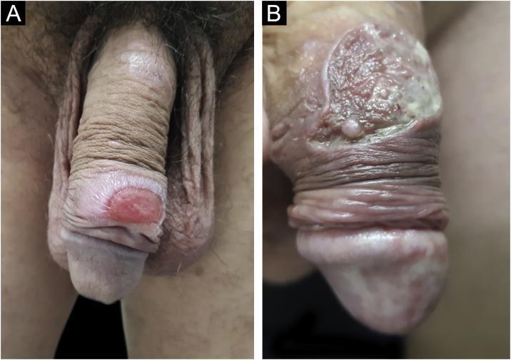
- Chancre: painless, indurated ulcer with firm base and raised margins at site of inoculation (usually genitalia, cervix, anal, or oral area)
- Firm, nonsuppurative, painless regional lymphadenopathy within 1 week
- Heals spontaneously in 4-6 weeks
SECONDARY SYPHILIS
- Appears 2-8 weeks after chancre (primary lesion may still be present)
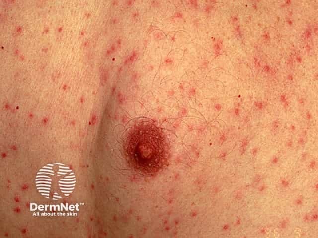
- Symmetric maculopapular rash - distributed on trunk, extremities, PALMS and SOLES (key exam feature), face
- Generalized nontender lymphadenopathy + fever + malaise
- Condylomata lata: painless mucosal warty erosions in warm moist areas (genitals, perineum)
- All lesions teeming with spirochetes - HIGHLY infectious
- Resolves spontaneously; 1/3 of patients resolve, 2/3 enter latent stage
LATENT SYPHILIS
- No clinical manifestations; positive serology only
- Early latency: relapses of secondary syphilis possible
- Late latency (>4 years): relapses cease; resistance to reinfection develops
- Mothers can still transmit to fetus throughout latency
TERTIARY SYPHILIS (appears 5-20 years later)
- Occurs in ~1/3 of untreated secondary cases
| Type | Manifestations |
|---|
| Neurosyphilis | Chronic meningitis, cortical degeneration (psychosis, hallucinations), tabes dorsalis (demyelination of posterior columns/dorsal roots - ataxia, wide-based gait, loss of sensation), paresis |
| Cardiovascular syphilis | Arteritis of vasa vasorum → medial necrosis of aorta → aortic aneurysm (ascending/transverse aorta), aortic valve incompetence |
| Gummatous syphilis | Localized granulomatous lesion (gumma) in skin, bone, joints, other organs |
Mnemonic for Paresis: P-A-R-E-S-I-S = Personality, Affect, Reflexes, Eyes, Sensorium, Intellect, Speech
CONGENITAL SYPHILIS
- Susceptibility after 4th month of gestation
- Analogous to secondary syphilis in adult
- Manifestations: rhinitis (snuffles), maculopapular rash, bone changes
- Routine serologic screening in early pregnancy (repeat in 3rd trimester in high-risk women)
Diagnosis
| Test | Category | Notes |
|---|
| VDRL / RPR | Non-treponemal (screening) | May be false positive (SLE, pregnancy, mono); titer correlates with disease activity |
| FTA-ABS / TPPA | Treponemal (confirmatory) | Specific; remains positive for life |
| Dark-field microscopy | Direct visualization | Requires fresh specimen from lesion |
Treatment
- Penicillin G remains drug of choice at ALL stages
- Primary/secondary/early latent: Benzathine penicillin G IM single dose
- Late latent/tertiary: Benzathine penicillin G IM x 3 doses
- Neurosyphilis: IV aqueous penicillin G
- Penicillin allergy (non-pregnant): doxycycline
- Congenital syphilis / neurosyphilis / pregnancy: penicillin G - NO substitute
- Sherris & Ryan's Medical Microbiology 8e
3. TUBERCULOSIS (Mycobacterium tuberculosis)
Classification
- Family: Mycobacteriaceae
- Weakly gram-positive, strongly acid-fast aerobic rods
- Lipid-rich cell wall (mycolic acids) = resistant to stains, disinfectants, detergents, many antibiotics, and host immune response
Key Trigger Words
Acid-fast, caseating granuloma, Ghon complex, Langhans giant cells, PPD/TST, IGRA, INH + rifampin + PZA + EMB, BCG vaccine, intracellular macrophage survival
Biology & Virulence
- Obligate intracellular pathogen (alveolar macrophages)
- Strictly pathogenic (unlike NTM)
- Humans are the only natural reservoir
- Spread: person-to-person via infectious aerosols (droplet nuclei <5 µm)
Pathogenesis & Immunity
Step 1 - Entry:
- Inhaled droplet nuclei reach alveoli
- Phagocytized by alveolar macrophages
Step 2 - Macrophage Survival (KEY):
- M. tuberculosis prevents phagosome-lysosome fusion by blocking EEA1 (early endosomal autoantigen 1)
- Phagosome can still fuse with other vesicles - organism accesses nutrients
- Result: intracellular survival and replication
Step 3 - Immune Response:
- T cell activation triggers IFN-γ release
- Activated macrophages form granulomas (tubercles)
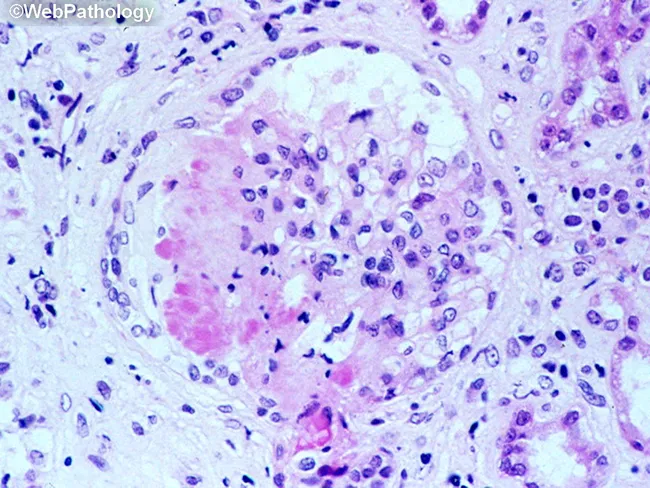
- Granuloma = epithelioid macrophages + Langhans giant cells + lymphocytic cuff + central caseous necrosis
Disease States:
| State | Description |
|---|
| Primary TB | Initial infection; Ghon focus (lower/mid lung) + hilar lymphadenopathy = Ghon complex; usually self-limited |
| Latent TB | Infection contained; TST/IGRA positive, no symptoms, non-infectious |
| Secondary (Reactivation) TB | Upper lobe cavitary disease; occurs when immunity wanes |
| Miliary TB | Hematogenous dissemination; millet-seed lesions on CXR; common in immunocompromised |
Epidemiology
- ~1/4 of world population infected (latent)
- ~10.4 million new active cases/year; ~1.6 million deaths/year
- Highest burden: India, Pakistan, sub-Saharan Africa, South Africa, China, Eastern Europe
- At-risk groups: foreign-born, HIV+ patients, immunocompromised, homeless, drug/alcohol abusers, those in close contact with active TB
Laboratory Diagnosis
Microscopy (Acid-Fast Staining):
| Stain | Method |
|---|
| Ziehl-Neelsen | Hot acid-fast stain (carbolfuchsin) |
| Kinyoun | Cold acid-fast stain |
| Truant fluorochrome | Auramine-rhodamine fluorescent dye (most sensitive) |
Culture:
- Löwenstein-Jensen (egg-based) or Middlebrook (agar-based): 4+ weeks
- Broth cultures: 2 weeks faster
- Specimens decontaminated with 2% NaOH before culture (kills normal flora, mycobacteria survive brief alkali)
Immunologic Tests:
- TST (Tuberculin Skin Test / Mantoux / PPD): intradermal injection; induration read at 48-72h; sensitive marker for exposure (not disease)
- IGRA (Interferon-γ Release Assays): QuantiFERON-TB Gold, T-SPOT.TB; in vitro; not affected by BCG vaccination
Nucleic Acid Tests:
- NAAT (nucleic acid amplification tests): important where culture unavailable or microscopy inaccurate
Identification Methods:
- Species-specific molecular probes, nucleic acid sequencing, mass spectrometry (MALDI-TOF), biochemical tests (slow, being replaced)
Treatment
| Phase | Drugs | Duration |
|---|
| Intensive phase | Isoniazid (INH) + Rifampin (RIF) + Pyrazinamide (PZA) + Ethambutol (EMB) | 2 months |
| Continuation phase | INH + Rifampin | 4-6 months |
| Total | | 6-9 months |
Mnemonic: RIPE = Rifampin, Isoniazid (INH), Pyrazinamide, Ethambutol
Prophylaxis (Latent TB):
- INH monotherapy for 6-9 months
- OR daily rifampin for 4 months
- Drug-resistant exposure: pyrazinamide + ethambutol or levofloxacin x 6-12 months
Why multiple drugs? Prevent emergence of drug-resistant strains (MDR-TB, XDR-TB)
Prevention
- BCG vaccine (Bacillus Calmette-Guérin): live attenuated M. bovis; used in endemic countries; protects children from severe disseminated disease but does NOT reliably prevent pulmonary TB in adults; makes TST positive
- Active surveillance, contact tracing, case monitoring
- Medical Microbiology 9e; Sherris & Ryan's Medical Microbiology 8e
QUICK COMPARISON TABLE
| Feature | VZV | Syphilis | TB |
|---|
| Organism | Herpesvirus (dsDNA) | T. pallidum (spirochete) | M. tuberculosis (acid-fast rod) |
| Transmission | Respiratory (aerosol) | Sexual/transplacental | Respiratory aerosol |
| Key lesion | Dermatome vesicles | Chancre → maculopapular rash → gumma | Caseating granuloma |
| Latency site | Dorsal root ganglia | Serology positive, no symptoms | Alveolar macrophages |
| Immunity | Cell-mediated | Humoral + cellular | Cell-mediated (T cells, IFN-γ) |
| Key test | PCR / FAMA | VDRL screen; FTA-ABS confirm | TST/IGRA; AFB smear; culture |
| Drug of choice | Acyclovir/valacyclovir | Penicillin G | RIPE regimen |
| Vaccine | Varivax (live), Shingrix (subunit) | None | BCG |
Good luck on your exam! These notes cover the key exam targets: classification, pathogenesis, clinical stages, diagnosis, and treatment for all three organisms.