

Man presents with breathlessness and dyspnea what could be the reason
giant pulmonary bulla chest X-ray mediastinal shift
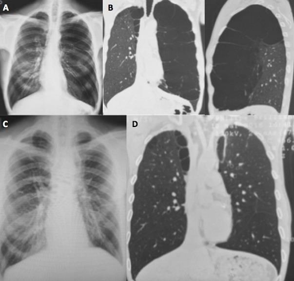
A multi-panel medical comparison chart displaying preoperative and postoperative imaging of a patient with giant bullous emphysema. Panels A and B present preoperative findings. Panel A is a posterior-anterior chest X-ray showing significant left-sided pulmonary hyperlucency and mediastinal shift. Panel B consists of coronal and sagittal chest CT views revealing a giant, thin-walled emphysematous bulla with internal septations replacing nearly the entire left upper lobe and compressing adjacent lung parenchyma. Panels C and D present postoperative findings following a thoracoscopic bullectomy. Panel C is a postoperative chest X-ray demonstrating complete pulmonary expansion and resolution of the previously seen hyperlucency. Panel D is a coronal chest CT confirming successful resection of the bulla, improved left lung volume, and more uniform parenchymal density. This clinical comparison illustrates the surgical management of vanishing lung syndrome and the radiographic restoration of lung architecture.
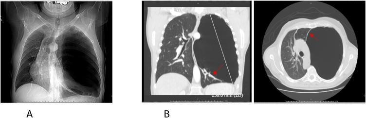
This composite of diagnostic images demonstrates a giant pulmonary bulla in the left lung, a key clinical finding in advanced emphysematous disease. Panel A is a preoperative posteroanterior chest X-ray showing a massive, hyperlucent, air-filled space in the left hemithorax. The bulla causes significant mass effect, leading to a rightward mediastinal shift and compression of the contralateral lung. Panel B presents thoracic CT images in coronal and axial planes. These scans better characterize the bulla as a large, thin-walled, air-containing structure measuring approximately 10.5 x 11 cm. Red arrows in the CT views highlight the 'double-wall sign,' where air within the bulla and air in the adjacent pleural space (if a pneumothorax is present) or another adjacent bulla outline the thin visceral wall. The CT illustrates how the bulla compresses the fairly normal surrounding lung parenchyma and deviates the trachea and heart toward the right. This visual sequence is intended to teach the differentiation between a giant bulla and a tension pneumothorax, utilizing specific radiographic signs to guide appropriate surgical or emergency management.
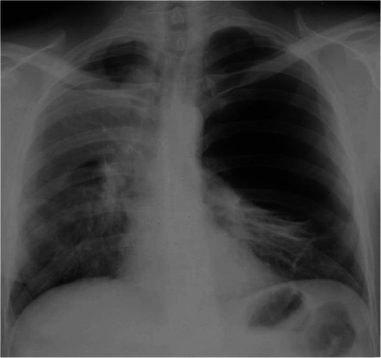
This diagnostic image is an anteroposterior (AP) chest plain film (X-ray) demonstrating advanced bullous emphysema, specifically illustrating features consistent with Vanishing Lung Syndrome. The primary finding is a giant, thin-walled bulla in the right hemithorax, which is significantly hyperextended and occupies approximately two-thirds of the hemithorax volume. This large, space-occupying lesion causes a visible mass effect, resulting in a shift of the mediastinum toward the right and substantial compression of the adjacent right lung parenchyma, which appears poorly expanded. The left lung also demonstrates hyperlucency and signs of bullous disease. Key clinical concepts illustrated include the radiographic presentation of pulmonary bullae, the compressive effects of giant bullae on mediastinal structures, and postoperative complications like secondary pneumothorax. This image serves as an educational example for pulmonology and thoracic surgery, particularly regarding the diagnosis and surgical management of end-stage bullous lung disease and COPD.
giant pulmonary bulla COPD emphysema management
Chest X-Ray Analysis - Mr. Raja Ram, 60 Years (PA View, 8/8/2026)

Primary Finding: Giant Pulmonary Bulla (Left Hemithorax)
Key Radiological Findings
| Finding | Description |
|---|---|
| Left hemithorax | Giant hyperlucent avascular space - lower 2/3 |
| Tracheal deviation | Shifted to the right (mediastinal mass effect) |
| Mediastinal shift | Cardiac silhouette displaced rightward |
| Left lung compression | Residual left upper lobe compressed superiorly |
| Left hemidiaphragm | Depressed and flattened (pushed down by bulla) |
| Right lung | Reticulonodular opacities, upper zone hyperinflation - consistent with COPD/emphysema |
Why is he Breathless?
-
Dead space ventilation - The bulla does not participate in gas exchange but occupies enormous volume, effectively "stealing" space from functional lung tissue.
-
Compression of adjacent lung - The left upper lobe is compressed superiorly/medially, reducing functional residual capacity. The right lung is also affected by underlying emphysema.
-
Mediastinal shift and cardiac compromise - Rightward displacement of the heart and great vessels can impair cardiac filling and output, worsening dyspnea.
Most Likely Underlying Diagnosis
Critical Differential: Tension Pneumothorax
- A thin curved wall/line is visible outlining the hyperlucent space (bulla wall) - in pneumothorax, you see a visceral pleural line with the lung edge separate
- The remaining compressed lung is still visible in the upper left zone
- The pattern of compression suggests a contained intraparenchymal process
Recommended Workup
- HRCT Chest - Confirms bulla anatomy, estimates remaining functional lung, excludes malignancy within/adjacent to bulla
- Spirometry / PFTs - Assess degree of obstructive deficit (expected: severe obstruction, reduced FEV1/FVC)
- ABG - Assess degree of hypoxemia/hypercapnia
- 6-minute walk test - Functional capacity
- Echo - Assess for cor pulmonale (right heart strain from chronic hypoxia)
Treatment Options
- Conservative / Medical: Bronchodilators, pulmonary rehab, oxygen therapy - for patients who are poor surgical candidates
- Surgical Bullectomy (VATS): If the bulla is compressing >50% of the hemithorax and functional lung is preserved elsewhere - can give dramatic symptomatic improvement
- Bronchoscopic techniques: Endobronchial valve placement or coil therapy (per recent 2024 review, [PMID 38448179])
Summary
This 60-year-old man's dyspnea and breathlessness are most likely due to a giant left-sided pulmonary bulla (likely from underlying COPD/emphysema) causing significant mass effect with rightward mediastinal shift, compression of functional lung parenchyma, and dead-space ventilation. Urgent HRCT chest and pulmonary function testing are the immediate next steps. Surgical bullectomy may offer substantial symptomatic benefit if functional lung reserve is adequate.
Important clinical note: This image analysis is for educational purposes. Clinical correlation by a treating physician is essential. The distinction from tension pneumothorax must be made urgently if the patient is acutely unwell.


Latest x ray ! Now whats the prognosis
bullous emphysema giant bulla prognosis survival
COPD emphysema acute exacerbation bilateral pneumonia prognosis mortality
bilateral interstitial opacities emphysema chest X-ray

This composite of six diagnostic images (two chest X-rays and four HRCT scans) illustrates the progression of combined pulmonary emphysema and fibrosis (CPFE) into acute exacerbation, potentially triggered by anticancer medication. Images (a-c) represent the baseline state: the chest X-ray (a) shows normal lung volumes with bilateral lower-zone reticular opacities, while HRCT slices (b, c) demonstrate centrilobular emphysema, subpleural cysts, and a Usual Interstitial Pneumonia (UIP) pattern characterized by reticular opacities abutting the chest wall. Images (d-f) depict the acute exacerbation phase: the follow-up chest X-ray (d) reveals new diffuse ground-glass opacities (GGO) in the left lung and a right-sided pneumothorax. The corresponding HRCT scans (e, f) confirm diffuse, bilateral GGO superimposed on the pre-existing emphysematous and fibrotic changes. This series serves as an educational example of monitoring interstitial lung disease and identifying radiological signs of acute respiratory decompensation, such as superimposed ground-glass attenuation and secondary pneumothorax in the context of underlying pulmonary fibrosis.
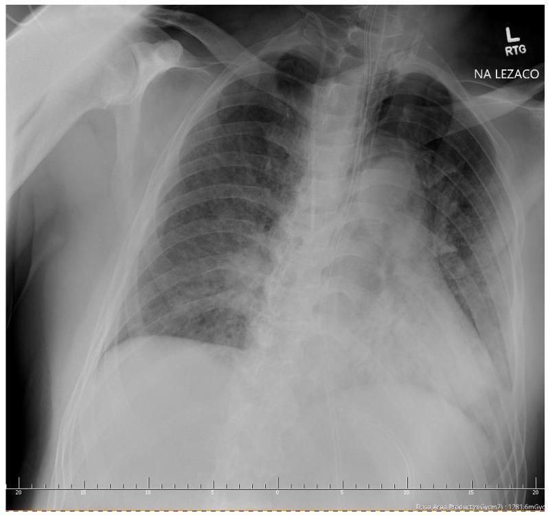
This diagnostic image is an anteroposterior (AP) chest X-ray of an adult patient, demonstrating severe acute respiratory pathology. The image reveals diffuse, bilateral interstitial and alveolar opacities, consistent with ground-glass opacities seen in severe viral pneumonia (e.g., COVID-19). These opacities are most dense in the lower lung zones, causing obscuration of the diaphragmatic borders. A prominent left-sided pneumothorax is visible, characterized by a thin, white visceral pleural line separated from the lateral chest wall by a hyperlucent peripheral space devoid of lung markings. This indicates significant pleural air accumulation. Additionally, signs of subcutaneous emphysema are present in the soft tissues of the left chest wall and neck. The image also depicts clinical monitoring and support equipment, including a central venous catheter and an endotracheal tube, suggesting an intensive care setting. The primary educational focus is the identification of secondary complications, such as barotrauma or spontaneous pneumothorax, in the context of severe diffuse interstitial lung disease.
Comparison: Previous X-Ray (Aug 8) vs. Latest X-Ray (Aug 10/11, Navodaya Hospital, Bareilly)
Patient: Mr. Raja Ram, now 62 years (note: age updated on new film), Male New study: Chest AP, 11-08-2026 at 00:20 AM, Navodaya Hospital, Bareilly
What Has Changed - The Critical Finding
| Feature | Previous X-Ray (Aug 8) | Latest X-Ray (Aug 10-11) |
|---|---|---|
| Left hemithorax | Giant hyperlucent bulla (lower 2/3) | Gone - now filled with dense bilateral opacities |
| Trachea/Mediastinum | Shifted right | Now midline or less shifted |
| Right lung | Emphysematous, hyperinflated | Diffuse bilateral opacities throughout |
| Both lung fields | Large avascular space left + emphysema right | Widespread bilateral reticulonodular/alveolar infiltrates |
| Lung markings | Absent in bulla region | Heavy interstitial marking throughout both lungs |
| Cardiac borders | Displaced right | Now obscured by bilateral infiltrates ("shaggy heart" sign) |
Radiological Interpretation of the New X-Ray
- Bilateral reticulonodular infiltrates - affecting all zones, worse mid-to-lower lung fields
- Superimposed alveolar/airspace consolidation - patchy, ground-glass type opacities
- Mediastinum now more central - suggests bulla may have partially deflated or the bilateral infiltrates are offsetting the mass effect
- Prominent bilateral hila - suggesting vascular congestion or reactive lymphadenopathy
- Shaggy cardiac borders - due to adjacent dense paracardiac infiltrates
Most Likely Diagnosis Explaining the Change
1. Superimposed Bilateral Pneumonia (Most Likely)
- Either community-acquired or hospital-acquired (given hospital admission)
- Patients with COPD/bullous emphysema have markedly impaired mucociliary clearance and are highly susceptible to bacterial pneumonia
- The bilateral symmetric pattern is classic for atypical organisms (Mycoplasma, viral) or PCP if immunocompromised
2. Acute Exacerbation of COPD (AECOPD) with Superimposed Infection
- A 2024 systematic review (PMID 39385140) found that pneumonia complicating COPD exacerbation significantly increases mortality, ICU admission rates, and need for mechanical ventilation compared to AECOPD alone
3. Acute Pulmonary Edema / Cardiogenic Component
- Bilateral symmetric opacities could suggest acute heart failure - common in COPD patients with cor pulmonale
- However, the reticulonodular pattern and lack of obvious vascular congestion pattern makes this less likely
4. Miliary Tuberculosis
- Given the setting (India, 60+ year male, known lung disease), miliary TB cannot be excluded - the diffuse nodular bilateral pattern is a classic presentation
- Tuberculosis is highly prevalent in this region and can rapidly disseminate in immunocompromised or COPD patients
5. Acute-on-chronic Interstitial Lung Disease / Combined Pulmonary Fibrosis-Emphysema
- Bilateral reticular opacities + known emphysema raises the possibility of combined pulmonary fibrosis and emphysema (CPFE)
Prognosis - Honest Assessment
Short-Term Prognosis (Days-Weeks): Guarded to Poor
- The rapid bilateral progression over 3 days is very concerning
- Bilateral pneumonia in a patient with pre-existing giant bullous emphysema carries 30-day mortality of 15-40% depending on causative organism and degree of underlying lung destruction
- The 2022 European Respiratory Journal data (PMID 35273031) confirms that mortality after admission with pneumonia in COPD patients is significantly higher than AECOPD alone
Key Prognostic Determinants:
| Factor | This Patient | Impact |
|---|---|---|
| Age | 62 years | Moderate risk |
| Underlying disease | Giant bullous emphysema + COPD | Very high risk |
| Bilateral lung involvement | Yes - both fields | Severe - limited reserve |
| Functional lung reserve | Markedly reduced (bulla + emphysema) | Very little room for compensation |
| Speed of deterioration | 3 days - rapid | Poor prognostic sign |
| Hospital level | Tertiary hospital (Navodaya, Bareilly) | Positive - access to care |
Longer-Term Prognosis (Months-Years): Dependent on immediate management
- If treatable cause (bacterial pneumonia) is identified and treated promptly with appropriate antibiotics + oxygen + NIV (non-invasive ventilation), recovery is possible
- Even if he recovers from this acute episode, his underlying bullous emphysema/COPD will progressively worsen without surgical bullectomy
- Without intervention, 5-year survival in COPD with giant bullae and recurrent exacerbations is poor (estimated 30-50%)
Immediate Management Priorities
- Sputum culture + blood culture - before starting antibiotics
- ABG urgently - assess CO2 retention (may need NIV/BiPAP)
- Broad-spectrum IV antibiotics - covering gram-negatives and atypicals (piperacillin-tazobactam + azithromycin or levofloxacin)
- HRCT chest - urgently differentiate infection vs. fibrosis vs. miliary TB
- Rule out TB - sputum AFB x3, GeneXpert
- Controlled oxygen - target SpO2 88-92% (avoid high-flow O2 in likely COPD - risk of CO2 narcosis)
- Consider ICU/HDU admission given rapid deterioration
Bottom line: The 3-day evolution from a giant isolated bulla to bilateral lung infiltrates is a medical emergency. The prognosis depends entirely on what is causing this bilateral pattern and how quickly effective treatment is started. The underlying lung reserve is critically reduced, leaving very little margin for error. Prompt HRCT, cultures, and empirical antibiotics + respiratory support are the immediate priorities.
⚠️ This analysis is for educational support only. The treating team at Navodaya Hospital must make all clinical decisions based on their direct assessment of the patient.
This x ray is taken after 1 day yesterday the hospital has inserted a ICD without prior hrct ! Now what this x ray suggests
This x ray is taken after 1 day yesterday the hospital has inserted a ICD without prior hrct ! Now what this x ray suggests
chest drain ICD giant bulla misdiagnosis pneumothorax complication
⚠️ CRITICAL FINDINGS - Post-ICD Insertion X-Ray Analysis
What the X-Ray Shows After ICD Insertion
1. Left-Sided ICD In Situ - MALPOSITIONED
- A left intercostal chest drain is visible entering via the left lateral chest wall, coursing superiorly and medially
- The drain tip is NOT in a free pleural space - it has been inserted directly into the giant bulla or into compressed lung tissue
- This is the core clinical error: the drain was placed thinking this was a pneumothorax - it was actually a giant bulla
2. EXTENSIVE SUBCUTANEOUS / SURGICAL EMPHYSEMA - NEW and Alarming
- There is extensive subcutaneous emphysema (air dissecting through chest wall tissue planes) that was NOT present on the prior X-ray
- This is tracking through the soft tissues of the left chest wall, axilla, and potentially the neck
- This is a direct complication of the ICD insertion into the bulla - puncturing the bulla wall has allowed air to track outside along the drain tract
3. Bilateral Lung Opacities Persist
- The diffuse bilateral interstitial and alveolar opacities from the previous film are unchanged or worse
- The underlying cause (pneumonia, infection, or AE-COPD) is still present and untreated
4. No Effective Lung Re-expansion
- Despite the drain, there is no meaningful lung re-expansion on the left
- This confirms the drain is not draining a true pneumothorax - it is draining (or more likely perforating) the bulla itself
- A true pneumothorax would show progressive lung re-expansion after ICD insertion
What Happened - The Medical Error Explained
| Step | What Occurred |
|---|---|
| Original X-ray (Aug 8) | Giant left bulla - correctly identified as bulla (not pneumothorax) |
| Follow-up X-ray (Aug 10-11) | Bilateral opacities developed - bulla less visible due to surrounding infiltrates |
| Clinical decision (without HRCT) | Team interpreted hyperlucent area as pneumothorax and inserted ICD |
| Actual pathology | The space was a giant bulla - NOT a free pneumothorax |
| Consequence | ICD punctured bulla → subcutaneous emphysema, air leak, no benefit, worsening |
Complications Already Present and At Risk
| Complication | Status |
|---|---|
| Subcutaneous emphysema | PRESENT - extensive, likely spreading |
| Persistent air leak | HIGH RISK - bulla punctured, air continuously leaking |
| Tension physiology | RISK - if ICD is clamped or kinked, air can accumulate under tension |
| Bulla infection | HIGH RISK - drain has now created a direct entry point for bacteria |
| Bronchopleural fistula | RISK - if drain has entered lung parenchyma |
| Worsening respiratory failure | ONGOING - bilateral pneumonia on destroyed lungs |
| Pneumomediastinum | RISK - subcutaneous air can track medially |
What Needs to Happen NOW
Immediate Actions:
- Do NOT clamp the ICD suddenly - could cause tension
- Urgent HRCT chest - this should have been done first, but now must be done to assess current anatomy, drain position, and degree of complication
- Do NOT insert another drain without HRCT guidance
- Senior pulmonologist + cardiothoracic surgeon review urgently - this patient likely needs transfer to a centre with thoracic surgery capability
- Continue treating underlying infection - IV antibiotics, respiratory support
- Monitor for tension pneumomediastinum / pneumopericardium - these are life-threatening secondary complications of extensive subcutaneous emphysema
Transfer Consideration:
- Cardiothoracic surgery
- Pulmonology / respiratory ICU
- Interventional radiology (for CT-guided drain repositioning if needed)
Prognosis - Updated (Significantly Worse)
| Scenario | Estimated Short-term Mortality |
|---|---|
| COPD + bilateral pneumonia alone | 20-35% |
| COPD + bilateral pneumonia + ICD-bulla complication | 50-70% |
| If tension develops or bronchopleural fistula forms | >80% |
⚠️ This is a medical emergency with a serious iatrogenic complication. Please ensure the treating team is fully aware and consider urgent transfer to a higher centre with thoracic surgery. The family should be counselled about the severity of the situation.
This analysis is for clinical support only and does not substitute for direct physician assessment.