
Microscopy/Anatomy/Powder Microscopy (Fennel, Cinnamon, Liquorice, Datura, Rauwolfia, Nux vomica, Senna, Ephedra)
-
Microscopy and anatomy
- Diagnostic transverse-section features
- Tissue systems, vascular arrangement, inclusions, and identifying characters
-
Powder microscopy
- Diagnostic fragments in powdered drugs
- Starch grains, sclereids, fibres, vessels, trichomes, crystals, calcium oxalate, and alkaloid-containing tissues
-
Individual crude drugs
- Fennel
- Cinnamon
- Liquorice
- Datura
- Rauwolfia
- Nux vomica
- Senna
- Ephedra
Create a comparison table for all eight drugs
medicinal plants AND microscopy
pharmacognosy powder microscopy fennel cinnamon liquorice datura rauwolfia nux vomica senna ephedra diagnostic characters
Comparison table: microscopy, anatomy, and powder characters
| Drug | Biological source and family | Key anatomy / transverse-section characters | Diagnostic powder microscopy | Major constituents |
|---|---|---|---|---|
| Fennel | Dried ripe fruits of Foeniculum vulgare; Apiaceae | Fruit is a cremocarp with five primary ridges and four secondary ridges. Mesocarp contains prominent vittae (oil ducts), usually 6: four dorsal and two commissural. Endosperm is large, oily, and has aleurone grains. | Polygonal epidermal cells; fragments of ridges; vittae with volatile oil; spiral/reticulate vessels; parenchyma containing fixed oil and aleurone grains. | Volatile oil, chiefly anethole, with fenchone and estragole. |
| Cinnamon | Dried inner bark of shoots of Cinnamomum verum; Lauraceae | Cork may be absent or scanty in commercial bark. Cortex has oil cells, mucilage cells, and groups of sclereids. Secondary phloem is broad with fibres and phloem parenchyma. Medullary rays are narrow. | Brown cork cells; thick-walled sclereids; lignified fibres; fragments of phloem; starch grains; oil cells, often containing yellowish-brown volatile oil. | Volatile oil, chiefly cinnamaldehyde; eugenol, tannins, mucilage. |
| Liquorice | Dried roots and stolons of Glycyrrhiza glabra; Fabaceae | Periderm externally. Cortex is parenchymatous, often with starch. Secondary phloem and xylem are well developed. Xylem has broad vessels and fibres. Medullary rays are distinct. No calcium oxalate crystals is a useful practical point. | Numerous simple and compound starch grains; pitted/reticulate vessels; lignified fibres; parenchyma; cork fragments. Characteristically lacks calcium oxalate crystals. | Glycyrrhizin or glycyrrhizic acid, flavonoids such as liquiritin, sugars and starch. |
| Datura | Dried leaves of Datura stramonium; Solanaceae | Dorsiventral leaf. Epidermis bears wavy-walled cells, anisocytic stomata, covering trichomes and glandular trichomes. Palisade tissue occurs below upper epidermis. Midrib contains a bicollateral vascular bundle. Calcium oxalate crystals may occur. | Epidermal fragments with anisocytic stomata; multicellular uniseriate covering trichomes; capitate glandular trichomes; spiral vessels; calcium oxalate crystals. | Tropane alkaloids: hyoscyamine, atropine, scopolamine. |
| Rauwolfia | Dried roots of Rauvolfia serpentina; Apocynaceae | Outer cork is multilayered. Cortex is broad and parenchymatous, containing starch. Phloem occurs in patches; secondary xylem has vessels, fibres and rays. Characteristic laticiferous tissue may be present. | Cork cells; abundant starch grains; lignified fibres; pitted and reticulate vessels; phloem parenchyma; occasional latex-containing elements. | Indole alkaloids: reserpine, rescinnamine, deserpidine, ajmaline and ajmalicine. |
| Nux vomica | Dried seeds of Strychnos nux-vomica; Loganiaceae | Seed coat has an outer epidermis with long, thick-walled, lignified unicellular hairs radiating from the surface, giving a silky appearance. Endosperm is large, horny, and contains fixed oil and aleurone grains. Embryo is small. | Characteristic long, thick-walled, lignified seed-coat hairs; fragments of endosperm with aleurone grains and fixed oil; no starch grains. | Indole alkaloids: strychnine and brucine. |
| Senna | Dried leaflets of Senna alexandrina or Cassia angustifolia; Fabaceae/Caesalpiniaceae | Dorsiventral leaf with upper and lower epidermis. Stomata are mainly paracytic. Palisade tissue is usually single layered. Midrib contains collateral vascular tissue. Covering trichomes are present; calcium oxalate crystals may occur. | Epidermal fragments with paracytic stomata; unicellular warty covering trichomes; palisade cells; spiral vessels; calcium oxalate crystals; fragments of veins. | Hydroxyanthracene glycosides, especially sennosides A and B. |
| Ephedra | Dried young stems of Ephedra gerardiana or Ephedra sinica; Ephedraceae | Stem shows a ridged outline. Epidermis has thick cuticle and sunken stomata. Hypodermis is sclerenchymatous. Cortex is chlorenchymatous. Vascular bundles are arranged in a ring; xylem contains vessels and fibres. Pith is large. | Epidermal fragments with sunken stomata; cuticular fragments; fibres; spiral, annular and pitted vessels; chlorenchymatous cells. | Protoalkaloids: ephedrine and pseudoephedrine. |
Rapid identification points
| Drug | Best spot character |
|---|---|
| Fennel | Vittae or oil ducts in fruit |
| Cinnamon | Sclereids plus oil cells in bark |
| Liquorice | Abundant starch with vessels and fibres, but no calcium oxalate crystals |
| Datura | Anisocytic stomata with covering and glandular trichomes |
| Rauwolfia | Root powder with starch, fibres, vessels and alkaloid-containing/laticiferous elements |
| Nux vomica | Long, lignified, radially arranged seed-coat hairs |
| Senna | Paracytic stomata and warty unicellular trichomes |
| Ephedra | Stem epidermis with sunken stomata and thick cuticle |
Explain with a neat labelled diagram the microscopy and powder microscopical characters of Cassia cinnamon for 10 m
Cinnamomum cassia cinnamon bark transverse section labelled microscopy oil cells sclereids phloem diagram
Cinnamomum cassia pharmacognosy microscopy
Cinnamomum cassia bark transverse section powder microscopy characters oil cells sclereids
A clean, exam-ready labelled botanical line diagram of a transverse section of Cassia cinnamon bark (Cinnamomum cassia). Show a curved bark section from outer to inner surface, with labels for cork/periderm, cortex with stone cells and mucilage cells, oil cells, primary phloem, secondary phloem, phloem fibres, and medullary rays. Include a separate inset of diagnostic powder microscopy fragments: thick-walled stone cell, lignified fibre, oil cell, starch grains, and pitted vessel. Use accurate black ink textbook line-art, clear labels, and no decorative elements.
Cassia cinnamon: microscopy and powder microscopy
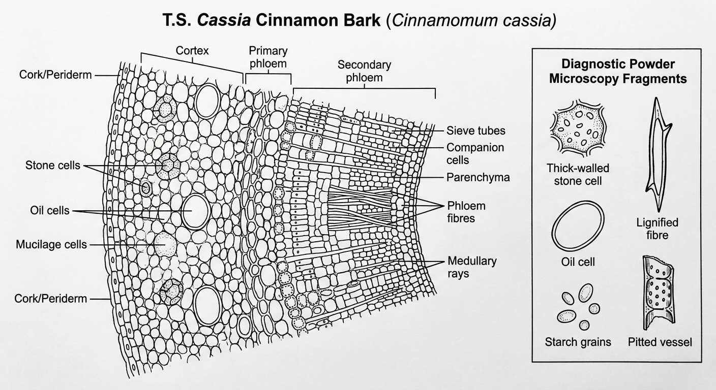
A. Microscopy of bark: transverse section
-
Cork / periderm
- Present as several layers of compact, rectangular to polygonal, brownish cork cells.
- This is a useful distinction from true cinnamon (C. verum), in which cork is commonly absent in commercial bark.
-
Cortex
- Broad, made up of parenchymatous cells.
- Contains many starch grains.
- Scattered oil cells are present. They are rounded or oval and contain volatile oil.
- Groups of large, thick-walled, lignified sclereids or stone cells occur in the cortex. They may appear elongated, U-shaped, or horseshoe-shaped in section.
-
Primary phloem
- Occurs as a narrow zone beneath the cortex.
- It may be crushed or compressed in older bark.
-
Secondary phloem
- Broad and made of phloem parenchyma, sieve elements, phloem fibres, and medullary rays.
- Sieve tubes are generally collapsed and arranged in tangential groups.
- Phloem parenchyma contains starch.
-
Phloem fibres
- Thick-walled and lignified.
- They occur in tangential bands or groups, often with about 4 to 5 fibres in a row.
-
Medullary rays
- Radial strips of parenchymatous cells.
- Usually 2 to 3 cells broad in Cassia cinnamon.
- They may contain starch.
-
Calcium oxalate crystals
- Needle-like crystals may occur in parenchymatous cells or near medullary rays.
Cork + broad cortex + large sclereids + few oil cells + many starch grains + 2 to 3 layered medullary rays.
B. Powder microscopical characters
| Character | Description / identifying feature |
|---|---|
| Cork cells | Brown, polygonal or rectangular cells arranged compactly in surface view. |
| Sclereids / stone cells | Large, thick-walled, lignified cells with narrow lumen; irregular, elongated, horseshoe-shaped, or U-shaped. These are prominent diagnostic elements. |
| Phloem fibres | Long, lignified fibres with pointed ends, thick walls and narrow lumen; may show a slit-like central lumen. |
| Starch grains | Numerous, small, simple and compound starch grains in cortical, phloem, and ray parenchyma. |
| Oil cells / oil globules | Rounded or oval cells containing yellowish-brown volatile oil or free oil droplets. |
| Phloem parenchyma | Thin-walled, elongated or polygonal cells, often packed with minute starch grains. |
| Medullary-ray fragments | Rows of rectangular parenchymatous cells, usually 2 to 3 cells wide. |
| Vessels | Fragments of pitted, reticulate, or spiral vessels may be present. |
| Calcium oxalate | Occasional needle-shaped crystals, especially in association with parenchyma or ray tissues. |
Important staining reactions
- Phloroglucinol and concentrated HCl: sclereids and fibres stain pink to red due to lignin.
- Iodine solution: starch grains stain blue.
- Sudan III/IV: oil cells or oil droplets stain orange-red.
Conclusion / identification
- Brown cork fragments
- Large lignified stone cells
- Thick-walled phloem fibres
- Abundant starch grains
- Few volatile oil cells
- Fragments of broad medullary rays