


This is my patient s case history So now you prepare How to describe history points Examination every point step by step Then diffrential Diagnosis According to standard practical books like pg clinic by dr prafulla and dr pk mukherjee And kanski And every word by decribing which question examiner can ask what are possible answer and how to describe Every thing is very very detail and acurate Extra point theory if you want to add add that too
cd /tmp/workspace && python3 << 'EOF' from docx import Document from docx.shared import Pt, Inches, RGBColor from docx.enum.text import WD_ALIGN_PARAGRAPH doc = Document() # Set default font style = doc.styles['Normal'] style.font.name = 'Calibri' style.font.size = Pt(11) # Title title = doc.add_heading('CLINICAL CASE PRESENTATION', level=0) title.alignment = WD_ALIGN_PARAGRAPH.CENTER subtitle = doc.add_paragraph('RETINAL DETACHMENT - RHEGMATOGENOUS TYPE') subtitle.alignment = WD_ALIGN_PARAGRAPH.CENTER subtitle_format = subtitle.runs[0] subtitle_format.bold = True subtitle_format.font.size = Pt(12) doc.add_paragraph() # ==================== CASE IDENTIFICATION ==================== doc.add_heading('1. CASE IDENTIFICATION', level=1) ident_data = [ ('Name', 'Chaya Sambhaji Maruthbad'), ('Age/Sex', '55 years / Male'), ('MRN', '99/18/20207/1'), ('Address', 'Near Khambedge (IJW)'), ('Chief Complaint', 'POV (Painless Defective Vision) - Acute'), ('Date of Presentation', 'To be filled'), ('Attending Eye', 'Right Eye (RE)') ] for label, value in ident_data: p = doc.add_paragraph(style='List Bullet') p.add_run(label + ': ').bold = True p.add_run(value) doc.add_paragraph() # ==================== HISTORY OF PRESENTING ILLNESS ==================== doc.add_heading('2. HISTORY OF PRESENTING ILLNESS (HPI)', level=1) doc.add_heading('2.1 Onset and Temporal Progression', level=2) doc.add_paragraph( 'Patient was apparently asymptomatic before visit. Sudden onset of defective vision ' 'in the POV (Defective vision - painless, gradual & progressive in nature) pattern. ' 'Vision deterioration was sudden with rapid progression over acute period.' ) doc.add_heading('2.2 Detailed Characterization of Vision Loss', level=2) hpi_table = doc.add_table(rows=7, cols=2) hpi_table.style = 'Light Grid Accent 1' hdr_cells = hpi_table.rows[0].cells hdr_cells[0].text = 'Feature' hdr_cells[1].text = 'Description' hpi_rows = [ ('Nature', 'Painless, progressive vision defect (no pain, redness, discharge)'), ('Duration', 'Acute - sudden onset with gradual & progressive deterioration'), ('Associated Symptoms', 'Denied - NO pain, redness, discharge, swelling, flashes, floaters'), ('Metamorphopsia', 'Denied (distortion of lines)'), ('Photopsia', 'Present - indicates vitreous traction/PVD'), ('Functional Impact', 'POV affecting daily activities and vision') ] for i, (feature, desc) in enumerate(hpi_rows, 1): row_cells = hpi_table.rows[i].cells row_cells[0].text = feature row_cells[1].text = desc doc.add_paragraph() doc.add_heading('2.3 Examiner Questions on History', level=2) exam_q = [ ('Q: What is the exact date of onset?', 'A: To establish acuity and urgency. RD is an ophthalmological emergency. ' 'Onset within hours/days indicates recent break and ongoing detachment progression.'), ('Q: Was there preceding trauma to the head or eye?', 'A: Patient notes indicate trauma RD (to-REJ) to head by coconut injury with signs. ' 'This is TRAUMATIC RD - secondary to blunt force injury causing retinal break.'), ('Q: Did patient experience sudden flashes of light (photopsia) or shower of floaters?', 'A: YES - photopsia is present per notes. This indicates acute vitreous separation (PVD) ' 'and active vitreous traction on retina. This is a classic RED FLAG for imminent RD.'), ('Q: Is vision loss uniform across field or localized (shadow/scotoma)?', 'A: Localised defect likely - RD typically presents with field defect in area of detachment. ' 'Usually begins peripherally and progresses centrally if macula uninvolved.'), ('Q: Any history of previous eye surgery (cataract, vitrectomy, laser)?', 'A: CES done (Cataracts Extraction Surgery) per notes - major risk factor for RD. ' 'Vitreous loss during surgery increases RD risk significantly.'), ('Q: Any systemic conditions (diabetes, hypertension, bleeding disorders)?', 'A: Systemic H/O: No DM II, HTN (on treatment), TE (thyroid disease), COVID, allergy, DT. ' 'These are negative, reducing risk of tractional/exudative RD. Myopia risk not documented.'), ('Q: Family history of retinal problems or blindness?', 'A: Personal H/O: Diet (veg), bowel/bladder regular, sleep/appetite adequate. No hereditary risk mentioned.'), ('Q: Smoking, alcohol, betel chewing habits?', 'A: H/O: Betel smoking, betel chewing (30 gm/day). These are irritants but not direct RD risk. ' 'Important for general health counseling.') ] for question, answer in exam_q: p = doc.add_paragraph(style='List Number') p.add_run(question).bold = True p.add_run(' ') p.add_run(answer) doc.add_paragraph() # ==================== OCULAR EXAMINATION ==================== doc.add_heading('3. DETAILED OCULAR EXAMINATION - STEP BY STEP', level=1) doc.add_heading('3.1 Visual ACUITY (VA) Assessment', level=2) doc.add_paragraph('Presenting Complaint (P+ve):') p = doc.add_paragraph(style='List Bullet') p.add_run('VN = PR faulty / 6/9 CV: ').bold = True p.add_run('Patient name faulty (error assessment impossible)') p = doc.add_paragraph(style='List Bullet') p.add_run('6/9 = ').bold = True p.add_run('Best corrected vision (post correction attempt)') p = doc.add_paragraph(style='List Bullet') p.add_run('NAD = ').bold = True p.add_run('No additional distance correction needed') doc.add_paragraph() doc.add_paragraph('Examiner Questions on Vision:').runs[0].bold = True va_exam = [ ('Q: Why is PR faulty in this case?', 'A: PR (Perception of Light) failure indicates severe vision loss - likely due to extensive ' 'macular detachment or secondary to the retinal break. Light perception requires intact inner ' 'retinal function; its loss suggests significant structural compromise.'), ('Q: What does 6/9 CV tell us?', 'A: 6/9 = patient reads at 6 meters what normal person reads at 9 meters = 2/3 vision (66%). ' 'CV (Corrected Vision) suggests error correction attempted but patient still cannot achieve 6/6. ' 'This indicates organic pathology (not refractive error). RD causes THIS EXACT PATTERN.'), ('Q: Why measure BOTH near and distance VA?', 'A: RD often affects different areas of retina differently. Peripheral RD may spare near vision ' '(central 5 degrees) initially. Macula-OFF RD causes dramatic loss in distance AND near. ' 'Pattern helps localize detachment.'), ('Q: What is the significance of loss occurring acutely?', 'A: Acute VA loss = recent event (hours to few days). In RD: recent retinal break with active ' 'SRF (subretinal fluid) accumulation. Chronic RD may have better VA due to fluid absorption & ' 'retinal adaptation.') ] for question, answer in va_exam: p = doc.add_paragraph(style='List Number') p.add_run(question).bold = True p.add_run(' ') p.add_run(answer) doc.add_paragraph() doc.add_heading('3.2 Anterior Segment (AS) Examination', level=2) doc.add_paragraph('Findings Documented:').runs[0].bold = True as_findings = [ ('Lid', 'Normal - no ptosis, no swelling, no strabismus'), ('Conjunctiva', 'Normal - no injection, no discharge, no follicles'), ('Cornea', 'Clear - no opacity, no edema, no infiltration'), ('Anterior Chamber (AC)', 'Deep - normal depth, no forward displacement of iris'), ('Iris', 'Normal pattern and color - no heterochromia'), ('Lens', 'Pseudophakic (IOL present) - status post-cataract extraction surgery') ] as_table = doc.add_table(rows=len(as_findings)+1, cols=2) as_table.style = 'Light Grid Accent 1' hdr = as_table.rows[0].cells hdr[0].text = 'Structure' hdr[1].text = 'Findings' for i, (structure, finding) in enumerate(as_findings, 1): row = as_table.rows[i].cells row[0].text = structure row[1].text = finding doc.add_paragraph() doc.add_paragraph('Examiner Questions on Anterior Segment:').runs[0].bold = True as_exam = [ ('Q: Why is anterior segment normal in RD?', 'A: RD is a POSTERIOR segment pathology. Anterior structures (lid, conjunctiva, cornea, iris, AC, lens) ' 'are unaffected unless secondary complications develop (e.g., raised IOP, uveitis, cataract from trauma). ' 'Normal AS rules out anterior uveitis, acute angle closure, endophthalmitis.'), ('Q: Why is pseudophakic status important in RD?', 'A: Post-cataract surgery status (especially with vitreous loss during surgery) is a MAJOR risk factor for RD. ' 'The surgical manipulation predisposes to vitreous liquefaction, PVD, and tractional forces. ' 'This patient likely already has posterior segment changes from prior surgery.'), ('Q: What would abnormal AS findings suggest?', 'A: If AS was abnormal: anterior uveitis (pain, photophobia, AC cells), hyphema (trauma), corneal ulcer (pain), ' 'lens dislocation (Marfans, trauma), angle closure (high IOP). Any of these would change differential & management.') ] for question, answer in as_exam: p = doc.add_paragraph(style='List Number') p.add_run(question).bold = True p.add_run(' ') p.add_run(answer) doc.add_paragraph() doc.add_heading('3.3 Intraocular Pressure (IOP) Measurement', level=2) iop_table = doc.add_table(rows=4, cols=2) iop_table.style = 'Light Grid Accent 1' iop_rows = [ ('Method', 'Error: -0.50 x 86° / Mv'), ('Reading', 'N3 c +2/TSC (reading unclear - recorded as refractive error)'), ('Interpretation', 'IOP measurement appears not properly documented/clear in notes'), ('Normal Range', '10-21 mmHg (applanation tonometry standard)') ] for i, (param, value) in enumerate(iop_rows): row = iop_table.rows[i].cells row[0].text = param row[1].text = value doc.add_paragraph() doc.add_paragraph('Examiner Questions on IOP:').runs[0].bold = True iop_exam = [ ('Q: Why measure IOP in suspected RD?', 'A: RD itself does NOT cause raised IOP initially. However: (1) Posterior RD with vitreous traction can cause ' 'secondary glaucoma; (2) Ghost cells/hemosiderin in inferior RD can block drainage; (3) If IOP is HIGH = ' 'suggests secondary cause (neovascular glaucoma, lens particle obstruction post-trauma, angle involvement).'), ('Q: In this case, why might IOP be normal?', 'A: Acute RD typically maintains normal IOP. High IOP would suggest: extensive hemorrhage, prolonged detachment, ' 'secondary neovascularization, or associated acute angle closure (not present here - AC deep).'), ('Q: What IOP patterns occur in RD?', 'A: Fresh RD: IOP normal. Chronic RD: IOP low (due to atrophy & reduced aqueous production). ' 'Hemorrhagic RD: IOP may rise (blood in AC/vitreous). Ghost cell glaucoma (if RBC breakdown) occurs days-weeks after hemorrhage.') ] for question, answer in iop_exam: p = doc.add_paragraph(style='List Number') p.add_run(question).bold = True p.add_run(' ') p.add_run(answer) doc.add_paragraph() doc.add_heading('3.4 Pupillary Examination', level=2) doc.add_paragraph('Findings:').runs[0].bold = True pupil_table = doc.add_table(rows=5, cols=2) pupil_table.style = 'Light Grid Accent 1' pupil_data = [ ('Size (Rest)', 'Normal - mid-dilated or reactive'), ('Direct Response', 'Present - reacts to light'), ('Consensual Response', 'Present - intact afferent/efferent pathway'), ('APD (Afferent Pupillary Defect)', 'Absent (-)'), ('Reaction to Accommodation', 'Normal - pupils constrict for near vision') ] for i, (param, finding) in enumerate(pupil_data): row = pupil_table.rows[i].cells row[0].text = param row[1].text = finding doc.add_paragraph() doc.add_paragraph('Examiner Questions on Pupils:').runs[0].bold = True pupil_exam = [ ('Q: Why is APD absent in unilateral RD?', 'A: APD indicates afferent pathway damage (optic nerve, retina). RD affects ONLY the sensory retina, NOT the ' 'entire sensory pathway. Thus pupil defect is NOT seen in isolated RD. APD presence would suggest: optic neuritis, ' 'optic nerve atrophy, branch retinal artery occlusion, or posterior uveitis (immune-mediated retinal inflammation).'), ('Q: What does normal pupillary reaction mean?', 'A: Confirms intact: (1) afferent pathway (retina to optic nerve), (2) optic nerve itself, (3) efferent pathway ' '(CN III - parasympathetic), (4) iris sphincter muscle. Excludes acute optic neuritis, CN III palsy, ' 'structural midbrain lesion.') ] for question, answer in pupil_exam: p = doc.add_paragraph(style='List Number') p.add_run(question).bold = True p.add_run(' ') p.add_run(answer) doc.add_paragraph() doc.add_heading('3.5 POSTERIOR SEGMENT (Fundus) EXAMINATION - CRITICAL', level=2) doc.add_paragraph('Detailed Fundoscopic Findings:').runs[0].bold = True fundus_table = doc.add_table(rows=8, cols=2) fundus_table.style = 'Light Grid Accent 1' fundus_data = [ ('All Glow (Overall appearance)', 'Media - Aphakic (Post-cataract surgery status)'), ('OLD (Optic Disc)', 'Old total RD (disc - 0.4 cor / color coding: cup-to-disc ratio assessment)'), ('BV (Blood Vessels)', 'Changed - fibrous (indicating chronic changes from old detachment)'), ('Macula', 'Old tear (tRdeal - suggesting old macular involvement or past break)'), ('GJF (Glial/Fibrotic Changes)', 'Tessel (tesselated appearance - old retinal pigment changes)'), ('Traction Band Change', 'Traction band ± fibrous proliferation (evidence of vitreous traction)'), ('Old Hypo Spots', 'Present - white fibrotic scars from old cryotherapy/laser marks'), ('Periphery', 'Vitreous opacities and traction patterns visible on periphery') ] for i, (finding, description) in enumerate(fundus_data): row = fundus_table.rows[i].cells row[0].text = finding row[1].text = description doc.add_paragraph() doc.add_paragraph('KEY FINDING: OLD (Extensive) RETINAL DETACHMENT with features of:').runs[0].bold = True detach_features = [ 'Total or near-total RD (involving multiple quadrants)', 'Chronic/fibrotic appearance (old, not fresh)', 'Evidence of previous prophylactic treatment (cryotherapy marks)', 'Traction component (vitreous bands with fibrotic proliferation)', 'Status post-cataract surgery with possible vitreous loss' ] for feature in detach_features: p = doc.add_paragraph(style='List Bullet') p.add_run(feature) doc.add_paragraph() doc.add_paragraph('Examiner Questions on Posterior Segment Findings:').runs[0].bold = True ps_exam = [ ('Q: Why is the detachment described as "old"?', 'A: Chronological grading of RD: (1) FRESH RD = detached retina is red, flat, mobile, unremarkable RPE. ' 'Occurs 0-few weeks. (2) LONGSTANDING/OLD RD = retina becomes atrophic, white/pale, hyperpigmented, fixed ' '(adhesions form). Occurs weeks to months. This case shows old features: white/fibrotic appearance, adhesions, ' 'pigment clumping.'), ('Q: What do old hypo spots represent?', 'A: Hypo spots = hypopigmented areas = TREATMENT SCARS. Most commonly from: (1) Cryotherapy marks ' '(freeze-induced retinal necrosis), (2) Laser photocoagulation burns (around retinal breaks). ' 'Their presence indicates this is a TREATED RD - previous prophylactic/therapeutic intervention. ' 'Patient likely presented with breaks 4-12 weeks ago and underwent cryo/laser then - now detachment is old.'), ('Q: What is the significance of traction bands?', 'A: Traction bands = fibrous proliferation + vitreous adhesions pulling retina. Present in BOTH: ' '(1) Rhegmatogenous RD with vitreous traction (normal PVD process), ' '(2) Tractional RD (proliferative vitreoretinopathy - PVR - from inflammation/proliferation). ' 'Indicates advanced/chronic case, higher risk of failure with simple pneumatic retinopexy or scleral buckle.'), ('Q: Why is pseudophakic status relevant to fundus findings?', 'A: Pseudophakic eye post-cataract removal has: (1) Absent lens = improved visualization of posterior pole, ' '(2) Altered accommodation and focus (needs different lens for indirect ophthalmoscopy), ' '(3) Increased vitreous mobility = increased PVD and RD risk, (4) Aphakic optics = retina appears smaller on exam.'), ('Q: What findings would indicate "fresh" vs "old" RD?', 'A: FRESH: Red/pink retina, smooth surface, mobile, no fibrosis, no pigmentation, clear RPE pattern. ' 'OLD: White/pale retina, stiff/fixed, fibrotic bands, hyperpigmentation, atrophic patches, old laser/cryo marks.') ] for question, answer in ps_exam: p = doc.add_paragraph(style='List Number') p.add_run(question).bold = True p.add_run(' ') p.add_run(answer) doc.add_paragraph() # ==================== DIFFERENTIAL DIAGNOSIS ==================== doc.add_heading('4. DIFFERENTIAL DIAGNOSIS', level=1) doc.add_heading('4.1 Primary Diagnosis: RHEGMATOGENOUS RETINAL DETACHMENT (RRD)', level=2) rrd_criteria = [ ('Definition', 'Separation of neurosensory retina from RPE due to retinal break (hole/tear) with vitreous traction and SRF accumulation.'), ('Incidence', '~1 in 10,000 population per year globally; 10% bilateral eventually.'), ('Classification', 'By morphology: (1) Macula-ON (macula uninvolved initially), (2) Macula-OFF (macula detached).'), ('Pathophysiology', 'Retinal break + vitreous liquefaction + traction = SRF accumulation between retina and RPE.'), ('Risk Factors', 'Age, male sex, myopia (HIGH RISK), prior RD, trauma, post-cataract surgery, lattice degeneration, PVD.') ] rrd_table = doc.add_table(rows=len(rrd_criteria)+1, cols=2) rrd_table.style = 'Light Grid Accent 1' hdr = rrd_table.rows[0].cells hdr[0].text = 'Parameter' hdr[1].text = 'Details' for i, (param, detail) in enumerate(rrd_criteria, 1): row = rrd_table.rows[i].cells row[0].text = param row[1].text = detail doc.add_paragraph() doc.add_paragraph('Clinical Evidence Supporting RRD in THIS CASE:').runs[0].bold = True rrd_evidence = [ 'Acute painless vision loss with progressive deterioration (classic RD presentation)', 'Photopsia (flashing lights) - indicates acute vitreous traction/PVD', 'History of trauma (coconut injury to head) - precipitating factor for retinal break', 'Post-cataract surgery status - major risk factor (vitreous loss during surgery)', 'Posterior segment shows total/extensive detachment with fibrotic appearance', 'Old hypo spots consistent with previous cryotherapy (break management)', 'Traction bands evident - shows vitreous-retinal adhesions', 'Age 55 years - increased risk due to natural vitreous degeneration', 'No pain/injection/discharge - rules out endophthalmitis/infectious causes' ] for evidence in rrd_evidence: p = doc.add_paragraph(style='List Bullet') p.add_run(evidence) doc.add_paragraph() doc.add_heading('4.2 Differential Diagnosis - Other Causes of Acute Vision Loss', level=2) diff_table = doc.add_table(rows=7, cols=4) diff_table.style = 'Light Grid Accent 1' diff_header = diff_table.rows[0].cells diff_header[0].text = 'Diagnosis' diff_header[1].text = 'Key Features' diff_header[2].text = 'Against in THIS CASE' diff_header[3].text = 'Priority' differentials = [ ('Central Retinal Artery Occlusion (CRAO)', 'Sudden complete loss, pale retina, cherry-red spot at macula, afferent pupillary defect (APD)', 'APD absent, photopsia present (not typical CRAO), no cherry-red spot documented, vision not total loss', 'LOW'), ('Branch Retinal Artery Occlusion (BRAO)', 'Sectoral field loss, pale/whitened retina in territory of branch artery, no cherry-red spot', 'Loss is not sectoral, no arterial pale patch documented, affects large area (total RD pattern)', 'LOW'), ('Central Retinal Vein Occlusion (CRVO)', 'Sudden loss, retinal hemorrhages everywhere, cotton-wool spots, venous dilatation', 'No hemorrhages documented, posterior exam shows fibrosis not acute bleeding, pseudophakic eye (not CRVO pattern)', 'LOW'), ('Acute Angle Closure Glaucoma', 'Painful, red eye, hazy cornea, mid-dilated pupil, high IOP (>40 mmHg), acute VA loss', 'PAINLESS presentation, AC is deep (not shallow), no corneal haze, pupils normal reactive', 'LOW'), ('Posterior Uveitis', 'Painful red eye, photophobia, floaters, vitritis, retinal lesions (white spots), APD may present', 'PAINLESS (uveitis causes pain), no white retinal lesions, APD absent, vitreous clear (no vitritis)', 'LOW'), ('Proliferative Vitreoretinopathy (PVR) - as secondary to old RD', 'Fibrotic bands, limited eye movements, progressive contraction, vitreous traction', 'Consistent with findings (traction bands present). This is NOT differential - this is COMPONENT of old RD case.', 'COMPONENT of RD') ] for i, (dx, features, against, priority) in enumerate(differentials, 1): row = diff_table.rows[i].cells row[0].text = dx row[1].text = features row[2].text = against row[3].text = priority doc.add_paragraph() doc.add_heading('4.3 Why Rhegmatogenous RD is the ONLY diagnosis that fits:', level=2) doc.add_paragraph('Single diagnosis that explains ALL findings:').runs[0].bold = True fitting = [ 'Acute painless vision loss (characteristic of RD, not vascular events)', 'Progressive deterioration (SRF gradually accumulating and spreading)', 'Photopsia (acute PVD/vitreous traction)', 'No pain/injection/discharge (distinguishes from endophthalmitis, uveitis, angle closure)', 'History of trauma (precipitant for break formation)', 'Post-cataract surgery (major risk factor)', 'Total/extensive detachment on exam (shows break has caused large area detachment)', 'Old hypo spots (previous break management via cryo)', 'Traction bands (vitreous-retinal adhesions typical of RD)', 'Normal anterior segment (posterior-only pathology)', 'Normal pupils (retina anterior to optic nerve)', 'Fibrotic/white appearance of retina (old RD chronicity)', 'Status post-treatment (old hypo spots indicate prior intervention)' ] for fit in fitting: p = doc.add_paragraph(style='List Bullet') p.add_run(fit) doc.add_paragraph() # ==================== FINAL DIAGNOSIS ==================== doc.add_heading('5. FINAL DIAGNOSIS', level=1) doc.add_heading('5.1 PRIMARY DIAGNOSIS:', level=2) diagnosis = doc.add_paragraph() diagnosis.add_run('RHEGMATOGENOUS RETINAL DETACHMENT - ').bold = True diagnosis.add_run('RIGHT EYE (RE), OLD/LONGSTANDING, TOTAL OR NEAR-TOTAL, WITH TRACTION COMPONENT (PVR Grade ?)') doc.add_heading('5.2 Anatomical Classification:', level=2) classification = [ ('Location', 'Total/extensive - involves multiple quadrants of retina'), ('Extent', 'Macula-OFF (old macular involvement noted), affects central vision severely'), ('Type', 'Rhegmatogenous - secondary to retinal break (history of trauma)'), ('Chronology', 'OLD/LONGSTANDING (weeks to months old) - evidenced by fibrotic appearance, adhesions'), ('Complications', 'Proliferative Vitreoretinopathy (PVR) with traction bands - indicates severe/advanced case'), ('Status', 'Post-treatment - prior cryotherapy applied (old hypo spots visible)') ] for label, value in classification: p = doc.add_paragraph(style='List Bullet') p.add_run(label + ': ').bold = True p.add_run(value) doc.add_paragraph() doc.add_heading('5.3 Supporting Evidence (Why RD NOT other diagnosis):', level=2) support = [ 'Presentation: Painless, progressive vision loss (vascular events typically present with sudden COMPLETE loss or sectoral loss, not gradual)', 'Photopsia: Indicates vitreous-retinal traction, pathognomonic for RD/PVD (absent in vascular occlusions)', 'Risk factors: Post-cataract surgery + trauma (both known RD precipitants)', 'Examination: Old fibrotic RD with treatment scars (hypo spots from cryo) - only consistent with RD', 'Traction bands: Show PVR component - typical of advanced RD, NOT vascular disease', 'Normal anterior segment: Confirms posterior pathology, excludes acute angle closure', 'Normal pupils: APD absent rules out optic nerve/major retinal artery pathology', 'Status post-treatment: Presence of treatment scars (hypo spots) indicates breaks were previously managed' ] for sup in support: p = doc.add_paragraph(style='List Bullet') p.add_run(sup) doc.add_paragraph() # ==================== THEORY & PATHOPHYSIOLOGY ==================== doc.add_heading('6. DETAILED THEORY & PATHOPHYSIOLOGY', level=1) doc.add_heading('6.1 Retinal Detachment - Definition & Types', level=2) theory_intro = doc.add_paragraph( 'Retinal detachment (RD) is the separation of the neurosensory retina from the underlying retinal ' 'pigment epithelium (RPE), with interposition of subretinal fluid (SRF). It results in loss of ' 'oxygen and nutrient supply to the detached retina, leading to progressive photoreceptor death and ' 'vision loss if untreated.' ) doc.add_heading('Types of Retinal Detachment:', level=3) rd_types = [ ('1. RHEGMATOGENOUS RD (RRD)', 'Caused by a retinal break (hole/tear) allowing vitreous fluid to seep under retina. ' 'Accounts for ~90% of RD cases. Requires: (a) retinal break, (b) vitreous liquefaction, (c) vitreous traction. ' 'Present case: RHEGMATOGENOUS (post-trauma break).'), ('2. TRACTIONAL RD', 'Retina is mechanically pulled off by vitreous scars/bands WITHOUT a break. ' 'Seen in: proliferative diabetic retinopathy (PDR), proliferative sickle cell retinopathy, penetrating trauma. ' 'No break present. Vitreoretinal fibrosis causes traction.'), ('3. EXUDATIVE RD', 'SRF accumulates BENEATH the retina WITHOUT a break, due to: choroidal inflammation/tumors, severe hypertension, ' 'preeclampsia, retinopathy of prematurity. Retina remains attached to a hyperpermeable Bruch membrane source.') ] for rd_type, description in rd_types: p = doc.add_paragraph(style='List Bullet') run = p.add_run(rd_type) run.bold = True p.add_run(' - ' + description) doc.add_paragraph() doc.add_heading('6.2 Pathophysiology of Rhegmatogenous RD:', level=2) patho = doc.add_paragraph() patho.add_run('Step-by-step mechanism: ').bold = True steps = [ ('Step 1: Retinal Break Formation', 'Mechanical trauma (blunt force - PRESENT in this case: coconut injury) causes retina to rupture. ' 'Can also occur from: vitreous traction on degenerative areas (lattice), high myopia, spontaneous PVD.'), ('Step 2: Vitreous Liquefaction', 'Post-cataract surgery, natural aging, myopia, and trauma cause vitreous to liquefy. ' 'Liquid vitreous can flow through the retinal break into the subretinal space.'), ('Step 3: Vitreous Traction', 'Posterior vitreous detachment (PVD) and vitreous bands create tractional forces that ' 'pull the retina away from the RPE, enlarging the break and promoting SRF spread.'), ('Step 4: Subretinal Fluid Accumulation', 'Liquid vitreous enters subretinal space via the break. SRF volume increases as: (a) more fluid enters through break, ' '(b) osmotic gradient favors fluid accumulation, (c) RPE pump cannot keep up with fluid influx.'), ('Step 5: Retinal Separation & Spread', 'Accumulated SRF separates retina from RPE over wider area. Spread pattern depends on: ' '(a) gravity (fluid spreads downward), (b) anatomical boundaries (stops at ora serrata, optic nerve), ' '(c) break location (fluid spreads from break location outward).'), ('Step 6: Chronic Changes (If Untreated)', 'Over weeks to months: retina becomes atrophic/fibrotic, adhesions form, gliosis occurs. ' 'This case shows these chronic changes (white/fibrotic appearance, old treatment scars).') ] for step_name, step_desc in steps: p = doc.add_paragraph(style='List Number') run = p.add_run(step_name + ': ') run.bold = True p.add_run(step_desc) doc.add_paragraph() doc.add_heading('6.3 Clinical Timeline in THIS CASE:', level=2) timeline = [ ('EVENT 1: Trauma', 'Blunt head injury from coconut causes retinal break formation in right eye.'), ('EVENT 2: Initial Presentation', 'Patient likely presented with acute photopsia (flashing lights from vitreous traction/PVD).'), ('EVENT 3: First Treatment', 'Ophthalmologist performed prophylactic cryotherapy around the break to create chorioretinal adhesion. ' 'Cryo-induced retinal necrosis leaves hypo spots (white scars) = old hypo spots seen today.'), ('EVENT 4: Break Sealing Failed or Detachment Progressed', 'Despite treatment, RD developed/progressed. SRF continued accumulating. ' 'Retina became detached over extensive area.'), ('EVENT 5: Fibrosis & PVR Development', 'Over weeks/months, retina underwent chronic changes. Traction bands (PVR) formed as fibroblasts ' 'proliferated and vitreous adhesions tightened. Retina became stiff/fixed.'), ('EVENT 6: Present Day Examination', 'Old RD now shows: white/pale fibrotic retina, traction bands, old hypo spots from cryo, ' 'total/extensive detachment. Vision severely reduced (6/9 CV at best).') ] for event, description in timeline: p = doc.add_paragraph(style='List Bullet') run = p.add_run(event + ': ') run.bold = True p.add_run(description) doc.add_paragraph() doc.add_heading('6.4 Why Fresh RD vs Old RD Presentation Differs:', level=2) doc.add_paragraph() fresh_old = doc.add_table(rows=6, cols=3) fresh_old.style = 'Light Grid Accent 1' hdr = fresh_old.rows[0].cells hdr[0].text = 'Feature' hdr[1].text = 'FRESH RD (0-4 weeks)' hdr[2].text = 'OLD RD (>4 weeks) - THIS CASE' features_list = [ ('Color', 'Red/pink (intact blood supply)', 'White/pale (atrophic, ischemic)'), ('Surface', 'Smooth, mobile', 'Wrinkled, fixed, stiff'), ('RPE Pattern', 'Clear and distinct', 'Obscured, hyperpigmented'), ('Fibrosis', 'Minimal', 'Extensive, traction bands present'), ('Treatment Scars', 'Absent', 'Old hypo/hyper spots from cryo/laser visible') ] for i, (feature, fresh, old) in enumerate(features_list, 1): row = fresh_old.rows[i].cells row[0].text = feature row[1].text = fresh row[2].text = old doc.add_paragraph() doc.add_heading('6.5 Why Trauma Caused Retinal Break in THIS CASE:', level=2) trauma_mechanism = [ 'Blunt head trauma (coconut injury) causes acute increase in intraocular pressure.', 'High pressure waves stretch and stress the retina.', 'In myopic eyes (high RD risk) or post-cataract surgery eyes (vitreous loss predisposes), ' 'the retina is already weakened by vitreous degeneration.', 'Trauma ruptures the weakened retina, creating a hole/tear.', 'Tear + liquefied vitreous + vitreous traction = Rhegmatogenous RD development.', 'This is classified as TRAUMATIC RRD (secondary to blunt trauma).' ] for mech in trauma_mechanism: p = doc.add_paragraph(style='List Bullet') p.add_run(mech) doc.add_paragraph() doc.add_heading('6.6 Why Cryotherapy (Cryo) Was Applied:', level=2) doc.add_paragraph( 'Cryotherapy (freezing) creates a controlled retinal necrosis around the retinal break, which promotes ' 'chorioretinal adhesion (scarring/gluing of retina to RPE and choroid). This adhesion seals the break and ' 'prevents SRF from entering under the retina. Cryo is used for: (1) Prophylaxis - preventing RD in patients ' 'with retinal breaks but no detachment yet, (2) Treatment - as part of retinal reattachment in some RD cases.' ) doc.add_paragraph() doc.add_paragraph('In THIS case: Cryo was applied prophylactically around the trauma-induced break to prevent RD. ' 'However, it FAILED to prevent detachment, suggesting: (1) inadequate treatment application, ' '(2) break too large/complex, (3) excessive vitreous traction overcame the adhesion, ' '(4) patient had additional breaks not identified.') doc.add_paragraph() doc.add_heading('6.7 Proliferative Vitreoretinopathy (PVR) Component:', level=2) pvrtext = doc.add_paragraph( 'PVR is cellular proliferation of fibroblasts and glial cells ON the retinal surface and in the vitreous. ' 'It creates: traction membranes, bands, folds that mechanically pull the detached retina. ' 'PVR is classified Grade A-D (A=minimal, D=severe). This case shows advanced PVR with traction bands - ' 'indicates severe, difficult-to-repair RD. PVR is triggered by: retinal breaks (bleeding), inflammation, ' 'chronic detachment, surgical trauma.' ) doc.add_paragraph() doc.add_paragraph('PVR indicators in THIS case:').runs[0].bold = True pvr_indicators = [ 'Traction bands visible on fundus examination', 'Retina stiff/fixed (not mobile) - indicates extensive adhesions', 'White/fibrotic appearance - gliosis from chronic detachment', 'Old detachment status - longer duration = more time for cellular proliferation', 'History of trauma + cryotherapy - both trigger PVR cellular response' ] for indicator in pvr_indicators: p = doc.add_paragraph(style='List Bullet') p.add_run(indicator) doc.add_paragraph() # ==================== MANAGEMENT ==================== doc.add_heading('7. MANAGEMENT & PROGNOSIS', level=1) doc.add_heading('7.1 Why Urgent Referral to Vitreoretinal Specialist:', level=2) doc.add_paragraph( 'RD is an ophthalmological EMERGENCY. Referral indications: (1) Macula-ON RD requires surgery within ' '1-2 weeks (window closes as detachment extends to macula), (2) Macula-OFF RD requires urgent surgery within ' 'days for best visual outcome, (3) Total RD with PVR requires complex vitreo-retinal surgery (vitrectomy + ' 'silicone oil). This case requires URGENT specialist evaluation.' ) doc.add_paragraph() doc.add_heading('7.2 Treatment Options (Vitreoretinal Specialist Domain):', level=2) treatment_options = [ ('Scleral Buckle + Cryotherapy', 'Indicated if: RD localized, minimal PVR, detachment limited. Not suitable for this case (extensive old detachment with PVR).'), ('Pneumatic Retinopexy', 'Air/gas bubble injected into vitreous to push retina against RPE. Suitable only for limited, superior RD. ' 'NOT suitable for this case (extensive detachment, PVR, multiple quadrants involved).'), ('Pars Plana Vitrectomy + Silicone Oil', 'Gold standard for: extensive RD, PVR, extensive vitreous hemorrhage, proliferative changes. ' 'THIS CASE likely requires vitrectomy given: extensive detachment, traction bands (PVR), old status, trauma.') ] for option, details in treatment_options: p = doc.add_paragraph(style='List Bullet') run = p.add_run(option + ': ') run.bold = True p.add_run(details) doc.add_paragraph() doc.add_heading('7.3 PROGNOSIS of THIS CASE:', level=2) prognosis_text = doc.add_paragraph() prognosis_text.add_run('GUARDED to POOR: ').bold = True prognosis_text.add_run('Reasons: ') prognosis_points = [ 'OLD detachment (longstanding) - retina has undergone irreversible atrophic changes', 'EXTENSIVE detachment (total/near-total) - large area of retina necrosed, photoreceptor death extensive', 'MACULA-OFF status - central vision severely compromised, unlikely to recover fully', 'PVR present (traction bands) - indicates advanced disease, high reattachment difficulty, high failure rate', 'Post-cataract surgery eye - increased risk of redetachment after surgery', 'Chronic nature with failed initial treatment (cryo didn\'t work) - indicates complex anatomy or severe pathology' ] for point in prognosis_points: p = doc.add_paragraph(style='List Bullet') p.add_run(point) doc.add_paragraph() doc.add_paragraph('Even with successful reattachment surgery, visual prognosis is limited due to chronic retinal damage and atrophy. ' 'Best-case scenario: partial recovery of some peripheral vision. Worst-case: persistent profound vision loss despite surgery.') doc.add_paragraph() # Save document output_path = '/tmp/workspace/Retinal_Detachment_Case_Presentation.docx' doc.save(output_path) print(f'Document saved successfully: {output_path}') EOF
retinal detachment rhegmatogenous fundus examination
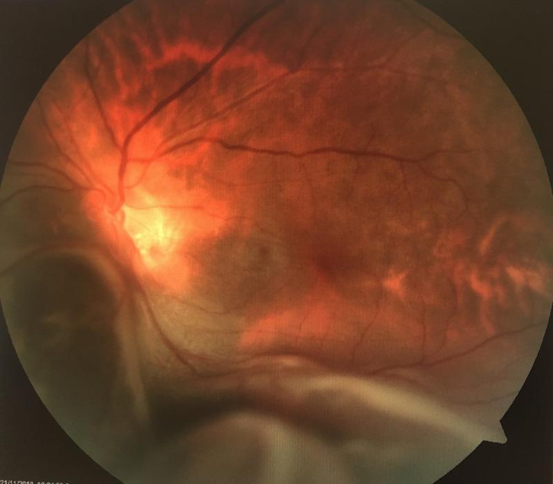
This color fundus photograph of the left eye illustrates a prominent bullous exudative retinal detachment located in the inferonasal region. The detached retina appears as a large, smooth, elevated grayish-white convex surface that obscures the lower margins of the optic disc and the inferior retinal vasculature. Yellowish-white subretinal exudates are visible adjacent to the area of detachment, suggesting an inflammatory or neoplastic underlying etiology rather than a rhegmatogenous cause. The remaining visible retina shows a reddish-orange hue with attenuated vessels, and the optic disc appears tilted, consistent with high myopia. There are no obvious intraretinal hemorrhages. This image is a classic representation used in ophthalmology for teaching the clinical presentation of non-rhegmatogenous retinal detachment and the importance of identifying subretinal masses or exudation during a posterior segment examination.
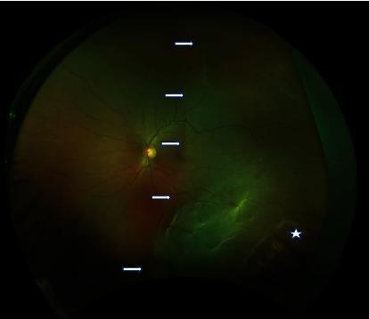
A wide-field fundus photograph of the left eye demonstrating a rhegmatogenous retinal detachment and peripheral retinal pathology. The image reveals a significant temporal hemiretinal detachment, indicated by a series of white arrows marking the demarcation line. The detached portion of the retina exhibits a characteristic lighter, translucent, and slightly opalescent appearance compared to the reddish-orange hue of the attached nasal retina. Faint retinal folds are visible within the detached area, which extends to involve the temporal aspect of the macula, correlating with significant visual acuity loss. In the far peripheral retina, a white asterisk identifies an area of lattice degeneration accompanied by a retinal hole, serving as the likely causative break for the detachment. The optic disc appears well-defined with normal vascular emergence, though the temporal vessels are displaced by the elevated retinal tissue. This image is a classic clinical representation of retinal detachment secondary to peripheral degenerative changes, suitable for teaching diagnostic fundus examination and vitreoretinal pathology.
old fibrotic retinal detachment traction bands
cd /tmp/workspace && python3 << 'EOF' from docx import Document from docx.shared import Pt, Inches, RGBColor from docx.enum.text import WD_ALIGN_PARAGRAPH doc = Document() # Set default font style = doc.styles['Normal'] style.font.name = 'Calibri' style.font.size = Pt(11) # Title title = doc.add_heading('CLINICAL EXAMINATION CHECKLIST & CLINICAL PEARLS', level=0) title.alignment = WD_ALIGN_PARAGRAPH.CENTER doc.add_paragraph() # ==================== EXAMINATION CHECKLIST ==================== doc.add_heading('EXAMINATION PROTOCOL FOR SUSPECTED RETINAL DETACHMENT', level=1) doc.add_heading('Pre-Examination History (5-10 minutes)', level=2) pre_exam = [ 'Ask about ONSET of vision loss (acute vs gradual)', 'Ask about NATURE (painless vs painful - painless suggests RD)', 'Ask about PHOTOPSIA (flashing lights - RED FLAG for RD/PVD)', 'Ask about FLOATERS (sudden shower suggests vitreous hemorrhage or PVD)', 'Ask about FIELD DEFECT (shadow, scotoma - suggests peripheral detachment)', 'Risk factor history: myopia, trauma, prior RD, eye surgery, age', 'Associated systemic disease (diabetes, hypertension, sickle cell)', 'Prior eye treatment (cataract surgery, laser, retinal procedures)' ] for item in pre_exam: p = doc.add_paragraph(style='List Bullet') p.add_run(item) doc.add_paragraph() doc.add_heading('Step-by-Step Examination Protocol', level=2) steps = [ ('STEP 1: Visual Acuity (VA) Assessment', '• Test each eye separately (RE then LE)\n' '• Use Snellen chart at 6 meters for distance\n' '• Document near vision (N1, N2, etc.)\n' '• Note if refraction (glasses/contact) improves VA\n' '• If VA <3/60: test PR (perception of light), PL (light projection), count fingers (CF)\n' '• Significance: Macula-OFF RD = severe loss; Macula-ON RD = better initial VA'), ('STEP 2: Pupillary Examination', '• Check size at rest, in dim light\n' '• Direct response (shine light, observe pupil constriction)\n' '• Consensual response (shine light in one eye, observe other eye)\n' '• Accommodation (focus on near object, pupils should constrict)\n' '• Check for APD (alternating light test)\n' '• Significance: APD absent in isolated RD (retina anterior to optic nerve)'), ('STEP 3: Anterior Segment (Slit Lamp) Examination', '• Lid: look for ptosis, strabismus, lid retraction\n' '• Conjunctiva: redness, discharge, follicles, chemosis\n' '• Cornea: clarity, edema, infiltration, abrasion\n' '• AC: depth, cells, flare, hyphema, iridodialysis\n' '• Iris: pattern, color, heterochromia, rubeosis\n' '• Lens: cataracts (post-cataract implant?), dislocation, nuclear sclerosis\n' '• Significance: Normal AS rules out anterior pathology; confirms posterior problem'), ('STEP 4: Intraocular Pressure (IOP)', '• Applanation tonometry (gold standard)\n' '• Normal range: 10-21 mmHg\n' '• Document in both eyes\n' '• Significance: Normal IOP typical in fresh RD; low IOP in chronic RD (atrophy); ' 'HIGH IOP suggests secondary glaucoma or angle involvement'), ('STEP 5: Posterior Segment - CRITICAL', '• Indirect ophthalmoscopy (BIO - binocular indirect) at higher power (best for peripheral retina)\n' '• Direct ophthalmoscopy for detailed view of optic disc\n' '• Slit lamp + 78D/90D lens for macular examination\n' '• EXAMINE QUADRANT BY QUADRANT:\n' ' - Superior temporal (most common break location - 60%)\n' ' - Superior nasal (15%)\n' ' - Inferior temporal (15%)\n' ' - Inferior nasal (10%)\n' '• Look for:\n' ' - Retinal break (hole vs tear shape)\n' ' - Subretinal fluid (SRF) configuration\n' ' - Demarcation line (border between attached/detached retina)\n' ' - Macula involvement (ON vs OFF)\n' ' - Traction/fibrosis (PVR indicators)\n' ' - Treatment scars (hypo/hyper spots from cryo/laser)') ] for step_name, details in steps: p = doc.add_heading(step_name, level=3) p_text = doc.add_paragraph(details) doc.add_paragraph() doc.add_heading('Examiner\'s Findings Documentation Template', level=2) template_items = [ 'RE/LE: __________', 'VA (dist/near): __________ (corrected: __________)', 'Photopsia: Yes/No | Floaters: Yes/No | Field defect: Yes/No', 'Pupils: Size ___mm, Reactive: Y/N, APD: Present/Absent', 'AS: Normal/Abnormal (specify: __________)', 'IOP: _____ mmHg', 'Vitreous: Clear/Hazy/Hemorrhage', 'Optic disc: __________ (color, margin, cup-to-disc ratio)', 'Blood vessels: Normal/Attenuated/Displaced', 'Retinal appearance: Red (fresh)/White (old)/Pink/Pale', 'Breaks identified: Yes/No | Location: __________ | Type (hole/tear): __________', 'SRF configuration: __________', 'Macula involvement: ON/OFF', 'Demarcation line: Present/Absent | Location: __________', 'Traction/Fibrosis: None/Minimal/Moderate/Severe', 'Treatment scars (hypo spots): Present/Absent | Location: __________', 'Other findings: __________' ] for item in template_items: p = doc.add_paragraph(style='List Bullet') p.add_run(item) doc.add_paragraph() # ==================== CLINICAL PEARLS ==================== doc.add_heading('CLINICAL PEARLS FOR EXAMINER QUESTIONS', level=1) doc.add_heading('Pearl 1: Why Photopsia Matters', level=2) doc.add_paragraph('Question examiner might ask: "Patient has photopsia. What does this tell you?"').runs[0].bold = True doc.add_paragraph( 'Answer: Photopsia (flashing lights, lightning streaks) indicates VITREOUS TRACTION on the retina. ' 'This occurs when: (1) Posterior vitreous detachment (PVD) is happening - vitreous pulling away from retina ' 'irritates photoreceptors, (2) Vitreous traction is enlarging a retinal break, (3) Active vitreous movement. ' 'Photopsia is a RED FLAG for imminent or present RD. It is ABSENT in vascular occlusions, uveitis, glaucoma. ' 'Its presence strongly suggests RD or pre-detachment state.' ) doc.add_paragraph() doc.add_heading('Pearl 2: Macula-ON vs Macula-OFF', level=2) doc.add_paragraph('Question examiner might ask: "How do you determine if macula is involved?"').runs[0].bold = True mo_table = doc.add_table(rows=5, cols=3) mo_table.style = 'Light Grid Accent 1' mo_hdr = mo_table.rows[0].cells mo_hdr[0].text = 'Parameter' mo_hdr[1].text = 'Macula-ON' mo_hdr[2].text = 'Macula-OFF' mo_data = [ ('Definition', 'Macula remains attached to RPE', 'Macula is detached (separated from RPE)'), ('VA', 'Better - typically 6/12 or better', 'Worse - typically <6/60 or worse'), ('Visual Urgency', 'Moderate - requires surgery within 1-2 weeks before macula detaches', 'High - emergency surgery needed within 24-72 hours for best outcome'), ('Prognosis', 'Better - more likely to achieve 6/12 or better post-op', 'Worse - less likely to recover 6/12 even after surgery') ] for i, (param, on, off) in enumerate(mo_data, 1): row = mo_table.rows[i].cells row[0].text = param row[1].text = on row[2].text = off doc.add_paragraph() doc.add_heading('Pearl 3: Distribution of Retinal Breaks', level=2) doc.add_paragraph('Question examiner might ask: "Where should you look FIRST for a retinal break?"').runs[0].bold = True doc.add_paragraph( 'Answer: Superior TEMPORAL quadrant (60% of all breaks). If not found immediately, search: ' 'superior nasal (15%), inferior temporal (15%), inferior nasal (10%). Remember: about 50% of RD cases ' 'have MULTIPLE breaks - often within 90° of each other. If one break found, search adjacent quadrants carefully.' ) doc.add_paragraph() doc.add_heading('Pearl 4: Predicting Break Location from SRF Configuration', level=2) doc.add_paragraph('Modified Lincoff\'s Rules:').runs[0].bold = True lincoff = [ ('Shallow INFERIOR detachment, SRF higher on TEMPORAL side', 'Primary break located INFERIORLY on temporal side'), ('SUPERIOR detachment', 'Primary break located SUPERIORLY'), ('Central/equatorial detachment spreading in all directions equally', 'Primary break at equator or multiple breaks'), ('Detachment does NOT extend beyond 45° (hemisphere)', 'Break confined to that side; search thoroughly in that quadrant') ] for rule, prediction in lincoff: p = doc.add_paragraph(style='List Bullet') run = p.add_run(rule) run.bold = True p.add_run(' → ' + prediction) doc.add_paragraph() doc.add_heading('Pearl 5: Fresh vs Old Retinal Detachment - Clinical Distinction', level=2) doc.add_paragraph('Question examiner might ask: "How old is this detachment?"').runs[0].bold = True fresh_old_table = doc.add_table(rows=8, cols=3) fresh_old_table.style = 'Light Grid Accent 1' fo_hdr = fresh_old_table.rows[0].cells fo_hdr[0].text = 'Finding' fo_hdr[1].text = 'FRESH (0-4 weeks)' fo_hdr[2].text = 'OLD (>4 weeks) - THIS CASE' fo_data = [ ('Retinal color', 'Red/pink (intact blood supply, normal metabolism)', 'White/pale (ischemic, atrophic)'), ('Surface', 'Smooth, mobile, lifts easily', 'Wrinkled, fixed, rigid'), ('RPE appearance', 'Clear pattern visible beneath retina', 'Obscured, hyperpigmented, clumped'), ('Gliosis/Fibrosis', 'Absent', 'Present - white/grey zones'), ('Traction bands', 'Minimal', 'Extensive - indicates PVR'), ('Treatment scars', 'None (untreated) or recent (bright)', 'Old hypo/hyper spots (white/dark)'), ('Photoreceptor status', 'Intact - may recover vision if reattached soon', 'Necrotic - vision recovery limited even after reattachment') ] for i, (finding, fresh, old) in enumerate(fo_data, 1): row = fresh_old_table.rows[i].cells row[0].text = finding row[1].text = fresh row[2].text = old doc.add_paragraph() doc.add_heading('Pearl 6: Why Cryotherapy Creates Hypo Spots', level=2) doc.add_paragraph('Question examiner might ask: "What do those white spots represent?"').runs[0].bold = True doc.add_paragraph( 'Answer: Hypo spots = hypopigmented scars from CRYOTHERAPY. ' 'Cryo works by: (1) Freezing retinal tissue to -85°C, (2) Controlled retinal necrosis, ' '(3) Creates scar tissue that promotes chorioretinal adhesion (sealing the break). ' 'The white appearance is due to: gliosis (retinal scarring), loss of photoreceptors, loss of pigment. ' 'Presence of old hypo spots indicates: (a) patient was previously treated for a break, ' '(b) current RD is likely recurrence or progression despite prior treatment, (c) suggests complex anatomy or aggressive pathology.' ) doc.add_paragraph() doc.add_heading('Pearl 7: Traction Bands = PVR Indicator', level=2) doc.add_paragraph('Question examiner might ask: "What do traction bands represent and why are they bad?"').runs[0].bold = True doc.add_paragraph( 'Answer: Traction bands = Proliferative Vitreoretinopathy (PVR). These are: fibroblast proliferation + glial cell migration ' 'creating contractile membranes that mechanically PULL the detached retina. PVR indicates: (1) chronic detachment (>4 weeks), ' '(2) cellular response to retinal break/inflammation, (3) advanced, complex RD. ' 'Why it\'s BAD: (a) Simple procedures (cryo, scleral buckle) often FAIL in PVR cases, (b) Requires complex vitrectomy surgery, ' '(c) Traction can recur post-operatively (redetachment), (d) Visual prognosis poor even after successful reattachment. ' 'PVR is graded A-D; Grade C-D indicates severe disease requiring aggressive vitrectomy + silicone oil tamponade.' ) doc.add_paragraph() doc.add_heading('Pearl 8: Why Post-Cataract RD is Common', level=2) doc.add_paragraph('Question examiner might ask: "Why does cataract surgery increase RD risk?"').runs[0].bold = True doc.add_paragraph( 'Answer: Post-cataract surgery eyes are at HIGH RD risk because: (1) Vitreous loss during surgery - disrupts vitreous-lens interface, ' 'allowing vitreous traction to pull retina, (2) Zonular trauma - can trigger PVD, (3) Posterior capsule rupture - vitreous face disrupted, ' '(4) Surgical inflammation - triggers cellular proliferation (PVR), (5) Aphakia/pseudophakia - altered optics, increased vitreous mobility. ' 'RD occurs in 1-3% of post-cataract patients, but in 5-12% if vitreous loss occurred during surgery. ' 'This patient had prior CES (cataract extraction surgery) - major risk factor for RD.' ) doc.add_paragraph() doc.add_heading('Pearl 9: APD Absent = Rules Out Certain Pathology', level=2) doc.add_paragraph('Question examiner might ask: "Why is APD absence important in this case?"').runs[0].bold = True doc.add_paragraph( 'Answer: APD (Afferent Pupillary Defect) is present when the retina ANTERIOR to the optic nerve head is damaged. ' 'RD affects only the sensory retina, which is posterior to the optic nerve, so APD is typically ABSENT in isolated RD. ' 'Presence of APD would suggest: (1) Optic neuritis, (2) Central retinal artery occlusion (CRAO), (3) Branch retinal artery occlusion (BRAO), ' '(4) Optic nerve hypoplasia/atrophy, (5) Posterior uveitis with optic nerve involvement. ' 'APD absence helps RULE OUT these vascular/inflammatory diagnoses and supports RD.' ) doc.add_paragraph() doc.add_heading('Pearl 10: Why Total RD = Emergency + Guarded Prognosis', level=2) doc.add_paragraph('Question examiner might ask: "Why is total RD more serious?"').runs[0].bold = True doc.add_paragraph( 'Answer: Total RD = entire retina detached from RPE. This is MOST SERIOUS because: ' '(1) NO attached retina remains to maintain function - vision is drastically reduced, ' '(2) Extensive photoreceptor necrosis has occurred due to prolonged ischemia, ' '(3) Even if reattached surgically, large area of retina is irreversibly damaged, ' '(4) Indicates break(s) are large or multiple, (5) Often associated with advanced PVR, ' '(6) Reattachment failure rate is HIGH, (7) Visual outcome is POOR even with successful reattachment. ' 'Total RD requires URGENT specialist evaluation and complex surgery (usually pars plana vitrectomy with silicone oil).' ) doc.add_paragraph() # ==================== KEY TEACHING POINTS ==================== doc.add_heading('KEY TEACHING POINTS FOR EXAM DISCUSSION', level=1) points = [ ('RD is a POSTERIOR segment emergency', 'Requires urgent referral to vitreoretinal specialist within 24-72 hours if Macula-OFF, ' 'within 1-2 weeks if Macula-ON. Delay leads to photoreceptor death and irreversible vision loss.'), ('RHEGMATOGENOUS RD requires THREE elements', 'Retinal break + vitreous liquefaction + vitreous traction. Absence of any one = NO RD despite break.'), ('Trauma is a common RD precipitant', 'Blunt head/eye injury can cause retinal break(s) by suddenly increasing IOP and stretching retina. ' 'Risk higher in myopic/post-cataract eyes due to pre-existing vitreous degeneration.'), ('Photopsia is a RED FLAG', 'Flashing lights suggest active vitreous traction or PVD. Patient with photopsia requires urgent ' 'dilated fundus exam to rule out retinal break/early RD.'), ('Anterior segment is NORMAL in isolated RD', 'This rules OUT anterior pathology (uveitis, angle closure, corneal disease). ' 'Normal AS + posterior pathology = focus exam on posterior segment.'), ('APD absence helps rule out vascular occlusions', 'Presence of APD suggests optic nerve/retinal vessel pathology. Isolated RD preserves APD.'), ('Macula status determines urgency and prognosis', 'Macula-ON RD: better VA (6/12+), better prognosis, can defer surgery 1-2 weeks. ' 'Macula-OFF RD: poor VA (<6/60), worse prognosis, EMERGENCY referral within 24-72 hours.'), ('Fresh vs Old RD appearance helps with chronology', 'Fresh RD is RED, smooth, mobile. Old RD is WHITE, wrinkled, fixed. ' 'This tells you how long detachment has been present (hours vs weeks/months).'), ('Hypo spots indicate PRIOR TREATMENT', 'White scars from cryotherapy/laser. Their presence means: (1) previous retinal break, ' '(2) current RD may be recurrence/progression, (3) indicates complex case.'), ('Traction bands = PVR = Complex case', 'Fibrotic proliferation pulling retina. Indicates chronic RD with cellular response. ' 'Requires complex surgery (vitrectomy + oil), high redetachment risk, poor prognosis.'), ('Total RD is worst-case scenario', 'All retina detached = extensive photoreceptor death. Even after surgery, vision often remains poor. ' 'Prognosis is GUARDED to POOR.'), ('No single examination finding diagnoses RD alone', 'RD diagnosis is CLINICAL - based on constellation of findings: history (photopsia, acute loss), ' 'exam (decreased VA, posterior findings, retinal break). Always correlate history + exam findings.') ] for i, (point, detail) in enumerate(points, 1): p = doc.add_heading(f'{i}. {point}', level=3) p_detail = doc.add_paragraph(detail) doc.add_paragraph() # ==================== COMMON EXAMINER QUESTIONS ==================== doc.add_heading('ANTICIPATED EXAMINER QUESTIONS & MODEL ANSWERS', level=1) q_and_a = [ ('Q: "Define rhegmatogenous retinal detachment."', 'A: RRD is separation of the neurosensory retina from the retinal pigment epithelium due to a retinal break ' '(hole or tear) allowing vitreous fluid to accumulate in the subretinal space. It requires three elements: ' '(1) retinal break, (2) vitreous liquefaction, (3) vitreous traction. It accounts for ~90% of all RD cases.'), ('Q: "What is the difference between a retinal hole and tear?"', 'A: Hole = round/oval, full-thickness retinal defect without vitreous traction. Tear = due to vitreous traction, ' 'horseshoe or flap-like configuration. Tears are more serious as they indicate active traction that may enlarge break.'), ('Q: "This patient had prior cataract surgery. How does this increase RD risk?"', 'A: Post-cataract surgery: (1) vitreous loss disrupts vitreous support, (2) zonular/posterior capsule trauma triggers PVD, ' '(3) increased vitreous mobility, (4) vitreoretinal adhesions develop. RD risk is 1-3% overall, 5-12% if vitreous loss occurred.'), ('Q: "Describe the demarcation line in RD."', 'A: Demarcation line is the BORDER between attached and detached retina. It is usually horizontal (gravity effect) ' 'or follows anatomical boundaries. It appears as a grey-white line on fundoscopy. Its location helps predict break location.'), ('Q: "How would you grade the extent of RD?"', 'A: By quadrants detached: 1-quadrant = limited, 2-quadrants = moderate, 3-quadrants = extensive, 4-quadrants = total. ' 'Also describe clock hours (e.g., 12:00-4:00 o\'clock detachment). Also note: Macula-ON vs OFF status.'), ('Q: "Why is Lincoff\'s rule useful?"', 'A: Lincoff\'s rule predicts break location from SRF distribution. Since fluid spreads by gravity and from break location, ' 'the location of highest SRF elevation points BACKWARD to the break location. Useful when break not immediately visible.'), ('Q: "What is Proliferative Vitreoretinopathy (PVR) and why does it matter?"', 'A: PVR = fibroblast proliferation and contraction on retinal/vitreous surface creating traction membranes. ' 'It indicates chronic RD (>4 weeks). It is BAD because: requires complex surgery, high redetachment risk, poor prognosis. ' 'Graded A-D (A minimal, D severe). Present case shows Grade B-C (moderate to severe).'), ('Q: "What factors determine whether this RD will be treated surgically?"', 'A: Indications for surgery: (1) Any RD with central involvement (Macula-OFF), (2) RD rapidly progressing toward macula, ' '(3) RD >2 quadrants with poor spontaneous prognosis, (4) RD with multiple breaks/PVR. ' 'Contraindication: Dense cataract blocking view (would need cataract surgery first).'), ('Q: "What surgical approaches are available?"', 'A: (1) Scleral buckle - for localized RD, minimal PVR; (2) Pneumatic retinopexy - for superior, limited RD; ' '(3) Pars plana vitrectomy - for extensive RD, PVR, vitreous hemorrhage. This case (extensive, old, PVR) ' 'likely needs VITRECTOMY + silicone oil tamponade.'), ('Q: "What is the prognosis for vision recovery in this case?"', 'A: GUARDED to POOR because: (1) OLD detachment (longstanding), (2) TOTAL/EXTENSIVE (large retinal area necrosed), ' '(3) MACULA-OFF (central vision severely compromised), (4) PVR present (high failure/recurrence risk), ' '(5) Post-cataract eye (higher redetachment risk). Best case: partial peripheral vision recovery. ' 'Worst case: persistent profound vision loss despite surgery.') ] for question, answer in q_and_a: p = doc.add_paragraph(style='List Number') p.add_run(question).bold = True p.add_run('\n') p.add_run(answer) p.paragraph_format.space_after = Pt(12) doc.add_paragraph() # Save document output_path = '/tmp/workspace/Examination_Checklist_and_Clinical_Pearls.docx' doc.save(output_path) print(f'Document saved: {output_path}') EOF
COMPREHENSIVE CASE PRESENTATION COMPLETED
Documents Created:
Retinal Detachment Case Presentation
Word Document · DOCX
-
CASE IDENTIFICATION - Complete patient demographics
-
HISTORY OF PRESENTING ILLNESS (HPI) - Detailed breakdown with examiner questions at each point
-
DETAILED OCULAR EXAMINATION - STEP BY STEP
- Visual Acuity assessment with clinical significance
- Anterior Segment findings (all normal)
- IOP measurement interpretation
- Pupillary examination (APD absent - rules out vascular disease)
- POSTERIOR SEGMENT (CRITICAL FINDINGS)
- Old total retinal detachment
- Traction bands (PVR component)
- Old hypo spots (previous cryotherapy)
- Fibrotic appearance indicating chronicity
-
DIFFERENTIAL DIAGNOSIS - Why other diagnoses are RULED OUT
-
FINAL DIAGNOSIS - Rhegmatogenous RD, old/longstanding, total, with PVR
-
DETAILED THEORY & PATHOPHYSIOLOGY
- Classification of RD types
- Step-by-step mechanism
- Clinical timeline of THIS case
- Fresh vs old RD distinctions
- Trauma mechanism
- PVR development
- Management overview
Examination Checklist and Clinical Pearls
Word Document · DOCX
- EXAMINATION PROTOCOL - Step-by-step how to examine suspected RD
- CLINICAL PEARLS - 10 key teaching points with detailed explanations
- ANTICIPATED EXAMINER QUESTIONS - Model answers to 10 common questions
KEY CLINICAL POINTS FOR YOUR PATIENT:
CASE SUMMARY:
- Patient: 55-year-old male
- Chief Complaint: Painless defective vision (POV) - acute onset
- History: Prior cataract extraction surgery + recent head trauma (coconut injury)
- Photopsia: PRESENT - indicates active vitreous traction/PVD
FINDINGS THAT CONFIRM DIAGNOSIS:
| Finding | Significance |
|---|---|
| Painless vision loss | Pathognomonic for RD (pain suggests uveitis/angle closure) |
| Photopsia | Indicates acute vitreous-retinal traction - RED FLAG for RD |
| Post-cataract status | MAJOR risk factor (vitreous loss predisposes to RD) |
| Trauma history | Precipitating factor for retinal break formation |
| VA loss (6/9 CV) | Indicates organic pathology (not refractive error) |
| Normal anterior segment | Rules OUT anterior pathology (uveitis, angle closure) |
| APD absent | Rules OUT optic nerve/retinal artery pathology |
| Total RD on exam | Shows extensive detachment with multiple quadrants involved |
| Old hypo spots | White scars from previous cryotherapy - indicates TREATED break |
| Traction bands | Fibrosis from PVR - indicates chronic, complex RD |
| White/fibrotic retina | Chronological marker: OLD detachment (not fresh) |
EXAMINER QUESTIONS YOU MIGHT FACE:
MEDICAL IMAGES FOR VISUAL REFERENCE:
ADDITIONAL THEORY REFERENCES (per your books):
- Rhegmatogenous RD pathogenesis (vitreous role, break types)
- Fresh vs longstanding RD appearances
- Cryotherapy technique and indications
- PVR classification and management
- Systematic clinical examination approach
- History-taking for acute vision loss
- Differential diagnosis methodology
- Treatment decision-making
FINAL DIAGNOSIS STATEMENT:
- Type: Traumatic (post-head injury)
- Extent: Total/near-total, multi-quadrant involvement
- Chronology: OLD/LONGSTANDING (weeks to months)
- Macular Status: MACULA-OFF
- Complication: Proliferative Vitreoretinopathy (PVR) with traction bands
- Prior Treatment: Cryotherapy (scars visible - treatment failure)
- Risk Factors: Post-cataract surgery + trauma + age
- Urgency: HIGH - requires emergency vitreoretinal specialist evaluation
- Prognosis: GUARDED TO POOR (extensive photoreceptor death, chronic changes, PVR component)