
Generate me a easiest summar of hemostasis including intrinsic and extrinsic pathway
Reading File
Now let me search the library for hemostasis content in parallel.
Finding Sources
Finding Sources
Robbins is the best source for this. Let me read the hemostasis section from Robbins and also the Tietz lab medicine section in parallel.
Reading File
Finding Sources
Excellent! I found a coagulation cascade diagram and key content. Let me read the Harrison's section on fibrin clot formation and the coagulation cascade diagram from Tietz, and also more of the Robbins content.
Reading File
Reading File
Loading Image
I also want to get the Harrison's diagram and read a bit more about the coagulation cascade detail. Let me grab one more section.
Reading File
Loading Image
I now have rich content from Robbins Pathology, Harrison's Principles, and Tietz Lab Medicine, plus two excellent diagrams. Here is the complete, easy summary:
Hemostasis - Easy Summary
Hemostasis is the process that stops bleeding after vascular injury. It happens in four overlapping steps.
Step 1 - Vasoconstriction
Immediately after injury, the blood vessel narrows (vasoconstiction) to reduce blood flow. This is driven by neurogenic reflexes and endothelin (a vasoconstrictor released by damaged endothelium). It is temporary - bleeding resumes without the next steps.
Step 2 - Primary Hemostasis (Platelet Plug)
- Injury exposes subendothelial collagen and von Willebrand Factor (vWF)
- Platelets bind vWF via GpIb receptors → platelet adhesion
- Platelets activate → change shape, release granules (ADP, TXA2)
- More platelets are recruited and aggregate via GpIIb-IIIa binding fibrinogen
- Result: a soft, loose primary platelet plug
Step 3 - Secondary Hemostasis (Coagulation Cascade)
This is where the intrinsic and extrinsic pathways come in. Both converge on a common pathway to generate thrombin.
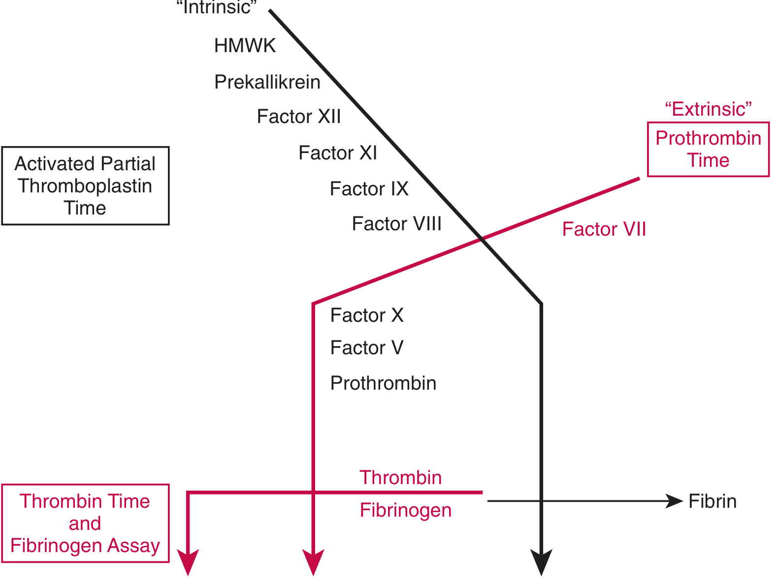
Here is the classic cascade diagram from Tietz Lab Medicine:
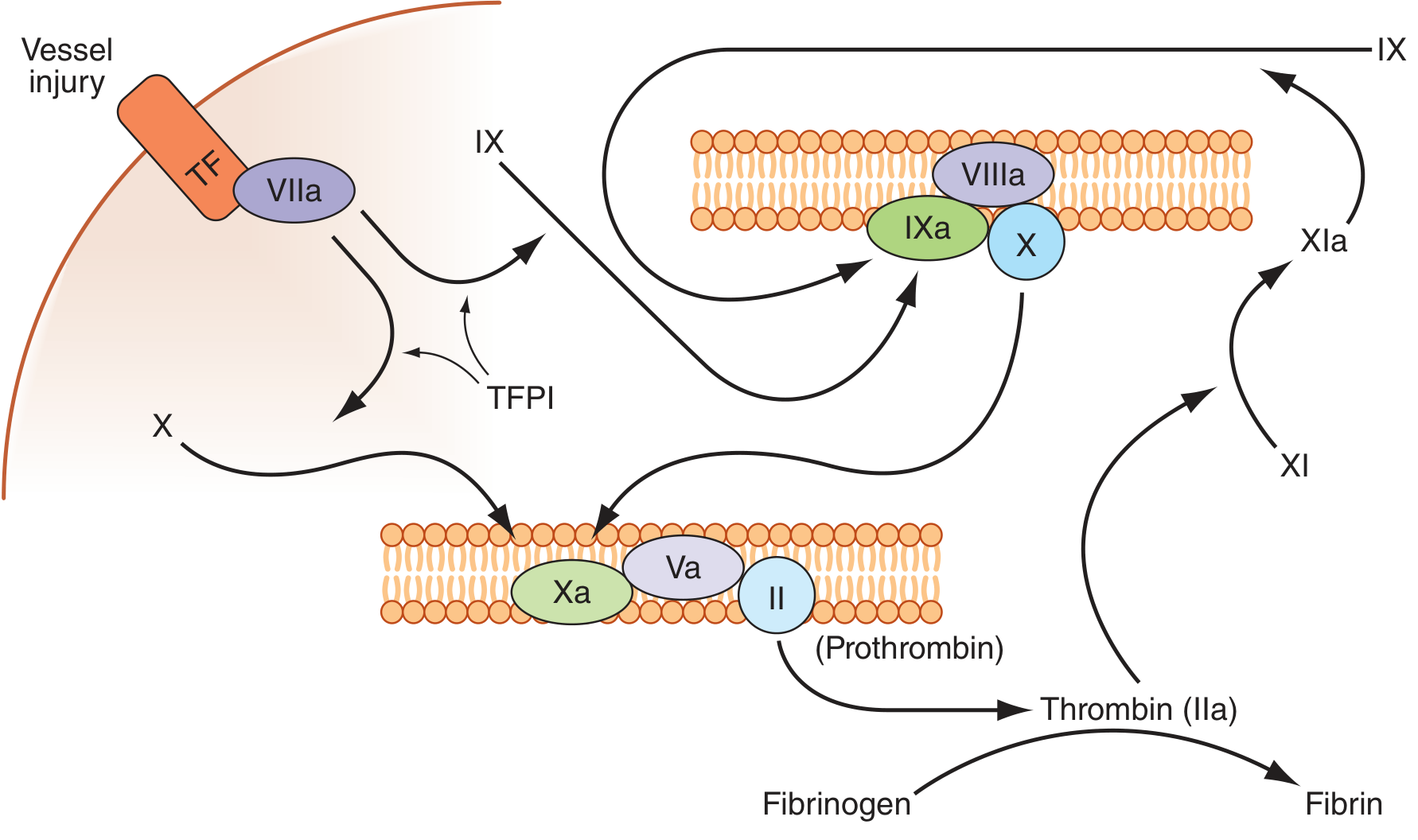
And the modern in-vivo view from Harrison's Principles (how it really works in the body):
Extrinsic Pathway ("Tissue Factor Pathway") - Fast, Dominant In Vivo
| Step | What happens |
|---|---|
| Vessel injury | Subendothelial Tissue Factor (TF) is exposed |
| TF + Factor VIIa | Form a complex on the cell surface |
| TF-VIIa complex | Activates Factor X → Xa (directly) |
| Also | TF-VIIa activates Factor IX → IXa (linking to intrinsic) |
| Xa + Va | Form prothrombinase complex → converts Prothrombin (II) → Thrombin (IIa) |
Lab test: Prothrombin Time (PT) measures the extrinsic pathway.
Intrinsic Pathway ("Contact Activation Pathway") - Slower, Important for Amplification
| Step | What happens |
|---|---|
| Contact with collagen | Activates Factor XII → XIIa |
| XIIa | Activates Factor XI → XIa (with HMWK, Prekallikrein) |
| XIa | Activates Factor IX → IXa |
| IXa + VIIIa | Form tenase complex → activates Factor X → Xa |
| Xa + Va | Form prothrombinase → Prothrombin → Thrombin |
Lab test: Activated Partial Thromboplastin Time (aPTT) measures the intrinsic pathway.
Modern understanding (Harrison's): In the body, coagulation is initiated by the extrinsic/TF pathway but amplified through the intrinsic pathway. Thrombin itself feeds back to activate Factor XI, VIII, and V, creating a powerful amplification loop. Factor XII is not essential for in-vivo hemostasis.
Common Pathway
Both pathways funnel into this:
Factor Xa + Va (prothrombinase complex)
       ↓
Prothrombin (II) → Thrombin (IIa)
       ↓
Fibrinogen → Fibrin (loose mesh)
       ↓
Factor XIII (activated by thrombin) crosslinks fibrin
       ↓
Stable fibrin clot
Step 4 - Clot Stabilization and Limitation
- Factor XIII covalently crosslinks fibrin strands → stable, solid clot
- Platelet contraction tightens the clot
- Anti-clotting mechanisms prevent runaway clotting:
- TFPI (Tissue Factor Pathway Inhibitor) - inhibits the TF/VIIa complex
- Antithrombin - neutralizes thrombin and Xa (heparin enhances this)
- Protein C/S - thrombin activates Protein C (via thrombomodulin on endothelium) → inactivates Factors Va and VIIIa
- tPA (tissue plasminogen activator) - triggers fibrinolysis (clot breakdown) via plasmin
Quick Reference Table
| Feature | Intrinsic | Extrinsic |
|---|---|---|
| Trigger | Contact with collagen/glass | Tissue Factor exposure |
| Key factors | XII, XI, IX, VIII | VII, TF |
| Speed | Slower | Faster |
| Lab test | aPTT | PT / INR |
| Role in body | Amplification | Initiation |
Memory Aid
"I Eat Chocolate" = Intrinsic, Extrinsic, Common
- Intrinsic: 12, 11, 9, 8 (counts down)
- Extrinsic: 7 (only one factor)
- Common: 10, 5, 2 (prothrombin), 1 (fibrinogen)
Sources: Robbins & Kumar Basic Pathology; Harrison's Principles of Internal Medicine 22E; Tietz Textbook of Laboratory Medicine, 7th Ed.
This is a shared conversation. Sign in to Orris to start your own chat.