
Stapedotomy, stapedectomy and reverse stapedectomy in detail, indications,contraindications,steps,complications,each and every single thing, according to ENT textbook and in simplified manner
stapedotomy stapedectomy stapes surgery procedure diagram
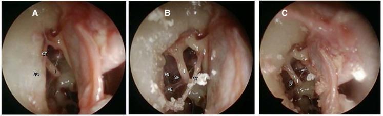
This set of three intraoperative endoscopic photographs (A, B, C) captures sequential stages of a stapes surgery (stapedotomy/stapedectomy) in a human middle ear. Image A shows the initial surgical field after elevation of the tympanomeatal flap, identifying the chorda tympani (CT) crossing the incus (I) and the bony scutum (SC). Image B depicts the field after scutum removal (atticotomy) to improve exposure, clearly revealing the facial nerve (FN) canal, the pyramidal eminence (PE), the long process of the incus (I), and the stapes footplate (SF). Image C illustrates the final stage with a stapes prosthesis (P) in situ, crimped to the long process of the incus and extending toward the oval window. These images serve as educational material for otolaryngology, demonstrating essential anatomical landmarks of the tympanic cavity and the technical progression of endoscopic ear surgery. The views highlight the spatial relationships between the ossicular chain, the facial nerve, and the chorda tympani required for successful surgical outcomes.
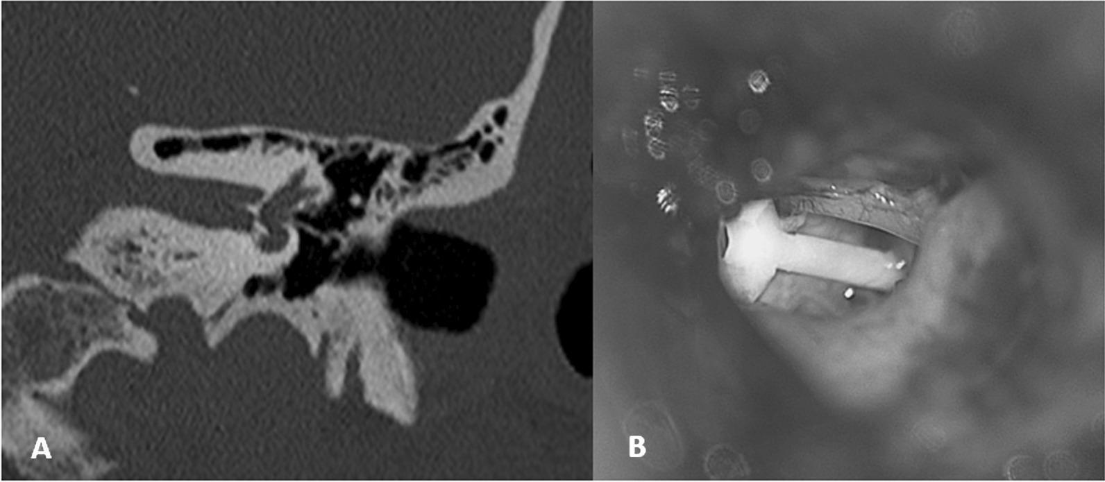
This dual-panel image demonstrates a stapedial prosthesis dislocation in the left ear via radiological and intraoperative perspectives. Panel A is a High-Resolution Computed Tomography (HRCT) scan of the temporal bone in coronal reconstruction. It displays the middle ear cavity, mastoid air cells, and the inner ear structures, specifically highlighting a high-density metallic/synthetic piston-style prosthesis that is dislocated from its anatomical position at the oval window. Panel B provides a per-operative endoscopic view during revision stapes surgery. It shows a T-shaped, light-colored stapedial piston (prosthesis) lying displaced within the surgical field, surrounded by moist soft tissue and surgical fluid. This comparison is used to illustrate the clinical diagnosis and surgical confirmation of prosthesis loop dislocation, a potential complication of stapedotomy or stapedectomy procedures in patients with otosclerosis. Key educational concepts include otologic imaging, prosthetic complications in middle ear surgery, and revision stapes surgery techniques.
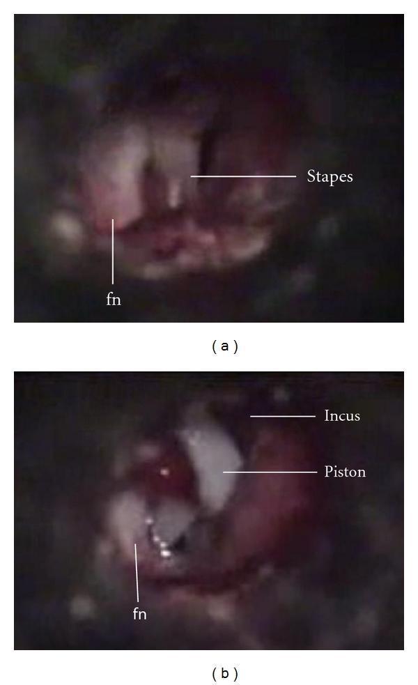
This image consists of two side-by-side intraoperative clinical photographs (a and b) documenting a stapes surgery in a patient with otosclerosis and an anatomical variant of the facial nerve. Image (a) displays the middle ear prior to reconstruction, showing the stapes and a completely exposed, dehiscent facial nerve (fn) running in an abnormal course anterior to the stapes. Image (b) shows the same middle ear after surgical intervention, demonstrating the placement of a stapes piston prosthesis. Key structures labeled include the long process of the incus, the piston prosthesis, and the exposed facial nerve (fn). The comparison illustrates the surgical management of otosclerosis in the presence of a facial nerve dehiscence, a critical surgical landmark for otolaryngologists to avoid iatrogenic nerve injury during stapedotomy or stapedectomy.
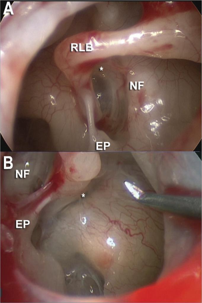
This composite diagnostic clinical photograph presents two endoscopic views (A and B) of the human middle ear, specifically the oval window niche, captured with a 30-degree endoscope during an otologic procedure. Panel A illustrates the anatomical relationship between the long process of the incus (RLB), situated superiorly, and the facial nerve canal (NF), which appears as a rounded prominence. The stapes superstructure is visible, featuring the pyramidal eminence (EP) and the anterior and posterior crura (marked with asterisks). Panel B provides a similar view in a different clinical case, demonstrating the same landmarks from a slightly different angle, with a surgical micro-instrument visible in the field for manipulation. The images emphasize the enhanced visualization of middle ear recesses and the stapes footplate provided by endoscopic approaches compared to traditional microscopy. These visuals serve as educational material for stapes surgery (stapedotomy/stapedectomy), highlighting the spatial orientation of the facial nerve relative to the ossicular chain and the pyramidal eminence.
Stapes Surgery: Stapedotomy, Stapedectomy & Revision Stapedectomy
Background: Why Is Stapes Surgery Done?
Anatomy Recap (Quick)
- A head (capitulum) - articulates with incus via incudostapedial joint
- Two crura (anterior and posterior)
- A footplate - sits in the oval window, held by the annular ligament
PART 1: STAPEDECTOMY
Definition
- KJ Lee's Essential Otolaryngology
Indications
- Conductive hearing loss with an air-bone gap of at least 25-30 dB, sufficient to produce a negative Rinne test with the 512-Hz tuning fork
- Normal otoscopic examination (intact tympanic membrane, no active middle ear disease)
- Acoustic reflex either biphasic or absent (indicating stapes fixation)
- Surgery is performed on the worse-hearing ear first. If both ears are similar, the patient identifies their subjectively better ear and the worse one is operated on
- Active otosclerosis confirmed clinically (progressive CHL in young adults, typically 20s-30s, often bilateral, female > male)
- Carhart's notch (dip at 2 kHz on audiogram) - pathognomonic of otosclerosis
- Patient unwilling or unable to use hearing aids
- KJ Lee's Essential Otolaryngology; Cummings Otolaryngology
Absolute Contraindications
- Only hearing ear - never operate on the sole functioning ear
- Active middle ear infection (otitis media)
- Tympanic membrane perforation (must be repaired first)
- Active external otitis
- Patient refusal or unrealistic expectations
- Inner ear hydrops / active Ménière disease - risk of saccule puncture causing profound SNHL
Relative Contraindications
- Obliterative (biscuit) footplate - entire footplate replaced by dense otosclerotic bone; surgery technically more difficult (requires drillout technique)
- Severe dehiscence of facial nerve over the oval window - may need to abort surgery
- Contralateral profound SNHL - if that ear has no hearing, extreme caution on the other ear
- Osteogenesis imperfecta - brittle bones risk tympanic ring fracture and incus fracture; proceed with extreme care
- Blind patient - poor compensation for vestibular deficits
- Contralateral peripheral vestibulopathy - caloric testing indicated before proceeding
- Mixed hearing loss - counsel that hearing aids may still be needed postoperatively
- Pregnancy - defer surgery
- Persistent stapedial artery - if not vestigial, abort surgery
- Cummings Otolaryngology; KJ Lee's Essential Otolaryngology
Pre-Operative Assessment
- Pure tone audiometry (PTA) with air-bone gap quantification
- Tympanometry (type As - shallow/stiff curve)
- Acoustic reflexes (absent or biphasic)
- 512-Hz tuning fork: negative Rinne (BC > AC), positive Weber lateralizing to worse ear
- CT temporal bone: anterior oval window lucency (early); "halo sign" around cochlea (cochlear involvement); late disease shows dense sclerotic bone
- Never operate on the better ear or only hearing ear
Operative Steps (Stapedectomy)
-
Canal injections - Four-quadrant canal block + vascular strip injection with 1% lidocaine and 1:100,000 epinephrine (just under the skin)
-
Incisions and tympanomeatal flap - Sickle knife incisions at the lateral process of malleus and inferiorly at the lower fourth of canal, extended laterally ~8 mm, connected with a House canal knife. Flap elevated evenly from lateral to medial. The annulus is carefully elevated out of its bony canal. The chorda tympani nerve is identified and protected.
-
Exposure (scutum removal / atticotomy) - The posterosuperior bony canal wall overhang is removed with a curette or microdrill until the facial nerve is visible superiorly and the pyramidal process is visible posteriorly.
-
Confirm stapes fixation - Palpate the malleus, incus, and stapes to confirm fixation at the footplate.
-
Measure incus-to-footplate distance - Using a measuring rod (usual distance: 4.5 mm from lateral surface of incus). Prosthesis size = 4.0 mm medial surface measurement minus 0.25 mm = typically 4.25 mm piston.
-
Divide stapedius tendon - Cut the tendon of stapedius muscle.
-
Separate incudostapedial joint - Using a joint knife.
-
Remove stapes superstructure - Fracture and remove the crura and head (down-fracture technique).
-
Remove footplate (stapedectomy-specific) - Remove all or most of the footplate using picks, hooks, or microdrill. This is the key step that distinguishes stapedectomy from stapedotomy.
-
Place oval window covering/graft - A connective tissue seal (vein graft, fat, or perichondrium) is placed over the open oval window to prevent perilymph leakage and protect the inner ear.
-
Place prosthesis - The piston prosthesis is positioned through the graft into the vestibule and hooked onto the long process of the incus.
-
Crimp prosthesis - The wire loop is mechanically crimped to the incus (or heat-activated for nitinol prostheses).
-
Test prosthesis mobility - Palpate and confirm movement.
-
Replace tympanomeatal flap - Return tympanic membrane to anatomic position. Place Gelfoam in canal.
- Cummings Otolaryngology; KJ Lee's Essential Otolaryngology
Prosthesis Types
- Piston style (wire loop + piston into oval window) - requires mechanical crimping
- Bucket handle style - flipped over the distal incus for stability
- Nitinol pistons - heat-activated, crimped with heating tool or laser
- Piston length: commonly 4.25 mm; diameter 0.4-0.6 mm
- Materials: Teflon, stainless steel, titanium, platinum, nitinol
- KJ Lee's Essential Otolaryngology
PART 2: STAPEDOTOMY
Definition
- Cummings Otolaryngology
Why Stapedotomy Replaced Stapedectomy
| Feature | Stapedectomy | Stapedotomy |
|---|---|---|
| Footplate | Removed completely | Small hole (0.6-0.8 mm) made |
| Oval window seal | Tissue graft required | Blood or fat around piston - no large graft needed |
| High-frequency hearing | Moderate gain | Better gain at 4-8 kHz |
| SNHL risk | ~1.5% | Lower (<0.6%) |
| Perilymph fistula | ~2% | ~0.6% |
| Post-op vertigo | More common | Less common |
| Revision need | Higher (7%) | Lower (3%) |
| Labyrinthine trauma | More | Less |
Stapedotomy Technique (Cummings - BOX 146.1)
- Canal injections (same as stapedectomy)
- Create and elevate tympanomeatal flap
- Remove posterosuperior canal wall bone (curette/microdrill) for facial nerve and pyramidal process visibility
- Palpate ossicular chain to confirm stapes fixation
- Measure incus-to-footplate distance
- Use laser to make a "rosette" in the center of the footplate (preparatory scoring)
- Use microdrill with 0.7-mm diamond bur to weaken posterior crus and create fenestra (the stapedotomy hole)
- Place piston prosthesis into the fenestra and onto the incus - piston now goes directly into the small hole
- Crimp prosthesis hook or use laser for nitinol prostheses
- Down-fracture stapes superstructure and remove (after prosthesis is secured)
- Place tissue graft or blood around prosthesis to form a seal (smaller seal than stapedectomy)
Laser vs Microdrill in Stapedotomy
- Argon, KTP, CO2, and Er:YAG lasers all used since the 1980s
- Potential risks: photothermal, photochemical, and photoacoustic inner ear damage
- CO2 and argon laser: comparable outcomes; no clear winner
- Laser may offer lower footplate fracture rate (5x lower than conventional picks in some studies)
- Microdrill vs handheld microperforator: no significant difference in outcomes
- Scott-Brown's Otorhinolaryngology Vol 2
Endoscopic Stapedotomy
- Safe with similar audiologic outcomes to microscopic stapes surgery
- Possible advantage: reduced need for bony annulus resection
- Growing evidence base but results are still evolving
- Scott-Brown's Otorhinolaryngology Vol 2
PART 3: REVERSE STAPEDECTOMY
What Is It?
Indications for Revision (Reverse) Stapedectomy
- Displaced/extruded prosthesis - most common cause (53% of failures)
- Incus erosion/necrosis - long process of incus eroded (26%)
- Bony regrowth over the fenestra / reosteogenesis (14%)
- Prosthesis too short - insufficient reach to oval window
- Inadequately crimped prosthesis - loose connection
- Perilymph fistula - ongoing CSF/perilymph leak
- Reparative granuloma - foreign body reaction 1-6 weeks post-op
- Persistent or recurrent CHL after primary surgery
- Malleus or incus ankylosis not recognized at primary surgery
- Unrecognized superior canal dehiscence syndrome
- Cummings Otolaryngology
Results of Revision Surgery
- Air-bone gap closure to within 20 dB in 71-96% of cases
- Risk of profound SNHL: 0-2%
- Worse overall than primary surgery
- Cummings Otolaryngology
Key Steps and Principles of Revision Stapedectomy
-
Preferred anesthesia: LOCAL - so the patient can be monitored for vertigo during the procedure. This is critical especially when manipulating a displaced prosthesis.
-
Use of laser is strongly recommended - to dissect adhesions and free the existing prosthesis safely.
-
Prosthesis removal - If the piston has migrated into the vestibule, extract it carefully while asking the patient about vertigo.
-
Assess the incus - Often eroded. Options if incus is no longer usable:
- Reinforce with hydroxyapatite bone cement
- Use a malleus-stapes prosthesis (incus replacement prosthesis)
- Kurz malleovestibulopexy Clip prosthesis
- TORP (Total Ossicular Replacement Prosthesis) from tympanic membrane to oval window
-
Remove or address the cause of failure (granuloma removal, adhesion lysis, new fenestration)
-
Place new prosthesis and secure
-
Seal oval window
- Cummings Otolaryngology
COMPLICATIONS (ALL Stapes Surgery)
Intraoperative Complications
| Complication | Detail | Management |
|---|---|---|
| Tympanic membrane perforation | During flap elevation | Repaired with underlay fascia graft |
| Markedly thickened footplate | Dense otosclerotic bone | Can be drilled out; prosthesis still placeable |
| Floating footplate | Footplate breaks free into vestibule during removal | Laser fenestration of floating plate, OR abort and return after refixation |
| Facial nerve injury | Dehiscent facial nerve over oval window; mild overhang - bend prosthesis wire; severe overhang - abort | Bend wire / abort |
| Persistent stapedial artery | Rare anomaly - artery between crura; vestigial = proceed with caution; non-vestigial = abort | Abort if significant |
| Intraoperative vertigo | Prosthesis too long; check immediately | Replace with shorter prosthesis |
| Chorda tympani injury | Stretching or tearing during flap elevation (30% transient irritation) | If stretched/torn: section it completely (paradoxically fewer symptoms) |
Early Postoperative Complications
| Complication | Timing | Detail |
|---|---|---|
| Serous labyrinthitis | Days-weeks | SNHL above 2 kHz + mild disequilibrium; usually resolves spontaneously |
| Reparative granuloma | 1-6 weeks | Initial hearing improvement then sudden/gradual deterioration; reddish discoloration in posterosuperior TM quadrant; foreign body reaction; treat with steroids + surgical removal |
| Transient facial palsy | 5 days post-op | Likely viral (HSV) reactivation; usually incomplete; treat with prednisone |
| Hyperacusis/phonophobia | Immediate post-op | Nearly universal; resolves over months |
| Taste disturbance (dysgeusia) | Immediate | ~1/3 of patients; metallic taste, dry mouth; usually resolves in 3-4 months |
| Perilymph fistula | Early-delayed | Mixed SNHL-CHL + unsteadiness; treat by removing prosthesis, grafting oval window, replacing prosthesis |
Late / Delayed Complications
| Complication | Detail |
|---|---|
| SNHL | Incidence ~1-2% primary surgery; higher in revision. Cause often unclear. Treat with immediate steroids. |
| Total/profound SNHL | ~0.2% (1 in 500); rare but devastating; patients must be counselled pre-op |
| Persistent CHL | Due to: unrecognized malleus/incus ankylosis, incus subluxation, short prosthesis, inadequate crimping, superior canal dehiscence |
| Recurrent CHL | Due to: incus long process necrosis, displaced prosthesis, reparative granuloma |
| Vertigo | Common for a few days (1 in 20 cases); persistent vertigo - suspect fistula or long prosthesis; re-explore |
| Tinnitus | Most existing tinnitus improves; rare new-onset tinnitus; related to serous labyrinthitis |
| Incus necrosis | Long-term; especially with wire loop pistons; causes recurrent CHL; requires revision |
Postoperative Care
- Head elevated ~30 degrees immediately post-op to reduce perilymph pressure
- Bed rest for 30-40 minutes, then mobilized if no vertigo
- Discharge 1-2 hours after surgery (outpatient procedure)
- Dry-ear precautions for 3 weeks
- Swimming/diving permitted 3 weeks post-surgery
- Flying permitted 1 week post-surgery
- First audiogram at 3 months post-op (not earlier)
- Tuning fork assessment at 3-week follow-up
- Cummings Otolaryngology
Results and Outcomes
- Air-bone gap closure to within 10 dB in 80-95% of cases for both stapedectomy and stapedotomy
- Stapedotomy has better high-frequency (4-8 kHz) outcomes and better speech discrimination in some studies
- Both techniques produce stable long-term hearing results
- SNHL risk: ~1-2% primary; higher for revision
- Total SNHL: ~0.2%
- In experienced hands, either technique gives excellent results
- Scott-Brown's Otorhinolaryngology Vol 2; Cummings Otolaryngology
Visual Reference
Quick Comparison Summary
| Feature | Stapedectomy | Stapedotomy | Revision (Reverse) |
|---|---|---|---|
| Footplate | Fully removed | Small fenestra only | Existing prosthesis removed, cause corrected |
| Graft needed | Yes (vein/fat) | Small seal only | Usually needed |
| Technique | Classic/historic | Modern gold standard | More complex |
| SNHL risk | ~1.5% | <0.6% | 0-2% |
| Fistula risk | ~2% | ~0.6% | Variable |
| High-freq hearing | Less gain | Better gain | Depends on cause |
| Preferred today | Less common | Standard of care | Only when primary fails |
| Anesthesia | Local preferred | Local preferred | Local mandatory |