
Explain burn in surgical subject
Reading File
Now let me search the medical library for burn-related surgical content in parallel.
Finding Sources
Finding Sources
The best surgical sources are Bailey & Love's Surgery, Sabiston, and Schwartz's. Let me read those in parallel.
Reading File
Reading File
Reading File
Excellent - I found the Bailey & Love diathermy burns content (surgical burns), and Sabiston's pediatric burns content. Now let me read the main Bailey & Love burns chapter and Schwartz's burns chapter for comprehensive surgical management.
Reading File
Reading File
Finding Sources
Let me now read the main Bailey & Love burns chapter (block6) around lines 1550-2200 and also the Schwartz chapter.
Reading File
Reading File
I have rich content from Bailey & Love's and Schwartz's. Let me now read the deeper surgical management sections from both.
Reading File
Reading File
I now have extensive content. Let me read the surgical treatment/excision and wound coverage sections from both books.
Reading File
Reading File
Excellent data collected. I now have very thorough content from both Bailey & Love's and Schwartz's covering the full surgical management of burns. Let me now fetch a key burn classification image and compile the comprehensive answer.
Reading File
Reading File
I now have comprehensive content. Let me compile the full, detailed answer.
Burns in Surgical Practice
A burn is a traumatic injury to skin and underlying tissues caused by thermal, chemical, electrical, or radiation agents. Burns remain a major cause of morbidity and mortality globally. Surgical care has evolved dramatically since the introduction of early burn excision and structured fluid resuscitation protocols.
1. Aetiology / Classification by Cause
| Type | Examples |
|---|---|
| Thermal | Scalds (hot liquids/steam), flame, contact with hot objects, flash burns |
| Chemical | Acid, alkali (alkali penetrates deeper due to liquefactive necrosis) |
| Electrical | Low-voltage, high-voltage, lightning; causes "entry and exit" wounds with internal damage disproportionate to surface appearance |
| Radiation | UV exposure (corneal burns/keratitis), nuclear, X-ray |
| Friction | Road rash, machinery injuries |
Diathermy burns (surgical context): Monopolar diathermy can cause unintended burns when:
- The indifferent (return) electrode has inadequate contact area
- The patient is earthed via contact with metal (Mayo table, stirrups)
- Faulty insulation of leads
- Accidental activation or contact of the active electrode with retractors/towel clips
- Channelling - when current is applied to a narrow tissue stalk (e.g., risk of penile/spermatic cord coagulation during circumcision), bipolar diathermy must be used instead
Alcohol-based skin preparation agents can ignite from diathermy sparks under theatre lights, making these fires difficult to detect early. - Bailey and Love's Short Practice of Surgery 28th Ed, p.133
2. Burn Depth Classification
This is the single most important determinant of surgical decision-making.
Superficial (First-Degree)
- Involves epidermis only
- Erythematous, painful, no blistering
- Not included in TBSA% calculations
- Heals in days with supportive care
Superficial Partial-Thickness (Second-Degree)
- Involves papillary dermis
- Pink, moist, blistering, brisk capillary return (blanches)
- Normal pinprick sensation
- Heals in 2 weeks without surgery, no significant scarring
Deep Partial-Thickness (Deep Dermal)
- Involves reticular dermis
- Dermis less moist, less erythematous; fixed capillary staining; does not blanch
- Reduced sensation (cannot distinguish sharp from blunt)
- Takes >3 weeks to heal and invariably leads to hypertrophic scarring without surgery
Full-Thickness (Third-Degree)
- Destroys entire dermis; leathery, insensate, white/brown/black
- No spontaneous healing - requires surgical grafting
- Fourth-degree: Involves underlying fascia, muscle, or bone
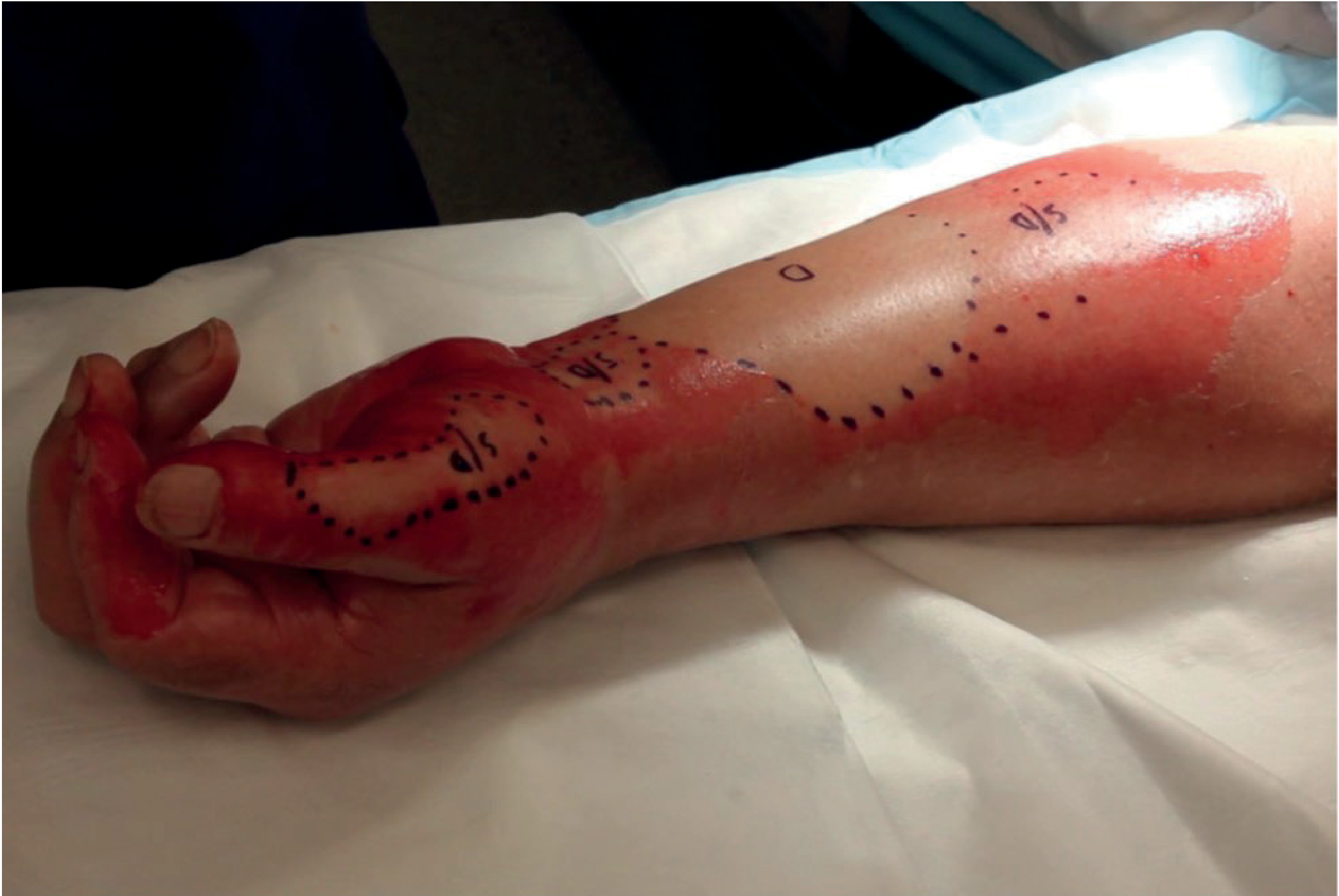
3. Assessment of Burn Size - TBSA
The Rule of Nines (Wallace Rule of Nines) for adults:
- Head and neck = 9%
- Each arm = 9%
- Anterior trunk = 18%
- Posterior trunk = 18%
- Each leg = 18%
- Perineum = 1%
The Lund and Browder chart is more accurate, especially in children, because it accounts for age-related changes in body proportions (head is larger relative to body in infants). The patient's palm (wrist crease to fingertips) = approximately 1% TBSA - useful for irregular burns.
Superficial (first-degree) burns are excluded from TBSA calculations for resuscitation purposes.
4. Admission Criteria (Burns Unit)
Burns meeting any of these criteria require admission to a burns unit (Bailey & Love's, p.688):
- Suspected airway or inhalational injury
- Any burn likely to require fluid resuscitation
- Any burn likely to require surgery
- Burns of significance to hands, face, feet, or perineum
- Suspicion of non-accidental injury
- Extremes of age
- High-tension electrical burns or concentrated hydrofluoric acid burns
- Social/psychiatric factors precluding home management
5. Major Determinants of Outcome
- Percentage TBSA involved
- Depth of burn
- Presence of inhalational injury
- Age and comorbidities
6. Initial Management - ATLS Approach
Management follows ABCDEF:
- A - Airway control (early intubation if airway burn suspected)
- B - Breathing/ventilation
- C - Circulation
- D - Disability (neurological status)
- E - Exposure with environmental control
- F - Fluid resuscitation
Airway Burns
Signs suggesting airway involvement:
- History of entrapment in smoke/hot gases
- Burns on palate or nasal mucosa; singed nasal hairs
- Deep burns around mouth/neck
- Hoarseness, stridor - these are late signs; intubation may already be impossible
The airway swells rapidly (4-24 hours post-burn). Early elective intubation is the treatment of choice. Cricotyroidotomy equipment must be available if delayed. Upper airway burns are managed by bypassing the obstruction with an ETT; lower airway/inhalational injury may progress to ARDS, usually after 48 hours - this determines the "anaesthetic window" for early surgery.

Inhalational Injury
Inhaled agents include CO (toxic/hypoxic), HCN (from burning synthetics), and direct thermal injury. Carboxyhaemoglobin >10% requires 100% O2 for 24 hours. Cyanide poisoning is treated with IV hydroxycobalamin (vitamin B12) - forms water-soluble cyanocobalamin. Nebulised heparin + N-acetylcysteine reduces fibrin cast formation in the lower airway. Bailey & Love's, p.689
Escharotomy
Full-thickness chest wall burns can cause a mechanical block to breathing (restricted chest expansion, CO2 retention, high ventilatory pressures). Treatment: escharotomy - scoring cuts through burned skin to allow chest expansion.
Escharotomy placement (Bailey & Love's, p.690):
| Site | Placement |
|---|---|
| Upper limb | Midaxial; anterior to elbow medially (avoid ulnar nerve) |
| Hand | Midline in digits; release muscle compartments if tight - best done in theatre |
| Lower limb | Midaxial; posterior to ankle medially (avoid long saphenous vein); anterior to head of fibula (avoid common peroneal nerve) |
| Chest | Lateral to nipples vertically; horizontal below clavicle and at xiphisternum level |
| General | Extend beyond the deep burn; diathermy bleeding vessels; apply haemostatic dressing and elevate limb |
7. Fluid Resuscitation
Fluid shifts occur due to the inflammatory capillary leak driven by the burn. Resuscitation is mandatory for:
- Children with burns >10% TBSA
- Adults with burns >15% TBSA
Parkland / Baxter Formula (crystalloid-based):
3-4 mL × kg body weight × %TBSA of Lactated Ringer's
- Half given in first 8 hours from time of burn
- Half given over subsequent 16 hours
The most recent ABA consensus formula recommends 2 mL/kg per %TBSA due to the tendency toward over-resuscitation with traditional formulas. (Schwartz's Principles of Surgery, p.282)
Muir and Barclay Formula (colloid-based, used in UK):
%TBSA × weight (kg) × 0.5 = one portion
- 6 portions given over 36 hours (4-hourly × 3 for first 12h; 6-hourly × 2 for next 12h; one portion over final 12h)
Monitoring of Resuscitation:
- Urine output is the primary endpoint: 0.5-1.0 mL/kg/h in adults
- Bolus 10 mL/kg if urine output drops with signs of hypoperfusion (tachycardia, cool peripheries, elevated lactate)
- Urine output >2 mL/kg/h should prompt reduction in infusion rate (avoid over-resuscitation)
- Complications of over-resuscitation: abdominal compartment syndrome, extremity compartment syndrome, intraocular compartment syndrome, pleural effusions
Patients with inhalational injury require significantly more fluid (average 5.76 mL/kg per %TBSA vs 3.98 without inhalation injury). (Schwartz's, p.282)
8. Burn Wound Treatment
Partial-Thickness Burns
Two key management goals:
- Prevent factors that cause the burn to "change group" (deepen) - infection is the primary culprit
- Control pain during dressing changes
Dressing options:
- Simple exposure (face only - painful, intensive nursing)
- Vaseline-impregnated gauze (± chlorhexidine), Mepitel (fenestrated silicone)
- Acticoat (silver nanocrystals) - antimicrobial, left for up to 7 days
- Hydrocolloids (Duoderm) - change every 3-5 days; high protease levels debride deeper areas; good evidence in burns
- Biosynthetic (Biobrane) / amniotic membranes - ideal for superficial burns, not for mixed-depth as they detach if applied to deep wounds
If the wound is heavily contaminated, formal wound cleaning under general anaesthetic is advised. Silver sulphadiazine cream is used for 2-3 days for contaminated wounds.
Full-Thickness Burns - Surgical Excision
The principle of early excision has transformed burn mortality. Two approaches:
1. Early Total Burn Excision:
- Excision of the entire burn as soon as possible after stabilisation
- Exploits three critical time windows:
- Anaesthetic window: Before inhalational injury progresses to ARDS (usually after 48h) - surgery must happen before this
- Haemodynamic window: Before progressive inflammatory vasodilation and coagulopathy reduce effectiveness of vasoconstrictors; removes the eschar that drives fluid shifts
- Bacterial window: Necrotic eschar is a bacterial culture medium; early removal reduces infection risk
2. Staged/Serial Debridement:
- Multiple operating sessions in the first week
- Shorter individual operating times, lower blood transfusion requirement
- Chosen when resources/facilities do not allow early total excision
Advantages of early excision (Bailey & Love's Summary Box 46.15, p.691):
- Reduces bacterial load
- Majority of surgery performed before substantial lung injury
- Allows effective use of vasoconstrictive tumescence fluids
- Enables earlier wound closure and rehabilitation
9. Wound Coverage
After excision, the wound requires coverage:
| Method | Indication |
|---|---|
| Split-thickness skin graft (STSG) | Most common; harvested from donor sites; meshed grafts allow coverage of larger areas |
| Full-thickness skin graft | Small areas, especially face/hands |
| Biological dressings (allograft, xenograft) | Temporary coverage while awaiting definitive grafting |
| Synthetic dressings (Integra, Biobrane) | Dermal substitutes for large burns |
| Cultured epithelial autografts (CEA) | Severe burns with limited donor sites; 3-week culture time required |
| Flap reconstruction | Areas over joints, specialised areas requiring bulk |
10. Nutrition
Hypermetabolism is one of the most profound responses to burn injury. Resting metabolic rate may rise by up to 200%, driving protein catabolism and loss of lean body mass. (Schwartz's, p.285)
- Early enteral feeding (within hours of admission) is safe for burns >20% TBSA; reduces hypermetabolism, preserves lean body mass, reduces ICU stay and wound infection rates
- Caloric need: Harris-Benedict × 2 or Curreri formula (25 kcal/kg/d + 40 kcal × %TBSA/d)
- Supplements: glutamine (immune modulation), antioxidant vitamins (C, E), trace minerals (selenium, zinc, copper)
- Oxandrolone (anabolic steroid): proven in paediatric burns to improve lean body mass, bone density, liver protein synthesis, and reduce length of stay
- Beta-blockers (especially propranolol in children): reduce heart rate, resting energy expenditure, and protein catabolism
11. Complications
| Complication | Notes |
|---|---|
| Infection / Sepsis | Most common cause of death in burns; surface wound colonisation vs. invasive infection |
| Inhalational ARDS | Develops 48h post-burn; drives ventilator management |
| Abdominal compartment syndrome | From over-resuscitation |
| Extremity compartment syndrome | From oedema or circumferential burns; requires escharotomy/fasciotomy |
| Acute renal failure | From myoglobinuria (electrical burns), hypovolaemia |
| Hypertrophic scarring | Deep dermal burns that heal spontaneously; treated with pressure garments, silicone, or surgical revision |
| Contractures | Especially over joints; require splinting and eventually surgical release |
| Curling's ulcer | Stress ulcer; prophylaxis with H2 blockers/PPIs |
| Heterotopic ossification | Rare; around joints in major burns |
12. Special Considerations
Chemical Burns
- Alkali burns penetrate deeply and cause ischaemic necrosis (more dangerous than acid)
- Immediate copious irrigation until pH is neutral; remove all particles from fornices
- Hydrofluoric acid: systemic fluoride toxicity; treat with topical/intralesional/IV calcium gluconate
Electrical Burns
- Entry and exit wounds with internal damage disproportionate to surface area
- Risk of deep muscle necrosis, rhabdomyolysis, renal failure
- Cardiac monitoring mandatory (arrhythmias, arrest)
- High-tension burns require burns unit admission
Burns in Children
- Thinner skin - deeper burns from the same exposure
- Larger surface area to body mass ratio - greater fluid losses; larger fluid volumes needed per kg
- Immature kidneys: urine output goal 1 mL/kg/h (vs. 0.5 in adults)
- Scald injuries most common in <2 years; flame burns more common in teenagers
- Carbon monoxide falsely elevates pulse oximetry - arterial blood gas mandatory
13. Prognosis
Mortality in burns correlates with:
- Increasing age
- Increasing %TBSA
- Presence of inhalational injury
The Baux score (age + %TBSA) is a simple prognostic tool; a score >140 historically carried near-certain mortality, though with modern burn centre care, even this has improved.
Sources:
- Bailey and Love's Short Practice of Surgery, 28th Edition - Ch. 46 Burns
- Schwartz's Principles of Surgery, 11th Edition - Ch. 8 Burns
- Sabiston Textbook of Surgery - Paediatric Burns
Explain me with the textbook of concise textbook of surgery
Finding Sources
Finding Sources
Finding Sources
Finding Sources
Finding Sources
Finding Sources
The library does not contain a book specifically titled "Concise Textbook of Surgery." The closest general surgery books available are S Das - A Manual on Clinical Surgery 13th Edition (a popular Indian surgery text), Bailey and Love's, Schwartz's, Sabiston, and others. Let me check what S Das covers on burns, as it is the most widely used concise surgical textbook in South Asian medical education.
Finding Sources
Finding Sources
S Das does not have a dedicated burns chapter. The library does not contain a book called "Concise Textbook of Surgery." The most likely textbook the user is referring to is "A Concise Textbook of Surgery" by S. Das (a different book from "A Manual on Clinical Surgery" by S. Das) - this specific title is not in the library. Let me inform the user and offer to use the best available surgical textbook.
The library does not contain a book titled "Concise Textbook of Surgery." Let me check if there are any other surgery books I may have missed that could be it.
Finding Sources
The library's full list of general surgery textbooks is confirmed. "A Concise Textbook of Surgery" by S. Das is not in the library. Here is my honest answer to the user:
The "Concise Textbook of Surgery" (by S. Das) is not available in the medical textbook library I have access to. The library contains the following general surgery textbooks:
| Book | Author/Edition |
|---|---|
| Bailey and Love's Short Practice of Surgery | 28th Edition |
| Schwartz's Principles of Surgery | 11th Edition |
| Sabiston Textbook of Surgery | Latest Edition |
| S Das - A Manual on Clinical Surgery | 13th Edition |
| Current Surgical Therapy | 14th Edition |
| Fischer's Mastery of Surgery | 8th Edition |
| Mulholland and Greenfield's Surgery | 7th Edition |
| Pye's Surgical Handicraft | 22nd Edition |
The S Das - A Manual on Clinical Surgery (13th Ed.) is available, but it does not contain a dedicated chapter on burns - only scattered references to burn contractures and keloids.
What I can offer instead:
-
Bailey and Love's Short Practice of Surgery (28th Ed.) - Has a comprehensive Burns chapter (Ch. 46) that is very similar in depth and style to S. Das's Concise Textbook. Bailey & Love is the gold standard reference for surgical exams in South Asia and the UK.
-
Schwartz's Principles of Surgery (11th Ed.) - Excellent American surgical text with a detailed burns chapter.
Would you like me to use Bailey and Love's to give you a detailed, exam-oriented explanation of burns? It covers all the same topics you would find in S. Das - classification, TBSA assessment, fluid resuscitation, wound management, surgical excision, and complications - and I already have the full chapter loaded. Just say yes and I will format it in the structured, point-wise style typical of concise surgery textbooks.
This is a shared conversation. Sign in to Orris to start your own chat.