
Steps of alt flap harvest
Reading File
Reading File
Reading File
Finding Sources
Searching the Web
anterolateral thigh free flap harvest surgical technique perforator dissection authoritative
Searching PubMed
"anterolateral thigh flap" AND harvest
Finding Sources
Reading Web Page
https://surgeryreference.aofoundation.org/cmf/reconstruction…
Searching Images
anterolateral thigh flap harvest anatomy descending branch lateral circumflex femoral artery perforators
Reading File
Reading File
Assuming ALT = anterolateral thigh free flap. This is an operative outline for trained reconstructive/microsurgical teams, with perforator anatomy and the final sequence tailored intraoperatively.
Steps of ALT flap harvest
-
Position and prepare
- Patient supine, with the whole thigh accessible.
- Prepare the recipient site concurrently where possible.
- Assess donor-site width for primary closure versus split-thickness skin graft.
-
Mark landmarks and perforators
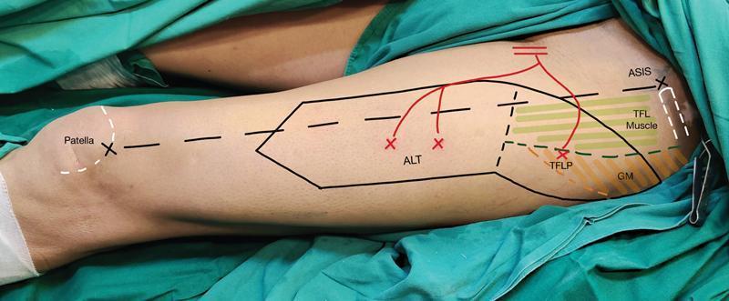
- Draw a line from the anterior superior iliac spine (ASIS) to the superolateral patella. This approximates the septum between rectus femoris and vastus lateralis.
- Identify perforators with handheld Doppler, usually around the midpoint of this line, often slightly lateral and distal.
- Design the skin paddle centered on a reliable perforator. A common design places roughly one-third of the flap medial and two-thirds lateral to the line. The vascular basis is usually the descending branch of the lateral circumflex femoral artery (LCFA). Campbell's Operative Orthopaedics, p. 3922.
-
Make the medial incision first
- Incise the medial edge of the planned paddle through skin, subcutaneous tissue, and deep fascia.
- Elevate the flap in a subfascial plane toward the intermuscular septum.
- Identify the yellow fat stripe between rectus femoris and vastus lateralis.
-
Identify suitable perforator(s)
- Look first for a septocutaneous perforator in the septum.
- If absent, continue laterally to identify a musculocutaneous perforator traversing the anterior vastus lateralis.
- Select one or more dominant pulsatile perforators with satisfactory caliber. Most ALT perforators are musculocutaneous. AO harvest technique
-
Perform retrograde perforator dissection
- Trace the selected perforator from the fascia back to the descending LCFA branch.
- For a musculocutaneous perforator, carefully split vastus lateralis fibers in the direction of the fibers, controlling small muscular branches as needed.
- Preserve motor nerve branches to vastus lateralis whenever feasible.
- Avoid traction, thermal injury, twisting, or skeletonizing the perforator excessively.
-
Dissect and lengthen the pedicle
- Retract rectus femoris medially to expose the descending LCFA branch and its venae comitantes.
- Dissect the pedicle proximally to obtain the required length and vessel caliber.
- Preserve the rectus femoris branch and its venous drainage. Do not extend dissection proximally in a way that compromises rectus femoris perfusion. Campbell's Operative Orthopaedics, p. 3922.
-
Complete flap elevation
- Once perforator perfusion is confirmed, make the lateral, distal, and posterior incisions.
- Elevate the flap completely while maintaining the selected perforator(s) and pedicle.
- If a sensate ALT is planned, include the lateral femoral cutaneous nerve as appropriate.
- For a chimeric or myocutaneous flap, include the needed cuff/segment of vastus lateralis while protecting the perforator supply.
-
Release tourniquet and assess flap
- Confirm dermal bleeding, flap color, capillary refill, perforator pulsatility, and venous outflow.
- Ensure the pedicle is not under tension, kinked, or compressed.
-
Divide pedicle only when recipient vessels are ready
- Systemic anticoagulation, pedicle division, ischemia-time recording, transfer, and microvascular anastomosis follow local protocol.
- The pedicle typically provides one artery and one or two venae comitantes; reported usable pedicle length is commonly about 8-16 cm. NCBI technique overview
-
Donor-site closure
- Achieve meticulous hemostasis.
- Insert a suction drain.
- Close primarily if tension-free; otherwise use skin grafting.
- Monitor for hematoma, seroma, wound dehiscence, sensory change, and quadriceps dysfunction.
Practical intraoperative points
- Medial-first elevation lets the surgeon identify perforators before committing to the remainder of the flap.
- In the absence of a septocutaneous vessel, do not abandon the flap prematurely: a musculocutaneous perforator through vastus lateralis is common.
- Final skin-paddle dimensions and flap choice should be adjusted after direct visualization of perforator anatomy.
- Do not divide the pedicle until recipient vessels are prepared and the flap is confirmed viable.
Recent reviews address donor-site closure and thin ALT flap outcomes, but they do not materially alter this core harvest sequence (PMIDs 40318362, 39793965).
Show corresponding pictures for the above
Reading File
Searching Images
Anterolateral thigh ALT free flap harvest steps markings medial incision perforator dissection
query_images_db
Searching the Web
anterolateral thigh flap harvest figures markings perforator dissection images
Searching PubMed
"anterolateral thigh flap" AND harvest
Loading Image
Loading Image
Loading Image
Loading Image
1. Surface marking and flap design
Mark the line from ASIS to superolateral patella, map Doppler perforators, and center the skin paddle over the selected perforator.
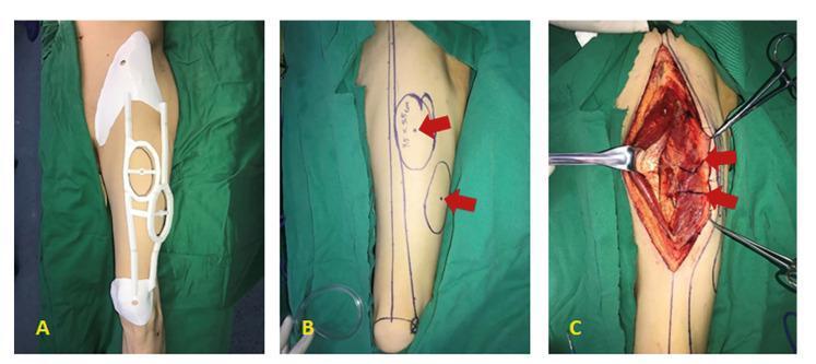
2. Perforator identification and intramuscular dissection
After a medial incision and subfascial elevation, identify the perforator and trace it retrogradely. The figure below shows marking, incision, and exposure of perforators from the descending LCFA branch.
3. Anatomy to recognize during harvest
The key structures are the rectus femoris, vastus lateralis, intermuscular septum, descending branch of the lateral circumflex femoral artery, venae comitantes, and either a septocutaneous or musculocutaneous perforator. The AO surgical reference illustrates the medial-first dissection, retrograde perforator skeletonization, pedicle mobilization, and closure sequence.
A stepwise operative demonstration is available here. It includes markings, medial subfascial incision, musculocutaneous perforator dissection through vastus lateralis, pedicle harvest, and donor closure.
These images are for surgical education. ALT harvest should be performed only by appropriately trained reconstructive/microsurgical teams.
This is a shared conversation. Sign in to Orris to start your own chat.