
Surrogate markers in CNS
"surrogate markers" AND "central nervous system"
PMID: 40937216
PMID: 42091269
surrogate markers CNS tumors neuropathology WHO 2021 classification IHC IDH H3K27M ATRX
https://www.frontiersin.org/journals/molecular-neuroscience/…
IDH1 R132H immunohistochemistry glioma surrogate marker
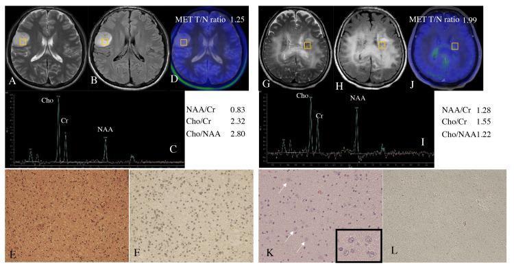
This composite figure presents a comparison between two representative cases of adult diffuse glioma, highlighting diagnostic imaging and histopathology used to determine IDH mutation status. Case 1 (A–F) demonstrates an IDH-mutant astrocytoma. Radiological findings include a well-circumscribed hyperintense lesion in the right frontal lobe on axial T2WI (A) and FLAIR (B). Magnetic resonance spectroscopy (C) shows an elevated Cho/NAA ratio (2.80), and MET-PET (D) indicates a T/N ratio of 1.25. Histology shows hypercellularity on H&E (E) and strong IDH1-R132H immunohistochemical positivity (F). Case 2 (G–L) depicts an IDH-wildtype astrocytoma with a gliomatosis cerebri pattern. Imaging (G, H) reveals diffuse, poorly demarcated bilateral involvement of the periventricular white matter. Spectroscopy (I) shows a lower Cho/NAA ratio (1.22), while MET-PET (J) shows a higher T/N ratio (1.99). H&E staining (K) reveals scattered ganglion-like cells (arrows/inset), and immunohistochemistry (L) confirms IDH-wildtype status via immune-negativity. This comparison illustrates the correlation between radiological margins, metabolic ratios, and molecular markers in glioma classification.
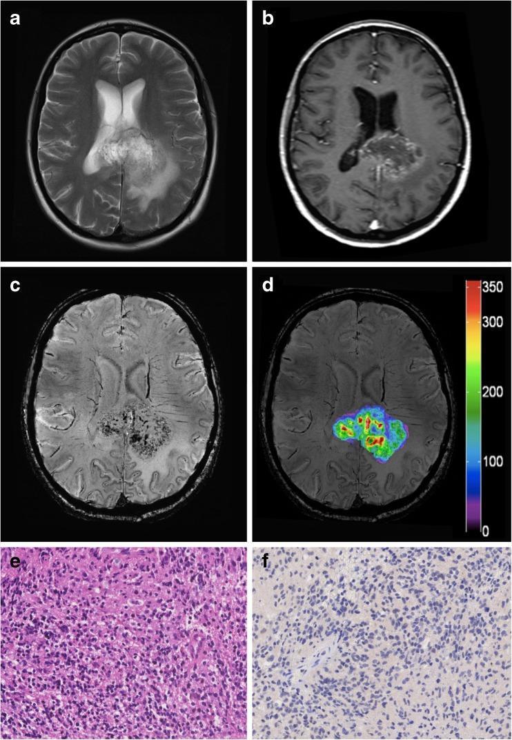
This composite figure illustrates a diagnostic workup of a high-grade glioma (HGG), specifically a glioblastoma (WHO grade IV) with negative IDH1-R132H mutational status. (A) Axial T2-weighted MRI displays a heterogeneous hyperintense lesion located in the posterior corpus callosum with surrounding vasogenic edema. (B) Contrast-enhanced T1-weighted MRI reveals significant ring-like enhancement, indicating blood-brain barrier disruption. (C) High-resolution 7 Tesla Susceptibility Weighted Imaging (SWI) shows numerous intratumoral hypointense foci, representing pathological microvasculature and microhemorrhages. (D) A Local Image Variance (LIV) map overlaid on the SWI correlates high variability (indicated by warm colors) with dense vascularity, providing a quantitative metric for tumor grading. (E) Histopathological H&E stain (200x) demonstrates hypercellularity, pleomorphism, and characteristics of a malignant glioma. (F) Immunohistochemistry (200x) confirms the negative IDH1-R132H mutational status. This multimodal comparison teaches the integration of advanced 7T imaging techniques with conventional radiology and pathology for precise brain tumor characterization.
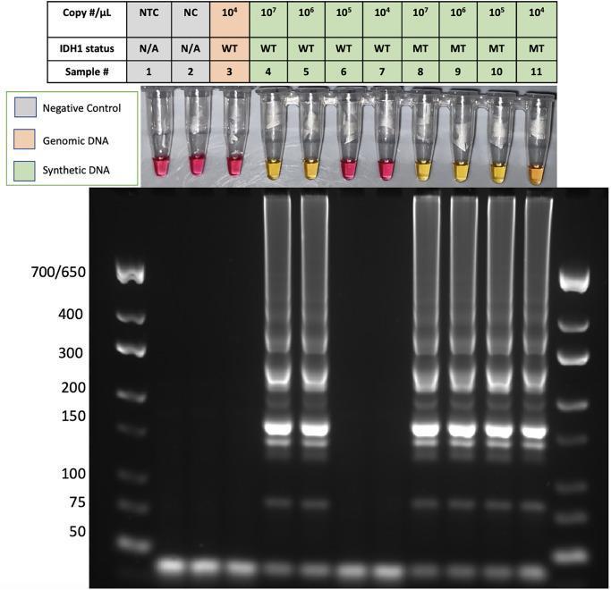
This composite diagnostic image demonstrates the specificity of a Peptide Nucleic Acid Loop-mediated Isothermal Amplification (PNA-LAMP) assay for detecting the IDH1-R132H mutation, a key biomarker in glioma. The top panel shows a colorimetric assay in eleven microtubes. A positive result for DNA amplification is indicated by a yellow color change (samples 4, 5, 8, 9, 10), while negative results remain pink (samples 1, 2, 3, 6, 7, 11). The samples include non-template (NTC) and negative controls (NC), human genomic DNA (hgDNA), and varying concentrations of synthetic wild-type (WT) and mutant (MT) DNA. The bottom panel displays the corresponding gel electrophoresis results. Positive reactions (yellow tubes) show a characteristic laddering pattern of concatemers across a wide range of molecular weights, confirming successful isothermal amplification. Negative reactions (pink tubes) show no such banding. The data illustrates how PNA successfully suppresses wild-type IDH1 amplification at concentrations ≤ 10^5, allowing for specific visual and molecular detection of the IDH1-R132H mutation.
Surrogate Markers in CNS Tumors
Major IHC Surrogate Markers - Organized by Marker
1. IDH1 R132H (Anti-IDH1 R132H Antibody)
- What it surrogates: The IDH1 R132H point mutation (the most common IDH mutation, accounting for >90% of IDH-mutant gliomas)
- Positive staining = IDH-mutant (cytoplasmic/nuclear staining of tumor cells)
- Diagnostic utility:
- Distinguishes IDH-mutant astrocytoma/oligodendroglioma from IDH-wildtype glioblastoma
- Separates reactive gliosis (IDH-negative) from low-grade glioma
- Screens for IDH mutation quickly; if negative in young patients (<55 years), sequencing for non-R132H IDH1 and IDH2 mutations is still required
- Limitation: Detects only the R132H variant; ~5-10% of IDH mutations (non-R132H IDH1 + all IDH2 mutations) are missed by IHC alone
2. ATRX (Loss of Nuclear ATRX Expression)
- What it surrogates: ATRX gene mutation/deletion
- Nuclear loss = ATRX-mutant
- Diagnostic utility:
- Loss of ATRX is characteristic of IDH-mutant astrocytoma (along with TP53 overexpression)
- ATRX is retained in oligodendroglioma (which has 1p/19q codeletion instead)
- Loss of ATRX rules OUT oligodendroglioma when 1p/19q status is not yet available
- Also seen in ~15% of diffuse midline gliomas (DMGs)
- Key point: ATRX loss supports astrocytic lineage; its retention in an IDH-mutant tumor raises the suspicion for 1p/19q codeletion (oligodendroglioma)
3. H3 K27M (Anti-H3K27M Antibody)
- What it surrogates: H3F3A or HIST1H3B gene mutation causing the K27M substitution in histone H3
- Positive nuclear staining = H3 K27M mutation
- Diagnostic utility:
- Defines Diffuse Midline Glioma, H3 K27-altered - a WHO CNS Grade 4 tumor
- Found in pediatric midline tumors (thalamus, brainstem/DIPG, spinal cord)
- Strong prognostic marker - associated with uniformly poor outcome
- Note: H3 K27M IHC does NOT detect EZHIP overexpression (another mechanism of H3K27 pathway alteration in PF ependymomas); for those, separate EZHIP IHC is needed
4. H3 K27me3 (Loss of H3 K27 Trimethylation)
- What it surrogates: Global loss of H3K27 trimethylation (whether due to H3K27M mutation, EZHIP overexpression, or EZH2 inhibitor effect)
- Loss of nuclear staining = altered H3K27 trimethylation (cutoff: >80% cells showing loss)
- Diagnostic utility:
- In DMGs: H3K27me3 loss is a highly specific marker for H3 K27M-mutant or EZHIP-overexpressing tumors
- In Posterior Fossa Ependymomas: Serves as a reliable IHC surrogate for DNA methylation profiling:
- PF-PFA (pediatric, aggressive): H3K27me3 lost
- PF-PFB (older, better prognosis): H3K27me3 retained
- In Meningiomas: H3K27me3 loss correlates with worse behavior (IAP classification notes)
- Practical point: Loss of H3K27me3 without H3K27M or EZHIP positivity mandates full molecular analysis
5. p53 (TP53 Protein Overexpression)
- What it surrogates: TP53 missense mutation (causes p53 protein accumulation)
- Strong diffuse nuclear staining (>10% cells) = TP53 mutation
- Diagnostic utility:
- Found alongside ATRX loss in IDH-mutant astrocytoma
- Helps differentiate astrocytoma (p53+, ATRX loss) from oligodendroglioma (p53-, ATRX retained)
- Caveat: p53 complete absence (null pattern) can also indicate truncating mutations - requires clinical context
6. BRAF V600E (VE1 Antibody)
- What it surrogates: BRAF V600E activating mutation
- Cytoplasmic positivity = BRAF V600E mutation
- Diagnostic utility:
- Positive in pleomorphic xanthoastrocytoma (PXA), ganglioglioma, papillary craniopharyngioma, and some pilocytic astrocytomas
- BRAF V600E-positive tumors are potential targets for vemurafenib/dabrafenib + trametinib therapy
- Helps distinguish PXA (BRAF+) from diffuse glioma (usually BRAF-)
- Specificity note: Some false positives exist; confirmatory molecular testing recommended before treatment
7. H3 G34R/V (Anti-H3 G34 Antibody)
- What it surrogates: H3F3A G34R or G34V mutations
- Diagnostic utility:
- Defines Diffuse Hemispheric Glioma, H3 G34-mutant - a pediatric-type high-grade glioma
- Located in cerebral hemispheres; distinct from H3K27M midline tumors
- Poor prognosis but different from midline DMG
8. Loss of INI1 (SMARCB1) / BRG1 (SMARCA4)
- What it surrogates: SMARCB1 or SMARCA4 gene deletion/mutation
- Loss of nuclear staining = mutation/deletion
- Diagnostic utility:
- Loss of INI1 = Atypical Teratoid/Rhabdoid Tumor (AT/RT) - critical diagnosis in young children
- Serves as a reliable IHC surrogate for the underlying SWI/SNF complex gene alterations
- Also lost in some epithelioid glioblastomas (SMARCB1) and chordomas
9. Vimentin
- What it surrogates: Mesenchymal/epithelial-to-mesenchymal transition marker
- Diagnostic utility in CNS:
- Highly expressed in DMGs and high-grade gliomas - reflects aggressive/invasive phenotype
- Usually absent in oligodendrogliomas - helps differentiate DMG from oligodendroglioma
- Correlates with poor prognosis
10. Synaptophysin
- What it surrogates: Neuronal differentiation
- Diagnostic utility:
- Frequently expressed in oligodendrogliomas but largely absent in DMGs
- Useful discriminatory marker between DMG and oligodendroglioma when molecular testing is limited
11. MYCN (IHC as surrogate for MYCN amplification)
- Diagnostic utility:
- Strong diffuse nuclear expression = likely MYCN amplification
- Used as a screening surrogate in MYCN-amplified spinal ependymomas (WHO CNS5 category)
- IHC finding should be confirmed with FISH for definitive amplification status
12. BCL-6 (in CNS Lymphoma context)
- Diagnostic utility:
- Expression of BCL-6 is a germinal-center B-cell surrogate in CNS lymphoma
- Occurs at lower frequency in primary CNS lymphoma than in nodal lymphoma
- Helps distinguish primary CNS lymphoma subtype - Quick Compendium of Clinical Pathology, 5th ed.
Summary Table
| Surrogate Marker | Gene/Alteration Detected | Tumor(s) Where Used | Key Significance |
|---|---|---|---|
| IDH1 R132H IHC | IDH1 R132H mutation | Astrocytoma, Oligodendroglioma | Distinguishes IDH-mutant from IDH-wildtype glioma |
| ATRX loss | ATRX mutation | Astrocytoma (IDH-mutant) | Differentiates from oligodendroglioma |
| H3 K27M | H3F3A/HIST1H3B K27M mutation | Diffuse Midline Glioma | Defines WHO Grade 4; poor prognosis |
| H3 K27me3 loss | H3K27 pathway alteration | DMG, PF Ependymoma, Meningioma | Replaces DNA methylation profiling in ependymoma PFA vs PFB |
| p53 overexpression | TP53 missense mutation | Astrocytoma | Supports astrocytic vs oligodendroglial lineage |
| BRAF V600E (VE1) | BRAF V600E mutation | PXA, Ganglioglioma, Craniopharyngioma | Therapeutic target; lineage differentiation |
| H3 G34R/V | H3F3A G34 mutation | Diffuse Hemispheric Glioma | Pediatric HGG; distinct from midline DMG |
| INI1 / BRG1 loss | SMARCB1/SMARCA4 deletion | AT/RT | Critical pediatric diagnosis |
| Vimentin | Mesenchymal transition | DMG vs Oligodendroglioma | High in DMG; absent in oligodendroglioma |
| Synaptophysin | Neuronal differentiation | Oligodendroglioma vs DMG | Present in OL; absent in DMG |
| MYCN IHC | MYCN amplification | Spinal Ependymoma, MYCN-amplified | Screening tool; needs FISH confirmation |
Important Limitations of Surrogate Markers
- IDH1 R132H IHC misses ~10% of IDH mutations - non-canonical IDH1 and all IDH2 mutations require sequencing
- H3 K27me3 loss without H3K27M/EZHIP - full molecular analysis mandatory
- p53 IHC - null pattern (complete loss) can indicate truncating mutation, not just missense
- BRAF V600E VE1 - some cross-reactivity with non-V600E tumors
- Methylome profiling remains the gold standard; IHC surrogates are approximations and are best used in a guided panel alongside clinical-radiological data
- Quick Compendium of Clinical Pathology, 5th ed. (sections 74.9 and 7.18)
- Kranendonk et al. (2026) - Surg Pathol Clin [PMID: 42091269]
- Qasim et al. (2025) - Vimentin, Synaptophysin, H3K27me as surrogate markers [PMID: 40937216]
- Louis DN et al. (2021) - WHO CNS5 Classification summary, PMC8328013
- Frontiers in Molecular Neuroscience (2024) - Pediatric CNS tumors and 2021 WHO classification