
Prostatic ureicles and mullerian emminance
prostatic utricle verumontanum anatomy diagram
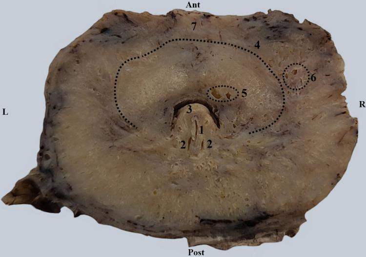
This clinical photograph displays a transverse anatomical section of a human prostate gland at the level of the seminal colliculus (verumontanum). Centrally, the prostatic urethra presents a characteristic semilunar shape with a posterior concavity. Key anatomical structures are labeled: the prostatic utricle (1) is positioned centrally on the seminal colliculus (3), flanked by the bilateral openings of the ejaculatory ducts (2). An arched dotted line (4) delineates the internal urethral sphincter (vesical sphincter), which serves as a structural boundary within the prostatic stroma. Small periurethral glands (5) are located medial to this muscular boundary, adjacent to the urethral lumen, while larger periurethral glands (6) are positioned lateral to the sphincter fibers. The anterior aspect shows the prostate isthmus (7). This section demonstrates the topographical relationship between the urinary and reproductive tracts within the male pelvic anatomy, highlighting the glandular distribution and the smooth muscle transition between the bladder neck and the prostatic urethra.
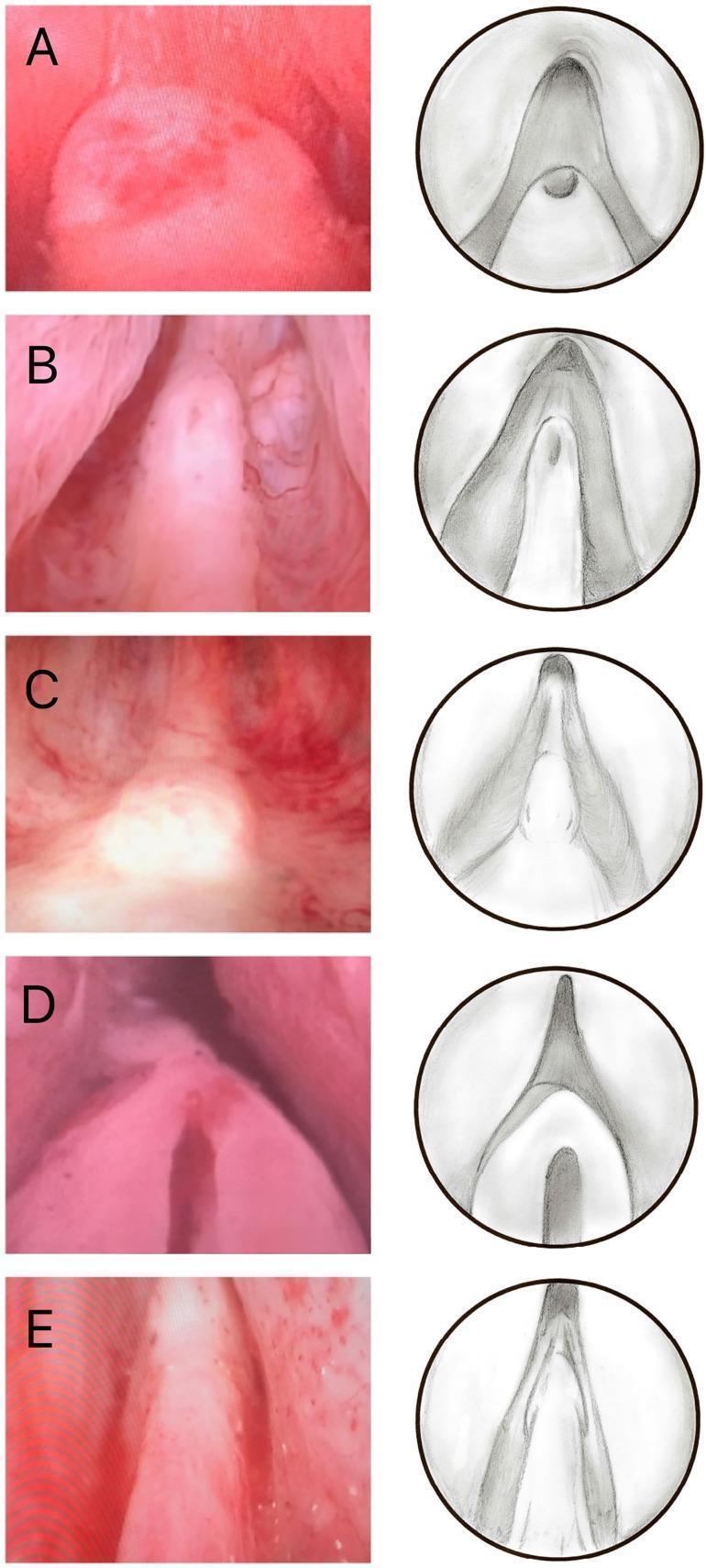
A medical comparison chart illustrating five morphological types of the verumontanum (seminal colliculus) as seen during cystoscopy. The image is organized into two columns: the left shows actual endoscopic clinical photographs, and the right presents corresponding illustrative diagrams highlighting structural anatomy. The five classified types are: (A) Volcano: a short, rounded structure with a superiorly located utricle; (B) Lighthouse: an elongated, more prominent colliculus with an anterior utricle; (C) Whale Tail: a flattened, broad organ with a prominent, split-appearing urethral crest; (D) Castle Door: a wide, short structure featuring a significantly enlarged prostatic utricle; (E) Hood: a tapered, elongated form that is continuous with the urethral crest. This visual classification is used in urology to standardize anatomical variations of the prostatic urethra, specifically for surgical planning in procedures like transurethral resection of the prostate (TURP). The images highlight key landmarks including the prostatic utricle and the urethral crest.
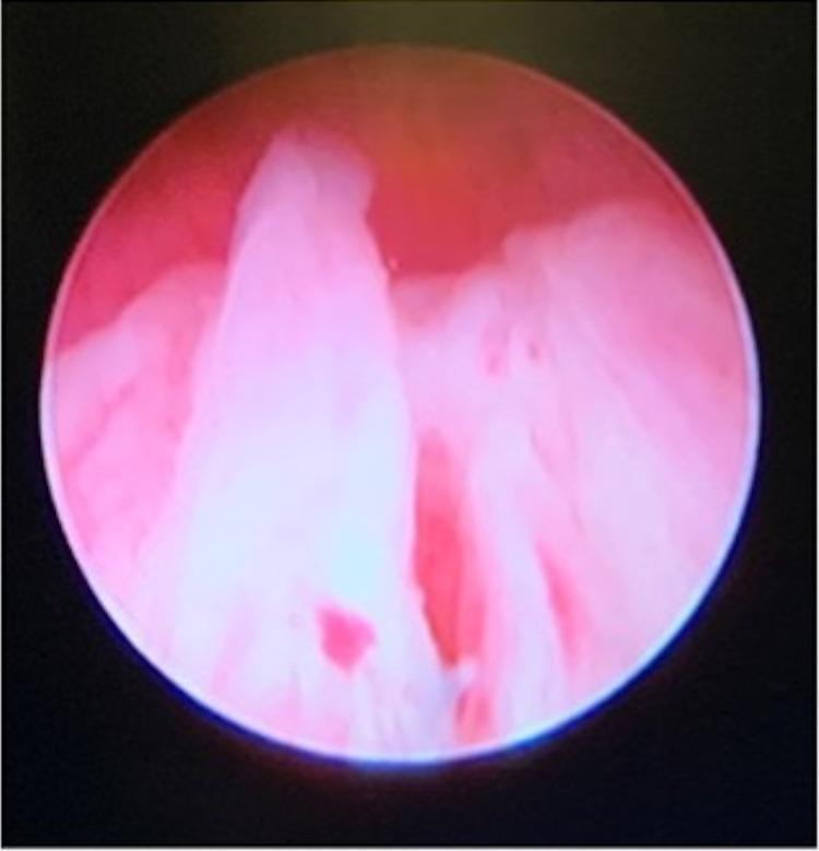
This cystoscopic image provides a procedural view of an undervirilized prostatic urethra in a patient with a Disorder of Sex Development (DSD). The primary anatomical focus is the urethral lumen, which appears underdeveloped and narrow. A prominent feature is the presence of a central tissue ridge or verumontanum-like structure. Adjacent to this, the opening to a large prostatic utricle or Mullerian remnant is visible as a dark, recessed cavity at the urethral apex. The surrounding mucosal tissue is characterized by a healthy pink-to-red color with smooth surfaces. This clinical photograph demonstrates the internal urogenital anatomy typical of complex ambiguous genitalia, specifically highlighting the confluence of a urogenital sinus and the persistence of Mullerian structures in a 46,XX individual with ovotesticular DSD.
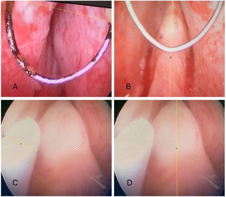
A set of four clinical endoscopic images (A-D) demonstrating the standardized measurement of the verumontanum (seminal colliculus) within the prostatic urethra using continuous-flow resectoscopy. Images A and B depict the use of a bipolar resectoscopic loop as a calibrated reference. In A, a yellow line measures the transverse distance between the loop's ends, while in B, a vertical yellow line measures the longitudinal diameter of the verumontanum. Images C and D show the control group methodology: C utilizes the known diameter of a ureteral catheter as a measurement scale, and D illustrates the resulting longitudinal diameter verification using ImageJ software. The images highlight anatomical variations in the verumontanum, which is a critical surgical landmark during transurethral resection of the prostate (TURP) to prevent damage to the external urinary sphincter. Key visible structures include the urethral crest and the prostatic utricle located on the apex of the colliculus. This educational content illustrates urological morphometric techniques used to assess prostatic changes related to age, BMI, or 5-alpha-reductase inhibitor therapy.
Prostatic Utricle and Mullerian Eminence
Terminology Clarification
- Prostatic utricle - the correct spelling (sometimes loosely written "uricle" in exam notes)
- Mullerian eminence - usually called the sinus tubercle or Mullerian tubercle in embryology; in adult anatomy it becomes the verumontanum (seminal colliculus)
Embryological Origin
| Mullerian Remnant in Males | Adult Structure |
|---|---|
| Cranial tip of paramesonephric duct | Appendix testis |
| Caudal fused end of paramesonephric duct | Prostatic utricle |
"The seminal colliculus, a small elevation in the posterior wall of the prostatic urethra, is the adult derivative of the sinus tubercle."
- The Developing Human: Clinically Oriented Embryology, p. 737
Anatomy of the Prostatic Utricle
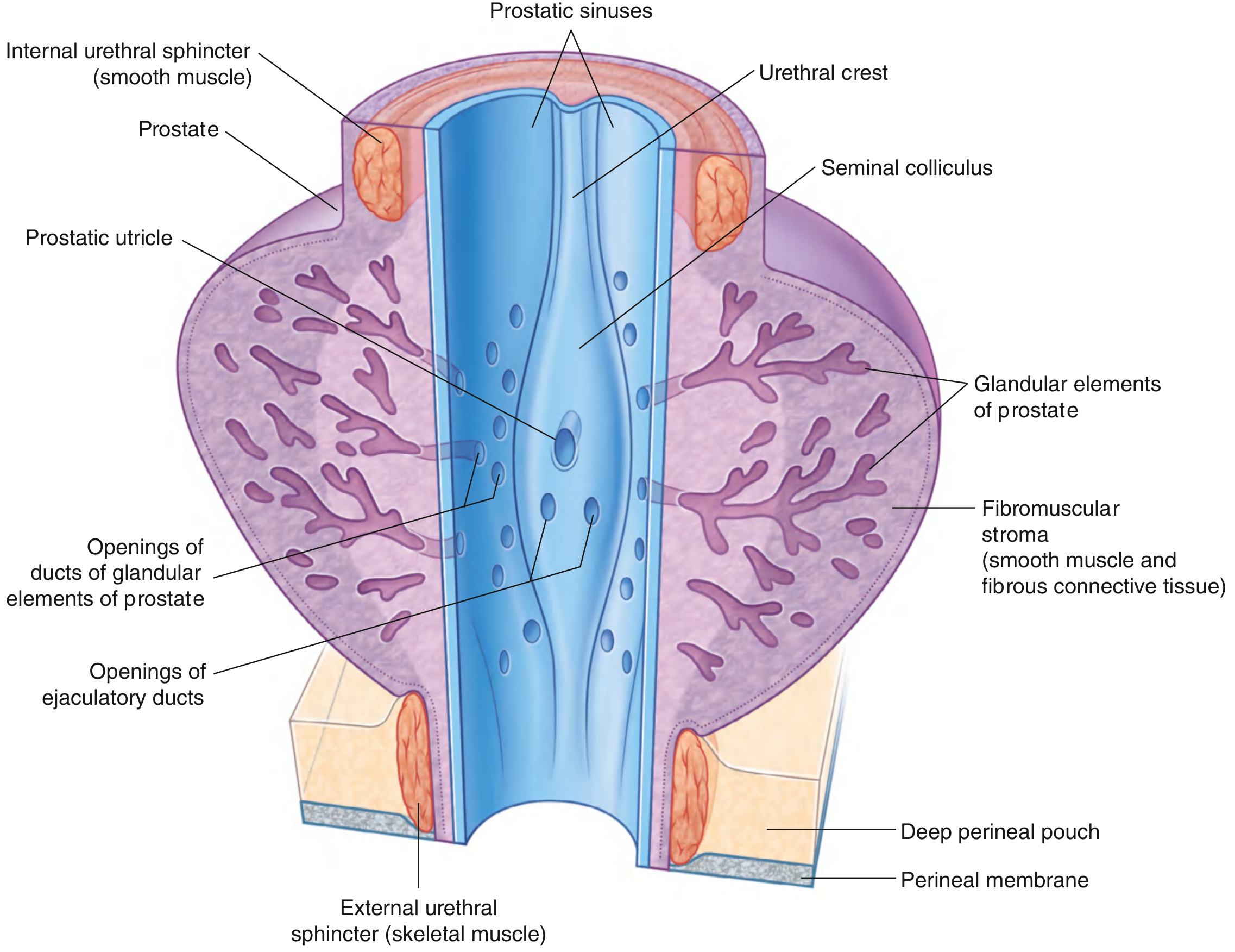
- A small, blind-ended sac-like pouch located in the posterior midline of the prostatic urethra
- Opens at the apex (center) of the verumontanum (seminal colliculus) as a slit-like orifice
- The orifice is cystoscopically visible and measures approximately 6 mm
- On either side of the utricular orifice lie the two ejaculatory duct openings
- Length is typically 6-8 mm but can be much larger in pathological states
"A small blind-ended pouch - the prostatic utricle (thought to be the homologue of the uterus in women) - opens onto the center of the seminal colliculus. On each side of the prostatic utricle is the opening of the ejaculatory duct."
- Gray's Anatomy for Students, p. 543
Homologue Relationship
| Female Structure | Male Homologue |
|---|---|
| Uterus + upper vagina | Prostatic utricle |
| Fallopian tubes | (Appendix testis - only cranial tip) |
| Ovary | Testis |
Developmental Table (from Campbell-Walsh Urology)
| Male | Embryonic Structure | Female |
|---|---|---|
| Prostatic utricle | Paramesonephric (Mullerian) duct (caudal fused portion) | Uterus + upper vagina |
| Verumontanum | Sinus tubercle (Mullerian tubercle) | Lower vagina |
| Appendix testis | Cranial tip of paramesonephric duct | Fimbriated end of fallopian tube |
Histology
Clinical Relevance
| Condition | Notes |
|---|---|
| Enlarged prostatic utricle | Associated with hypospadias, undescended testis, and disorders of sex development (DSD); the degree of enlargement correlates with the severity of undermasculinization |
| Prostatic utricle cyst | Can cause urinary obstruction, UTI, epididymitis, hematuria; visible on TRUS or MRI |
| DSD/Intersex | A large Mullerian remnant (persistent Mullerian duct syndrome) results from failure of MIS action or MIS receptor defect |
| TURP landmark | The verumontanum (where the utricle sits) is the critical inferior landmark during TURP - resection must stop here to protect the external urethral sphincter |
Summary Mnemonic
- Utricle = opens at the apex of Verumontanum = remnant of fused Mullerian (paramesonephric) ducts
- Verumontanum = adult form of the sinus tubercle (Mullerian eminence/tubercle)
- Ejaculatory ducts open on either side of the utricle (not at its apex)